Scanning Electron Microscope Collection (page 9)

"Unveiling the Hidden World

Background imageScanning Electron Microscope Collection: Biology, Bacteria, Cocci seen through scanning electron microscope

Biology, Bacteria, Cocci seen through scanning electron microscope
Biology - Bacteria - Cocci seen through the scanning electron microscope

Background imageScanning Electron Microscope Collection: Blood clot, SEM C016 / 9751

Blood clot, SEM C016 / 9751
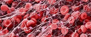
Blood clot, coloured scanning electron micrograph (SEM). Red blood cells (erythrocytes) are trapped within a fibrin protein mesh (beige)

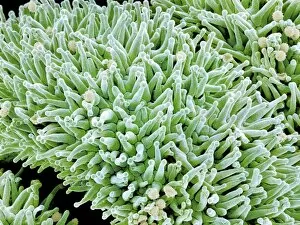
Background imageScanning Electron Microscope Collection: Carnation flower pistil, SEM C016 / 9762

Carnation flower pistil, SEM C016 / 9762
Carnation (Dianthus sp.) pistil, coloured scanning electron micrograph (SEM). Pistils hold a plants female reproductive structures

Background imageScanning Electron Microscope Collection: Blood clot, SEM C016 / 9746

Blood clot, SEM C016 / 9746
Blood clot, coloured scanning electron micrograph (SEM). Red blood cells (erythrocytes) are trapped within a fibrin protein mesh (beige)

Background imageScanning Electron Microscope Collection: Carnation flower pistil, SEM C016 / 9759

Carnation flower pistil, SEM C016 / 9759
Carnation (Dianthus sp.) pistil, coloured scanning electron micrograph (SEM). Pistils hold a plants female reproductive structures

Background imageScanning Electron Microscope Collection: Rust fungus on a rose leaf, SEM C017 / 7132

Rust fungus on a rose leaf, SEM C017 / 7132
Rust fungus on a rose leaf. Coloured scanning electron micrograph (SEM) of rust fungus (Phragmidium sp.) spores emerging from a rose (Rosa sp.) leaf (brown)

Background imageScanning Electron Microscope Collection: Blood clot, SEM C016 / 9749

Blood clot, SEM C016 / 9749
Blood clot, coloured scanning electron micrograph (SEM). Red blood cells (erythrocytes) are trapped within a fibrin protein mesh (beige)

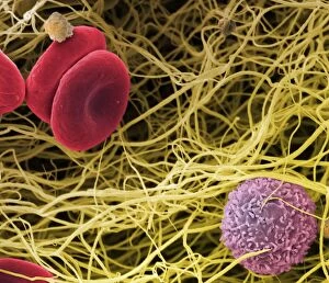
Background imageScanning Electron Microscope Collection: Blood clot, SEM C017 / 7141

Blood clot, SEM C017 / 7141
Blood clot, coloured scanning electron micrograph (SEM). Red blood cells (erythrocytes) and a white blood cell (purple) trapped within a fibrin protein mesh (cream)

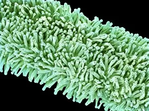
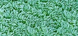
Background imageScanning Electron Microscope Collection: Trachea lining, SEM C016 / 9756

Trachea lining, SEM C016 / 9756
Trachea lining. Coloured scanning electron micrograph (SEM) of the lining of the trachea (wind pipe). The trachea links the larynx to the lungs

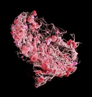
Background imageScanning Electron Microscope Collection: Blood clot, SEM C016 / 9750

Blood clot, SEM C016 / 9750
Blood clot, coloured scanning electron micrograph (SEM). Red blood cells (erythrocytes) are trapped within a fibrin protein mesh (beige)

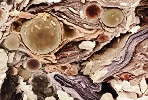
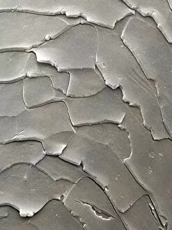
Background imageScanning Electron Microscope Collection: Freeze-fractured baby cream, SEM C017 / 7133

Freeze-fractured baby cream, SEM C017 / 7133
Freeze-fractured baby cream, coloured scanning electron micrograph (SEM). Mangification: x1680 when printed at 10 centimetres wide

Background imageScanning Electron Microscope Collection: Carnation flower pistil, SEM C016 / 9760

Carnation flower pistil, SEM C016 / 9760
Carnation (Dianthus sp.) pistil, coloured scanning electron micrograph (SEM). Pistils hold a plants female reproductive structures

Background imageScanning Electron Microscope Collection: Blood clot, SEM C016 / 9753

Blood clot, SEM C016 / 9753
Blood clot. Coloured scanning electron micrograph (SEM) of a blood clot from the inner wall of the left ventricle of a heart

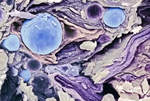
Background imageScanning Electron Microscope Collection: Freeze-fractured baby cream, SEM C017 / 7134

Freeze-fractured baby cream, SEM C017 / 7134
Freeze-fractured baby cream, coloured scanning electron micrograph (SEM). Mangification: x1680 when printed at 10 centimetres wide

Background imageScanning Electron Microscope Collection: Freeze-fractured baby cream, SEM C017 / 7135

Freeze-fractured baby cream, SEM C017 / 7135
Freeze-fractured baby cream, coloured scanning electron micrograph (SEM). Mangification: x1680 when printed at 10 centimetres wide

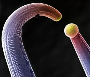
Background imageScanning Electron Microscope Collection: Blown tungsten filament, SEM C017 / 7142

Blown tungsten filament, SEM C017 / 7142
Blown tungsten filament. Coloured scanning electron micrograph (SEM) of a blown tungsten filament from the electron gun of a scanning electron microscope

Background imageScanning Electron Microscope Collection: Blood clot, SEM C016 / 9752

Blood clot, SEM C016 / 9752
Blood clot, coloured scanning electron micrograph (SEM). Red blood cells (erythrocytes) are trapped within a fibrin protein mesh (beige)

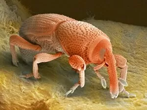
Background imageScanning Electron Microscope Collection: Grain weevil

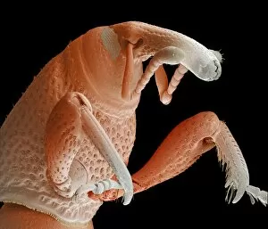
Grain weevil. Coloured scanning electron micrograph of the thorax and head of a grain weevil (Sitophilus granarius). This beetle infests and damages stored grain

Background imageScanning Electron Microscope Collection: Domestic cat hair, coloured SEM Z934 / 0486

Domestic cat hair, coloured SEM Z934 / 0486
Cat hair. Coloured scanning electron micrograph (SEM) of the hair from a domestic cat (Felis domesticus). Magnification: x370 when printed 10cm high

Background imageScanning Electron Microscope Collection: Fruit fly compound eye, SEM

Fruit fly compound eye, SEM
Fruit fly compound eye. Coloured scanning electron micrograph (SEM) of the compound eye of a male fruit fly (Drosophila melanogaster)

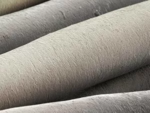
Background imageScanning Electron Microscope Collection: Red deer hair, coloured SEM Z952 / 0094

Red deer hair, coloured SEM Z952 / 0094
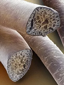
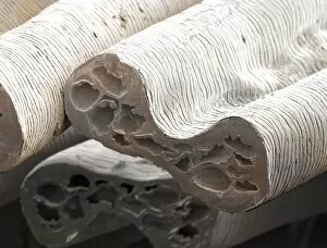
Red deer hair. Coloured scanning electron micrograph (SEM) of a cross section through the hairs of a red deer (Cervus elaphus). Magnification: x110 when printed at 10cm high

Background imageScanning Electron Microscope Collection: Brassica weevil, SEM

Brassica weevil, SEM
Brassica weevil (Ceutorhynchus sp.), coloured scanning electron micrograph (SEM). This beetle, which grows up to 3 millimetres in length, is a major pest of cabbages (Brassica sp.)

Background imageScanning Electron Microscope Collection: Grey squirrel hair, coloured SEM

Grey squirrel hair, coloured SEM
Grey squirrel hair. Coloured scanning electron micrograph (SEM) of the hair from a Grey squirrel (Sciurus carolinensis). Magnification: x730 when printed 10cm wide

Background imageScanning Electron Microscope Collection: Wild rabbit hair, coloured SEM

Wild rabbit hair, coloured SEM
Wild rabbit hair. Coloured scanning electron micrograph (SEM) of the hair from a wild rabbit (Oryctolagus cuniculus). Magnification: x360 when printed 10cm high

Background imageScanning Electron Microscope Collection: Camel hair, coloured SEM Z949 / 0010

Camel hair, coloured SEM Z949 / 0010
Camel hair. Coloured scanning electron micrograph (SEM) of the hair from a dromedary camel. These short dense hairs allow the camel to survive the extreme temperatures of the desert by reflecting

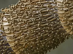
Background imageScanning Electron Microscope Collection: Fruit fly sex comb, SEM

Fruit fly sex comb, SEM
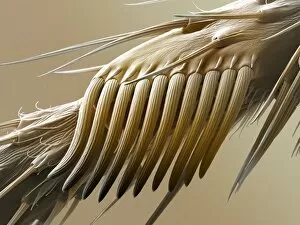
Fruit fly (Drosophila melanogaster) sex comb, coloured scanning electron micrograph (SEM). The sex comb is found only on the front legs of males

Background imageScanning Electron Microscope Collection: Rabbit hair, coloured SEM Z938 / 0037

Rabbit hair, coloured SEM Z938 / 0037
Rabbit hair. Coloured scanning electron micrograph (SEM) of the transverse section of hair from a rabbit (Oryctolagus cuniculus). Magnification: x670 when printed 10cm high

Background imageScanning Electron Microscope Collection: Horse body hair, coloured SEM

Horse body hair, coloured SEM
Horse hair. Coloured scanning electron micrograph (SEM) of hair from the body of a horse (Equus caballus). Magnification: x300 when printed 10cm high

Background imageScanning Electron Microscope Collection: Fruit fly antenna, SEM

Fruit fly antenna, SEM
Fruit fly antenna. Coloured scanning electron micrograph (SEM) of the head of a male fruit fly (Drosophila melanogaster). Seen here is an antenna (left) and part of a compound eye (upper right, red)

Background imageScanning Electron Microscope Collection: Fruit fly head, SEM

Fruit fly head, SEM
Fruit fly head. Coloured scanning electron micrograph (SEM) of the head of a male fruit fly (Drosophila melanogaster). Its two compound eyes (red) are seen on either side of the head

Background imageScanning Electron Microscope Collection: Fruit fly foot, SEM

Fruit fly foot, SEM
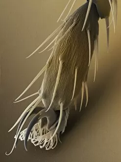
Fruit fly foot. Coloured scanning electron micrograph (SEM) of the foot of a male fruit fly (Drosophila melanogaster). The two claws are used to grip rough surfaces

Background imageScanning Electron Microscope Collection: Fruit fly spiracle, SEM

Fruit fly spiracle, SEM
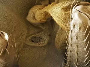
Fruit fly (Drosophila melanogaster) spiracle. Coloured scanning electron micrograph (SEM) of the spiracle (air pore) of a male fruit fly

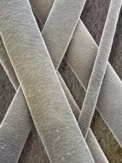
Background imageScanning Electron Microscope Collection: Daubentons bat hairs, SEM

Daubentons bat hairs, SEM
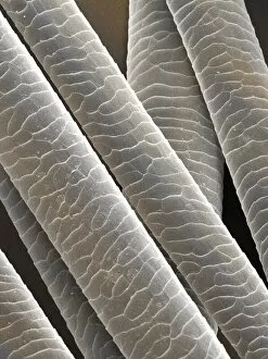
Bat hairs. Coloured scanning electron micrograph (SEM) of hairs from Daubentons bat (Myotis daubentonii). Each hair shaft has an outer cuticle made up of overlapping scales, or plates

Background imageScanning Electron Microscope Collection: Red squirrel hair, coloured SEM

Red squirrel hair, coloured SEM
Red squirrel hair. Coloured scanning electron micrograph (SEM) of the hair from a red squirrel (Sciurus vulgaris). Magnification: x370 when printed 10cm high

Background imageScanning Electron Microscope Collection: Horse mane hair, coloured SEM

Horse mane hair, coloured SEM
Horse hair. Coloured scanning electron micrograph (SEM) of hair from the mane of a horse (Equus caballus). Magnification: x2000 when printed 10cm high

Background imageScanning Electron Microscope Collection: Mouse hair, coloured SEM Z918 / 0198

Mouse hair, coloured SEM Z918 / 0198
House mouse hair. Coloured scanning electron micrograph (SEM) of hair from a house mouse (Mus musculus). Magnification: x900 when printed 10cm wide

Background imageScanning Electron Microscope Collection: Heathland ants antenna, SEM

Heathland ants antenna, SEM
Ants antenna. Coloured scanning electron micrograph (SEM), of sensory hairs on the antenna of a heathland ant (Formica fusca). Magnification: x400 when printed at 10 centimetres wide

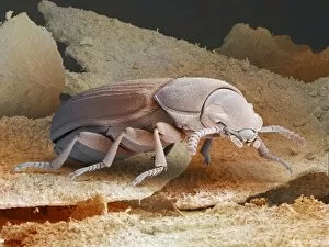
Background imageScanning Electron Microscope Collection: Red flour beetle

Red flour beetle. Coloured scanning electron micrograph (SEM) of a red flour beetle (Tribolium castaneum). One of the most significant pests of dried foods and the milling industry

Background imageScanning Electron Microscope Collection: Grey seal hair, coloured SEM

Grey seal hair, coloured SEM
Grey seal hair. Coloured scanning electron micrograph of the hair from a grey seal (Halichoerus grypus). Magnification: x161 when printed at 10cm height

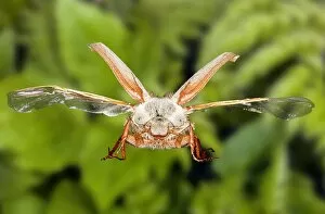
Background imageScanning Electron Microscope Collection: Cockchafer beetle, SEM composite

Cockchafer beetle, SEM composite
Common European cockchafer. Composite image of a common European cockchafer (Melolontha melolontha) in flight. This beetle, which makes a loud buzzing sound during flight

Background imageScanning Electron Microscope Collection: Fruit fly proboscis, SEM

Fruit fly proboscis, SEM
Fruit fly proboscis. Coloured scanning electron micrograph (SEM) of the coiled-up proboscis (lower centre) of a male fruit fly (Drosophila melanogaster)

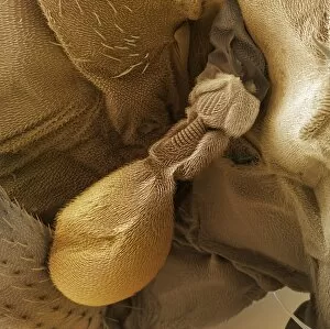
Background imageScanning Electron Microscope Collection: Fruit fly balance organ, SEM

Fruit fly balance organ, SEM
Fruit fly balance organ. Coloured scanning electron micrograph (SEM) of the balance organ (haltere) of a male fruit fly (Drosophila melanogaster). The haltere is the oval structure at lower left

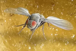
Background imageScanning Electron Microscope Collection: Fruit fly, SEM Z340 / 0662

Fruit fly, SEM Z340 / 0662
Fruit fly. Coloured scanning electron micrograph (SEM) of a fruit fly (Drosophila busckii) on a banana. Its compound eyes (red) are seen and its wings are outstretched

Background imageScanning Electron Microscope Collection: Hedgehog hair, coloured SEM

Hedgehog hair, coloured SEM
Hedgehog hair. Coloured scanning electron micrograph of the hairs from a hedgehog (Erinaceus europaeus). Magnification: x130 when printed 10cm high

Background imageScanning Electron Microscope Collection: Horse tail hair, coloured SEM

Horse tail hair, coloured SEM
Horse hair. Coloured scanning electron micrograph (SEM) of hair from the tail of a horse (Equus caballus). Magnification: x200 when printed 10cm high

Background imageScanning Electron Microscope Collection: Fruit fly, SEM Z340 / 0663

Fruit fly, SEM Z340 / 0663
Fruit fly. Coloured scanning electron micrograph (SEM) of a fruit fly (Drosophila busckii) on a banana. Its compound eyes (red) are seen and its wings are outstretched

Background imageScanning Electron Microscope Collection: Grain weevil Z330 / 0408

Grain weevil Z330 / 0408
Grain weevil. Coloured scanning electron micrograph of a grain weevil (Sitophilus granarius). This beetle infests and damages stored grain, using the mandibles at the end of its elongated snout

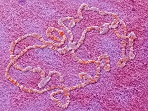
Background imageScanning Electron Microscope Collection: Giant chromosomes, SEM

Giant chromosomes, SEM
Giant chromosomes. Colured scanning electron micrograph (SEM) of giant (polytene) chromosomes from a fruit fly (Drosophila busckii)


For sale as Licensed Images

Choose your image, Select your licence and Download the media

"Unveiling the Hidden World: Exploring with a Scanning Electron Microscope" The scanning electron microscope (SEM) is an incredible tool that allows us to delve into the microscopic realm, revealing intricate details and unlocking secrets of the unseen. With its high magnification capabilities, it offers a glimpse into fascinating aspects of life and nature. In one captivating image, we witness the beauty of gender determination as X and Y chromosomes are captured under the SEM's lens. The intricate patterns and structures within these chromosomes showcase the complexity of our genetic makeup. Moving on to another astonishing discovery, a Scanning Electron Micrograph presents us with an up-close view of a Praying Mantis at 30 times magnification. Every tiny detail becomes visible, from its delicate limbs to its mesmerizing eyes – reminding us of nature's remarkable diversity. Shifting focus to plant life, lavender pollen grains come alive in another SEM image. These minuscule particles take center stage as their unique shapes and textures become apparent under intense scrutiny. Zooming in even further reveals a Tardigrade or 'Water Bear' at an astounding 1250 times magnification. This resilient creature captures our imagination as we explore its alien-like features through this powerful imaging technique. Delving deeper into biological wonders, T lymphocytes battling cancer cells are showcased in yet another breathtaking SEM capture. This visual representation highlights the ongoing fight within our bodies against diseases like cancer – emphasizing both hope and resilience. Calcareous phytoplankton takes center stage next as we uncover their intricate forms through SEM imagery. These microscopic organisms play crucial roles in marine ecosystems while leaving us awe-inspired by their stunningly detailed structures. Transitioning to neural marvels, nerve cells reveal their intricacy under the watchful eye of an SEM. Their branching extensions create complex networks that enable communication throughout our bodies – showcasing nature's ingenuity at work. Venturing into insect realms once again, a fruit fly is immortalized under the SEM's lens.