
Spongy Mesophyll Collection
The spongy mesophyll, a fascinating feature found in various plant species, plays a crucial role in their survival and growth
For sale as Licensed Images
Choose your image, Select your licence and Download the media
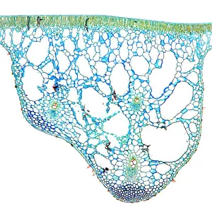
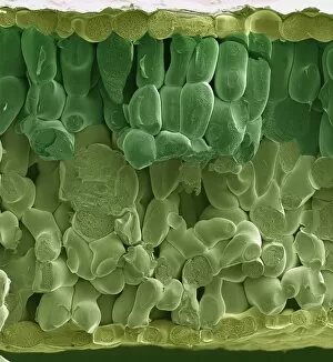
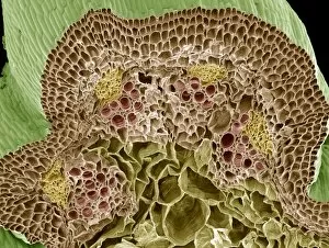
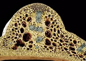
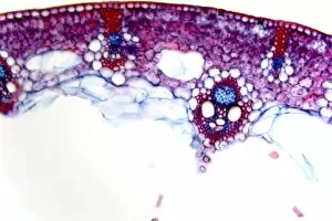
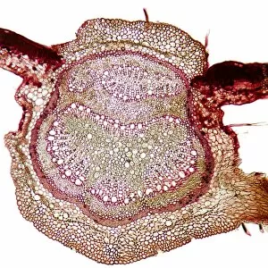
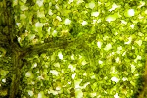
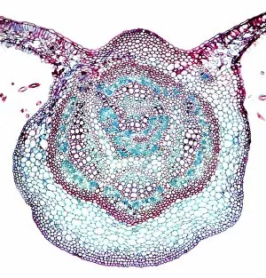
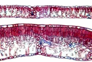
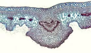
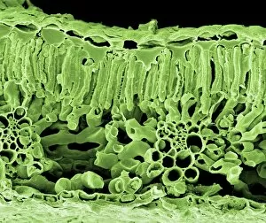
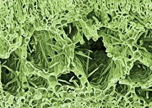
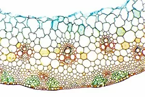
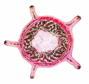
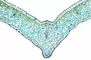
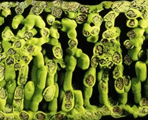
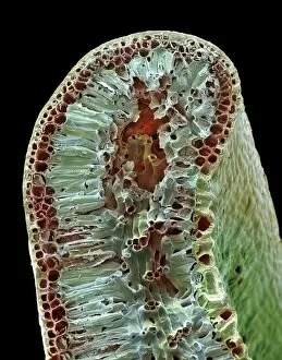
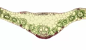
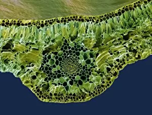
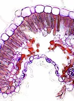
The spongy mesophyll, a fascinating feature found in various plant species, plays a crucial role in their survival and growth. This intricate network of cells can be observed through different microscopic techniques, providing us with valuable insights into its structure and function. In the water lily leaf, when viewed under a light micrograph, the spongy mesophyll appears as a delicate arrangement of loosely packed cells. These specialized cells allow for efficient gas exchange between the leaf and its surroundings. Similarly, an SEM image of a spinach leaf reveals the spongy mesophyll's sponge-like appearance. The interconnected air spaces within this tissue facilitate rapid diffusion of gases necessary for photosynthesis. Moving on to the nasturtium stem captured by SEM imaging, we witness another example of the spongy mesophyll's presence outside leaves. Here it forms part of the stem's internal structure and aids in nutrient transport throughout the plant. The water lily stem also showcases this unique tissue organization when examined under SEM. Its presence here suggests that even stems benefit from having these specialized cells for gas exchange purposes. A light micrograph depicting common rush stems further emphasizes how prevalent spongy mesophyll is among diverse plant species. The intricate cellular arrangements seen here highlight nature's ingenious design to optimize photosynthesis efficiency. Leaves continue to demonstrate their reliance on spongy mesophyll as shown by beech tree leaves imaged using light microscopy. The tightly packed yet porous cell arrangement allows for optimal carbon dioxide uptake while minimizing water loss through evaporation. Horse-chestnut leaves exhibit similar characteristics when observed at high magnification using light microscopy. The well-organized layers contribute to effective gas exchange within these broadleaf trees' foliage. Oleander leaves provide another captivating view under light microscopy where multiple layers of densely arranged spongy mesophyll cells are visible. This structural adaptation enables these plants to thrive in arid environments.