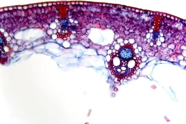
Common rush stem, light micrograph
![]()

Wall Art and Photo Gifts from Science Photo Library
Common rush stem, light micrograph
Common rush stem. Light micrograph of a section through the stem of a common rush (Juncus conglomeratus) plant, showing stellate cells. The stem has scattered vascular bundles suspended by stellate dead cells (pink). There is an outer ring of solid supporting tissue (epidermis) with a thick cuticle (red). Within this, the cortex contains spongy mesophyll with chloroplasts (red ovals), with vascular bundles in-between. The oval vascular bundles have supporting fibres (red) surrounding the outside. Facing the outside of the stem is the circular phloem (blue), and on the inner side the larger xylem vessels (red). Magnification: x70 when printed 10 centimetres wide
Science Photo Library features Science and Medical images including photos and illustrations
Media ID 6354269
© DR KEITH WHEELER/SCIENCE PHOTO LIBRARY
Bundle Cell Biology Chloroplast Chloroplasts Cortex Cuticle Cytological Cytology Dead Epidermis Histological Histology Microscopy Monocot Monocots Monocotyledon Monocotyledons Phloem Spongy Mesophyll Stain Stained Stem Structural Structures Supportive Tissue Tissue Vascular Bundles Vessel Xylem Vessels Cells Common Rush Light Micrograph Light Microscope Section Sectioned
EDITORS COMMENTS
This print showcases the intricate structure of a common rush stem, providing a glimpse into the fascinating world of plant biology. The image reveals an array of vibrant colors and distinct cellular features that highlight the complexity and beauty found within nature. At first glance, one is immediately drawn to the scattered vascular bundles suspended by stellate dead cells, painted in a delicate shade of pink. Surrounding these bundles is an outer ring of solid supporting tissue known as the epidermis, distinguished by its rich red hue and thick cuticle. Delving deeper into the stem's core, we encounter the cortex containing spongy mesophyll with chloroplasts - depicted as striking red ovals - responsible for photosynthesis. The oval-shaped vascular bundles are encased by supportive fibers in a matching crimson tone, ensuring their stability and functionality. On one side lies the circular phloem colored in serene blue tones, while on the other side we find larger xylem vessels portrayed in vivid shades of red. This light micrograph provides invaluable insight into various aspects of plant anatomy such as cell structure, tissue composition, and vascular organization. It serves as a testament to both scientific curiosity and artistic appreciation for nature's wonders.
MADE IN THE USA
Safe Shipping with 30 Day Money Back Guarantee
FREE PERSONALISATION*
We are proud to offer a range of customisation features including Personalised Captions, Color Filters and Picture Zoom Tools
SECURE PAYMENTS
We happily accept a wide range of payment options so you can pay for the things you need in the way that is most convenient for you
* Options may vary by product and licensing agreement. Zoomed Pictures can be adjusted in the Cart.

