Leaf midrib, SEM
![]()

Wall Art and Photo Gifts from Science Photo Library
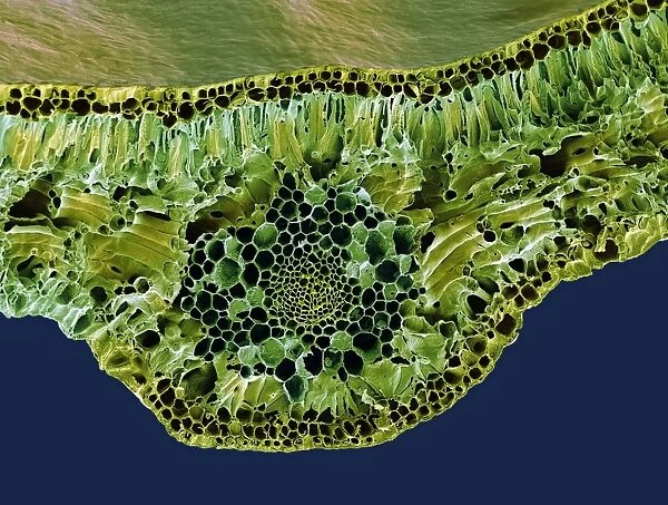
Leaf midrib, SEM
Leaf midrib. Coloured scanning electron micrograph (SEM) of a section through the midrib of a leaf from the Common Box (Buxus sempervirens). The midrib (midvein) is the continuation of a leafs stem along the centre of the leaf. At lower centre is a vascular bundle, which consists of xylem (small light green circles) and phloem (large dark circles) tissues. Xylem transports water and mineral nutrients from the roots throughout the plant and phloem transports carbohydrate and hormones around the plant. The surface (epidermis) of the leaf is covered in a waxy cuticle (top) that helps to prevent water loss. Magnification: x200 when printed 10 centimetres wide
Science Photo Library features Science and Medical images including photos and illustrations
Media ID 6287283
© STEVE GSCHMEISSNER/SCIENCE PHOTO LIBRARY
Axial Cellular Cross Section Cuticle Epidermis False Colour Histological Histology Mid Rib Phloem Spongy Mesophyll Stem Tissue Transverse Vascular Bundle Waxy Xylem Buxus Sempervirens Cells Common Box False Coloured Section Sectioned
EDITORS COMMENTS
This print showcases the intricate beauty of a leaf midrib from the Common Box plant. In this coloured scanning electron micrograph (SEM), we are granted a glimpse into the hidden world of cellular structures that make up this essential part of a leaf's anatomy. The midrib, or midvein, acts as the central axis running through the leaf, extending from its stem. At the center of attention lies a vascular bundle, composed of xylem and phloem tissues. The xylem transports water and vital nutrients throughout the plant, while the phloem carries carbohydrates and hormones to various parts. The leaf's surface is covered with a waxy cuticle on top, serving as nature's shield against excessive water loss. This microscopic view reveals an array of fascinating details such as palisade parenchyma cells and spongy mesophyll cells that contribute to photosynthesis. With a magnification factor of 200 when printed at 10 centimeters wide, this SEM image allows us to appreciate both structure and beauty in equal measure. It serves as a testament to how science can uncover remarkable wonders within even seemingly ordinary objects like leaves. This stunning photograph is brought to you by Science Photo Library – an invaluable resource for those fascinated by botany, biology, histology, and all things related to understanding our natural world at its most fundamental level.
MADE IN THE USA
Safe Shipping with 30 Day Money Back Guarantee
FREE PERSONALISATION*
We are proud to offer a range of customisation features including Personalised Captions, Color Filters and Picture Zoom Tools
SECURE PAYMENTS
We happily accept a wide range of payment options so you can pay for the things you need in the way that is most convenient for you
* Options may vary by product and licensing agreement. Zoomed Pictures can be adjusted in the Cart.

