Transmission Electron Micrograph Collection (#8)

"Exploring the Microscopic World: Unveiling Intricate Structures through Transmission Electron Micrograph" In the realm of science

Background imageTransmission Electron Micrograph Collection: Large intestine, TEM

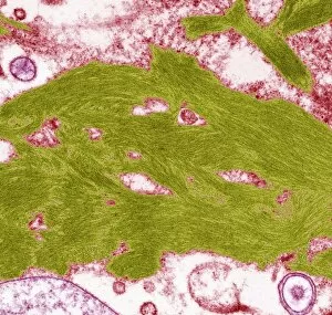
Large intestine, TEM
Large intestine. Coloured transmission electron micrograph (TEM) of a section through the wall of the large intestine. The surface consists of many small hair-like absorptive cells (green)

Background imageTransmission Electron Micrograph Collection: Alveolar cell, TEM

Alveolar cell, TEM
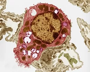
Alveolar cell. Coloured transmission electron micrograph (TEM) of a section through a type II alveolar cell. This cell is found in the alveoli (air sacs) of the lungs

Background imageTransmission Electron Micrograph Collection: Myelin surrounding a nerve axon, TEM

Myelin surrounding a nerve axon, TEM
Myelin surrounding a nerve axon, coloured transmission electron micrograph (TEM). The concentric round rings are the sheets of a Schwann cells myelin membrane (brown rings)

Background imageTransmission Electron Micrograph Collection: Osteoclast, TEM

Osteoclast, TEM
Osteoclast. Coloured transmission electron micrograph (TEM) of an osteoclast (blue and purple). Osteoclasts are found in cavities, known as Howslips lacunae, on the surface of bones

Background imageTransmission Electron Micrograph Collection: Skeletal muscle, TEM

Skeletal muscle, TEM
Skeletal muscle. Coloured transmission electron micrograph (TEM) of a longitudinal section through skeletal, or striated, muscle

Background imageTransmission Electron Micrograph Collection: Gut muscle cells, TEM

Gut muscle cells, TEM
Gut muscle cells, coloured transmission electron micrograph (TEM). Nuclei are pink. The involuntary contraction of this smooth muscle is responsible for peristalsis

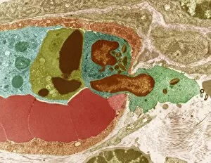
Background imageTransmission Electron Micrograph Collection: Damaged blood vessel, TEM

Damaged blood vessel, TEM
Damaged blood vessel. Coloured transmission electron micrograph (TEM) of a red blood cell (circular) leaking through the wall of a damaged blood vessel

Background imageTransmission Electron Micrograph Collection: T-lymphocyte, TEM

T-lymphocyte, TEM
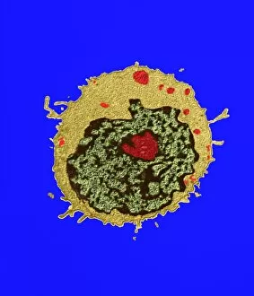
T-lymphocyte. Coloured transmission electron micrograph (TEM) of a section through a T- lymphocyte white blood cell. The cell contains a large nucleus (green/black)

Background imageTransmission Electron Micrograph Collection: Contracted artery, TEM

Contracted artery, TEM
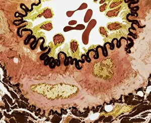
Contracted elastic artery. Coloured transmission electron micrograph (TEM) of a section through an artery. Red blood cells (erythrocytes) are seen in the lumen (top centre)

Background imageTransmission Electron Micrograph Collection: Coronary artery, TEM

Coronary artery, TEM
Coronary artery. Coloured transmission electron micrograph (TEM) of a cross-section through the wall of a coronary artery

Background imageTransmission Electron Micrograph Collection: Ducks and bird flu virus particles

Ducks and bird flu virus particles
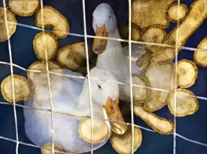
Ducks and avian influenza virus particles, composite image. The virus particles (brown) have been imaged using a transmission electron microscope (TEM)

Background imageTransmission Electron Micrograph Collection: Cluster of influenza viruses

Cluster of influenza viruses
Influenza virus. Computer manipulation of Transmission Electron Micrograph (TEM) of a cluster of five spherical-shaped influenza (flu) viruses

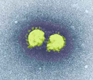
Background imageTransmission Electron Micrograph Collection: SARS virus particles, TEM

SARS virus particles, TEM
SARS virus particles. Coloured transmission electron micrograph (TEM) of two SARS virus particles (green). Severe acute respiratory syndrome (SARS)

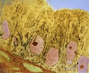
Background imageTransmission Electron Micrograph Collection: Uterus cells, TEM

Uterus cells, TEM
Uterus cells. Coloured transmission electron micrograph (TEM) of endometrial cells (yellow) from the uterus. These cells form the endometrium, the epithelial tissue lining inside the uterus

Background imageTransmission Electron Micrograph Collection: Liver cells, TEM

Liver cells, TEM
Liver cells. Coloured transmission electron micrograph (TEM) of cells in a liver, showing their tessellating boundaries (red) and nuclei (beige). Magnification: x9000 when printed 10 centimetres wide

Background imageTransmission Electron Micrograph Collection: Goblet cell

Goblet cell. Coloured transmission electron micrograph (TEM) of a section through a goblet cell in the lining of the small intestine, part of the digestive tract

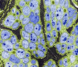
Background imageTransmission Electron Micrograph Collection: Nerve fibres, TEM

Nerve fibres, TEM
Nerve fibres. Coloured transmission electron micrograph (TEM) of a section through non- myelinated nerve fibres (blue). The fibres are arranged into bundles surrounded by the cytoplasm of Schwann

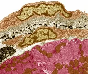
Background imageTransmission Electron Micrograph Collection: Pericardium, TEM

Pericardium, TEM
Pericardium. Coloured transmission electron micrograph (TEM) of a section through the serous pericardium. The pericardium is a double-walled sac surrounding the heart

Background imageTransmission Electron Micrograph Collection: Neutrophil and trapped bacteria, TEM

Neutrophil and trapped bacteria, TEM
Neutrophil and trapped bacteria. Coloured transmission electron micrograph (TEM) of Neisseria gonorrhoeae bacteria (blue) trapped by a neutrophil (orange), a type of white blood cell

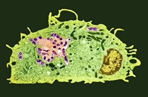
Background imageTransmission Electron Micrograph Collection: Macrophage and tuberculosis vaccine, TEM

Macrophage and tuberculosis vaccine, TEM
Macrophage and tuberculosis vaccine. Coloured transmission electron micrograph (TEM) of phagocytosed (engulfed) Mycobacterium bovis bacteria (purple) in a macrophage (green) white blood cell

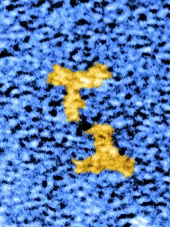
Background imageTransmission Electron Micrograph Collection: Immunoglobulin G antibodies, TEM

Immunoglobulin G antibodies, TEM
Immunoglobulin G antibody molecules, coloured transmission electron micrograph (TEM). IgG is the most abundant human immunoglobulin, and is found in all body fluids

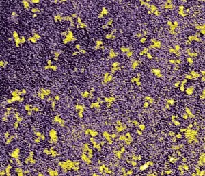
Background imageTransmission Electron Micrograph Collection: Human antibodies, TEM

Human antibodies, TEM
Human antibodies (yellow), coloured transmission electron micrograph (TEM). The Y-shaped structures are molecules of the immunoglobulin G (IgG) antibody

Background imageTransmission Electron Micrograph Collection: Blood vessel leak, TEM

Blood vessel leak, TEM
Blood vessel leak. Coloured transmission electron micrograph (TEM) of a white blood cell (centre, green and brown) leaking out of a blood vessel

Background imageTransmission Electron Micrograph Collection: Paramyxovirus particle, TEM

Paramyxovirus particle, TEM
Paramyxovirus. Transmission electron micrograph (TEM) of a paramyxovirus particle. The internal structure of the virus has been revealed

Background imageTransmission Electron Micrograph Collection: Rotavirus particle, artwork and TEM

Rotavirus particle, artwork and TEM
Rotavirus particle. Computer artwork (left) and coloured transmission electron micrograph (TEM) of the geometric structure of a rotavirus particles capsid shell

Background imageTransmission Electron Micrograph Collection: Kidney disease, TEM

Kidney disease, TEM
Kidney disease. Coloured transmission electron micrograph (TEM) of a section through the glomerulus of a kidney affected by membranous neuropathy

Background imageTransmission Electron Micrograph Collection: Abnormal blood clot, TEM

Abnormal blood clot, TEM
Abnormal blood clot. Coloured transmission electron micrograph (TEM) of a section through a blood vessel that is blocked by an abnormal blood clot (thrombus)

Background imageTransmission Electron Micrograph Collection: AIDS pneumonia infection, TEM

AIDS pneumonia infection, TEM
AIDS pneumonia infection. Coloured transmission electron micrograph (TEM) of a section through Pneumocystis carinii protozoa causing pneumonia. The protozoa are crescent-shaped (red/yellow)

Background imageTransmission Electron Micrograph Collection: Amyloidosis, TEM

Amyloidosis, TEM
Amyloidosis. Coloured transmission electron micrograph (TEM) of a section through tissue with abnormal deposoits of amyloid protein (green)

Background imageTransmission Electron Micrograph Collection: Avian influenza virus, TEM

Avian influenza virus, TEM
Avian influenza virus, type A strain H5N1, coloured transmission electron micrograph (TEM). This virus was isolated in Vietnam, during the avian flu outbreak in early 2004

Background imageTransmission Electron Micrograph Collection: Budding HIV particles, TEM

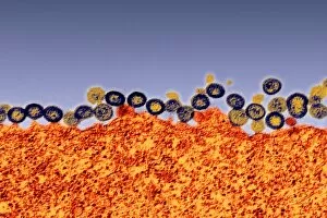
Budding HIV particles, TEM
Budding HIV particles. Coloured transmission electron micrograph (TEM) of HIV particles (blue and yellow) budding from the membrane of the host cell (orange)


For sale as Licensed Images

Choose your image, Select your licence and Download the media

"Exploring the Microscopic World: Unveiling Intricate Structures through Transmission Electron Micrograph" In the realm of science, transmission electron micrographs (TEM) have revolutionized our understanding of various biological entities. These powerful images provide a glimpse into the hidden intricacies that make up our world at an unimaginably small scale. One captivating TEM showcases regenerating nerve cells, offering hope for those seeking to understand and treat neurological disorders. The image captures the delicate process of nerve cell regrowth, highlighting their remarkable ability to heal and restore function. Another fascinating TEM reveals fat cells in all their glory - spherical structures filled with lipid droplets that play crucial roles in energy storage and insulation within our bodies. This microscopic view sheds light on how these adipocytes contribute to overall health and metabolism. Moving on, we encounter an E. coli bacterium captured by TEM, showcasing its distinctive rod-shaped structure. This notorious bacterium serves as both a model organism for research purposes and a cause of concern due to its potential pathogenicity. Delving deeper into the microscopic world, plasma cells come into focus through another mesmerizing TEM image. These specialized white blood cells produce antibodies vital for immune defense against invading pathogens - a testament to the intricate mechanisms at work within our bodies. The hauntingly beautiful transmission electron micrograph of influenza virus particles provides insight into one of humanity's greatest challenges - infectious diseases. Studying such images aids scientists in developing effective vaccines and antiviral treatments against these elusive viral foes. Hepatitis C viruses also reveal themselves under TEM scrutiny, reminding us of the ongoing battle against this chronic liver disease affecting millions worldwide. Understanding their structure helps researchers devise strategies for prevention and treatment. Shifting gears slightly from infections to anatomy, an eye muscle is showcased through TEM imagery – revealing its unique fiber arrangement responsible for precise eye movements essential for vision coordination. A Purkinje nerve cell takes center stage next; this neuron found in the cerebellum plays a crucial role in coordinating movement and balance.