Transmission Electron Micrograph Collection (#7)

"Exploring the Microscopic World: Unveiling Intricate Structures through Transmission Electron Micrograph" In the realm of science

Background imageTransmission Electron Micrograph Collection: Kidney glomerulus, TEM C016 / 5831

Kidney glomerulus, TEM C016 / 5831
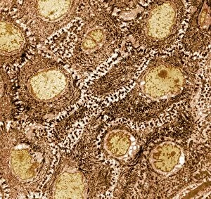
Kidney glomerulus. Transmission electron micrograph (TEM) of a section through a renal glomerulus. At centre left is a capillary with a red blood cell (black) in its lumen

Background imageTransmission Electron Micrograph Collection: Alveolar soft part sarcoma, TEM C016 / 5826

Alveolar soft part sarcoma, TEM C016 / 5826
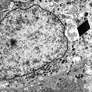
Alveolar soft part sarcoma. Transmission electron micrograph (TEM) of a section through an alveolar soft part sarcoma (ASPS) showing a characteristic rhomboid crystal (black, centre)

Background imageTransmission Electron Micrograph Collection: Alveolar soft part sarcoma, TEM C016 / 5816

Alveolar soft part sarcoma, TEM C016 / 5816
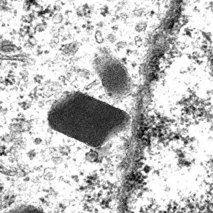
Alveolar soft part sarcoma. Transmission electron micrograph (TEM) of a section through an alveolar soft part sarcoma (ASPS) showing a characteristic rhomboid crystal (black)

Background imageTransmission Electron Micrograph Collection: Mitochondrial inclusions, TEM C016 / 5807

Mitochondrial inclusions, TEM C016 / 5807
Mitochondrial inclusions. Transmission electron micrograph (TEM) of a section through a mitochondrion showing crystalline inclusions (thick parallel rods) in the intermembrane spaces

Background imageTransmission Electron Micrograph Collection: Mitochondrial inclusions, TEM C016 / 5806

Mitochondrial inclusions, TEM C016 / 5806
Mitochondrial inclusions. Transmission electron micrograph (TEM) of a section through a mitochondrion showing crystalline inclusions (thick parallel rods) in the intermembrane spaces

Background imageTransmission Electron Micrograph Collection: Unmyelinated nerve, TEM C016 / 5805

Unmyelinated nerve, TEM C016 / 5805
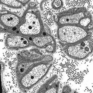
Unmyelinated nerve. Transmission electron micrograph (TEM) of a section through axon (nerve fibre) bundles of unmyelinated nerves

Background imageTransmission Electron Micrograph Collection: Unmyelinated nerve, TEM C016 / 5804

Unmyelinated nerve, TEM C016 / 5804
Unmyelinated nerve. Transmission electron micrograph (TEM) of a section through axon (nerve fibre) bundles of unmyelinated nerves

Background imageTransmission Electron Micrograph Collection: Myelinated nerve, TEM C016 / 5448

Myelinated nerve, TEM C016 / 5448
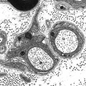
Myelinated nerve. Transmission electron micrograph (TEM) of a section through a myelinated nerve fibre and Schwann cell. Myelin (black)

Background imageTransmission Electron Micrograph Collection: Myelinated nerve, TEM C016 / 5370

Myelinated nerve, TEM C016 / 5370
Myelinated nerve. Transmission electron micrograph (TEM) of a section through a myelinated nerve fibre and Schwann cell (centre)

Background imageTransmission Electron Micrograph Collection: Pig retrovirus, TEM C016 / 4246

Pig retrovirus, TEM C016 / 4246
Pig retrovirus. Coloured transmission electron micrograph (TEM) of porcine endogenous retrovirus (PERV) particles (round) in infected tissue

Background imageTransmission Electron Micrograph Collection: Vesicular stomatitis virus, TEM C016 / 4244

Vesicular stomatitis virus, TEM C016 / 4244
Vesicular stomatitis virus. Coloured transmission electron micrograph (TEM) of a particle of the rhabdovirus vesicular stomatitis virus (VSV). The bullet shape is characteristic of rhabdoviruses

Background imageTransmission Electron Micrograph Collection: Vesicular stomatitis virus, TEM C016 / 4245

Vesicular stomatitis virus, TEM C016 / 4245
Vesicular stomatitis virus, TEM

Background imageTransmission Electron Micrograph Collection: Avian influenza virus, TEM C016 / 2354

Avian influenza virus, TEM C016 / 2354
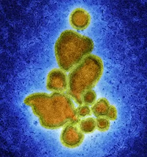
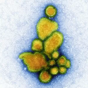
Avian influenza virus, type A strain H7N9, coloured transmission electron micrograph (TEM). This virus first emerged in the human population in China, in March 2013

Background imageTransmission Electron Micrograph Collection: Avian influenza virus, TEM C016 / 2353

Avian influenza virus, TEM C016 / 2353
Avian influenza virus, type A strain H7N9, coloured transmission electron micrograph (TEM). This virus first emerged in the human population in China, in March 2013

Background imageTransmission Electron Micrograph Collection: Avian influenza virus, TEM C016 / 2352

Avian influenza virus, TEM C016 / 2352
Avian influenza virus, type A strain H7N9, coloured transmission electron micrograph (TEM). This virus first emerged in the human population in China, in March 2013

Background imageTransmission Electron Micrograph Collection: Avian influenza virus, TEM C016 / 2351

Avian influenza virus, TEM C016 / 2351
Avian influenza virus, type A strain H7N9, coloured transmission electron micrograph (TEM). This virus first emerged in the human population in China, in March 2013

Background imageTransmission Electron Micrograph Collection: Retinal rod cell, TEM C013 / 4805

Retinal rod cell, TEM C013 / 4805
Retinal rod cell. Transmission electron micrograph (TEM) of a section through a rod cell from the retina of an eye, showing the inner segment (bottom) filled with mitochondria (green)

Background imageTransmission Electron Micrograph Collection: Retinal rod cell, TEM C013 / 4804

Retinal rod cell, TEM C013 / 4804
Retinal rod cell. Transmission electron micrograph (TEM) of a section through a rod cell from the retina of an eye, showing the inner segment (bottom) filled with mitochondria (green)

Background imageTransmission Electron Micrograph Collection: Brain cells, TEM C013 / 4801

Brain cells, TEM C013 / 4801
Brain cells. Transmission electron micrograph (TEM) of a section through oligodendrocytes (dark) in human brain tissue, showing free ribosomes (dark green dots), golgi apparatus (curved brown lines)

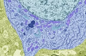
Background imageTransmission Electron Micrograph Collection: Macrophage engulfing a nerve cell, TEM C013 / 4803

Macrophage engulfing a nerve cell, TEM C013 / 4803
Macrophage engulfing a nerve cell. Transmission electron micrograph (TEM) of a section through a macrophage white blood cell (blue) that has engulfed a nerve cell (neuron, centre)

Background imageTransmission Electron Micrograph Collection: Brain cells, TEM C013 / 4800

Brain cells, TEM C013 / 4800
Brain cells. Transmission electron micrograph (TEM) of a section through oligodendrocytes in human brain tissue, showing free ribosomes (dark pink dots), golgi apparatus (curved brown lines)

Background imageTransmission Electron Micrograph Collection: Nerve cell, TEM C013 / 4797

Nerve cell, TEM C013 / 4797
Nerve cell. Transmission electron micrograph (TEM) of a section through a neuron (nerve cell), showing characteristic Nissl body (dark blue lines), numerous golgi apparatus (curved green lines)

Background imageTransmission Electron Micrograph Collection: Brain cell, TEM C013 / 4799

Brain cell, TEM C013 / 4799
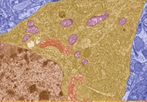
Brain cell. Transmission electron micrograph (TEM) of a section through an oligodendrocyte in human brain tissue, showing free ribosomes (dark brown dots), golgi apparatus (curved orange lines)

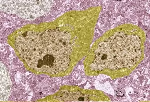
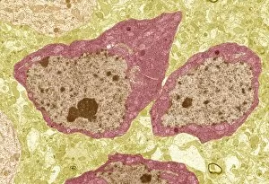
Background imageTransmission Electron Micrograph Collection: Nerve cell, TEM C013 / 4796

Nerve cell, TEM C013 / 4796
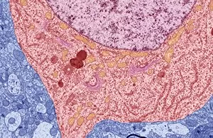
Nerve cell. Transmission electron micrograph (TEM) of a section through a neuron (nerve cell), showing characteristic Nissl body (dark red lines), numerous golgi apparatus (curved pink lines)

Background imageTransmission Electron Micrograph Collection: Brain cell, TEM C013 / 4798

Brain cell, TEM C013 / 4798
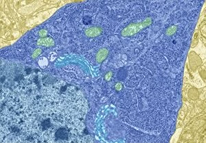
Brain cell. Transmission electron micrograph (TEM) of a section through an oligodendrocyte in human brain tissue, showing free ribosomes (dark blue dots), golgi apparatus (curved light blue lines)

Background imageTransmission Electron Micrograph Collection: Skin cell, TEM

Skin cell, TEM
Skin cell. Coloured transmission electron micrograph (TEM) of a section through a keratinocyte skin cell, which is found in the epidermis layer of the skin

Background imageTransmission Electron Micrograph Collection: Sperm in the testis, TEM

Sperm in the testis, TEM
Sperm cells. Coloured transmission electron micrograph (TEM) of a section through the testis, showing sperm cells. The heads of the sperm, which contain the genetic material, are red

Background imageTransmission Electron Micrograph Collection: Sperm tails, TEM

Sperm tails, TEM
Sperm tails. Coloured transmission electron micrograph (TEM) of cross sections through sperm tails. A sperms tails ability to move is partly due to the arrangement of microtubules

Background imageTransmission Electron Micrograph Collection: Mitotic cell division, TEM

Mitotic cell division, TEM
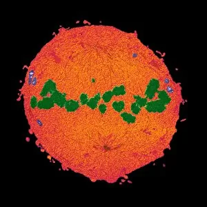
Mitotic cell division. Coloured transmission electron micrograph (TEM) of a cell during the metaphase stage of mitosis (cell division)

Background imageTransmission Electron Micrograph Collection: Mitosis, TEM

Mitosis, TEM
Mitosis, coloured transmission electron micrograph (TEM). Longitudinal section through a human T cell in metaphase. During mitosis two daughter nuclei are formed from one parent nucleus

Background imageTransmission Electron Micrograph Collection: Skin cells, TEM

Skin cells, TEM
Skin cells. Coloured transmission electron micrograph (TEM) of a section through keratinocyte skin cells, which are found in the epidermis layer of the skin

Background imageTransmission Electron Micrograph Collection: Thyroid gland follicle, TEM

Thyroid gland follicle, TEM
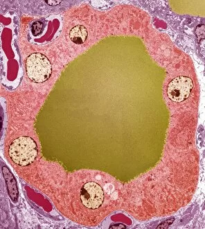
Thyroid gland follicle. Coloured transmission electron micrograph (TEM) of a follicle from the thyroid gland. The follicle consists of a layer of epithelial cells (pink)

Background imageTransmission Electron Micrograph Collection: Pituitary gland, TEM

Pituitary gland, TEM
Pituitary gland. Coloured transmission electron micrograph (TEM) of cells in the anterior pituitary gland, a hormone-secreting gland at the base of the brain

Background imageTransmission Electron Micrograph Collection: Large intestine, TEM

Large intestine, TEM
Large intestine. Coloured transmission electron micrograph (TEM) of a section through the wall of the large intestine. The surface consists of many small hair-like absorptive cells (green)

Background imageTransmission Electron Micrograph Collection: Alveolar cell, TEM

Alveolar cell, TEM
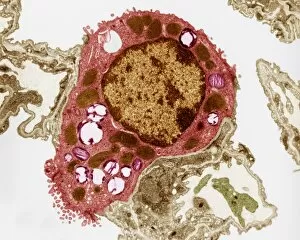
Alveolar cell. Coloured transmission electron micrograph (TEM) of a section through a type II alveolar cell. This cell is found in the alveoli (air sacs) of the lungs

Background imageTransmission Electron Micrograph Collection: Myelin surrounding a nerve axon, TEM

Myelin surrounding a nerve axon, TEM
Myelin surrounding a nerve axon, coloured transmission electron micrograph (TEM). The concentric round rings are the sheets of a Schwann cells myelin membrane (brown rings)

Background imageTransmission Electron Micrograph Collection: Osteoclast, TEM

Osteoclast, TEM
Osteoclast. Coloured transmission electron micrograph (TEM) of an osteoclast (blue and purple). Osteoclasts are found in cavities, known as Howslips lacunae, on the surface of bones

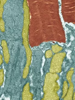
Background imageTransmission Electron Micrograph Collection: Skeletal muscle, TEM

Skeletal muscle, TEM
Skeletal muscle. Coloured transmission electron micrograph (TEM) of a longitudinal section through skeletal, or striated, muscle

Background imageTransmission Electron Micrograph Collection: Gut muscle cells, TEM

Gut muscle cells, TEM
Gut muscle cells, coloured transmission electron micrograph (TEM). Nuclei are pink. The involuntary contraction of this smooth muscle is responsible for peristalsis

Background imageTransmission Electron Micrograph Collection: Damaged blood vessel, TEM

Damaged blood vessel, TEM
Damaged blood vessel. Coloured transmission electron micrograph (TEM) of a red blood cell (circular) leaking through the wall of a damaged blood vessel

Background imageTransmission Electron Micrograph Collection: T-lymphocyte, TEM

T-lymphocyte, TEM
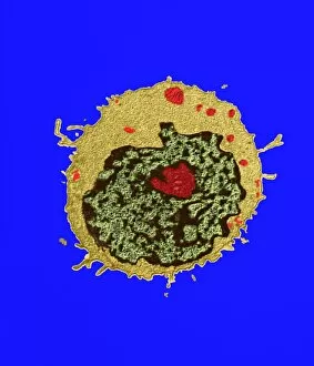
T-lymphocyte. Coloured transmission electron micrograph (TEM) of a section through a T- lymphocyte white blood cell. The cell contains a large nucleus (green/black)

Background imageTransmission Electron Micrograph Collection: Contracted artery, TEM

Contracted artery, TEM
Contracted elastic artery. Coloured transmission electron micrograph (TEM) of a section through an artery. Red blood cells (erythrocytes) are seen in the lumen (top centre)

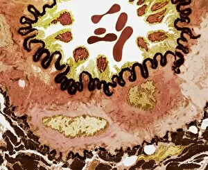
Background imageTransmission Electron Micrograph Collection: Coronary artery, TEM

Coronary artery, TEM
Coronary artery. Coloured transmission electron micrograph (TEM) of a cross-section through the wall of a coronary artery

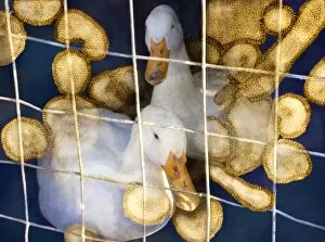
Background imageTransmission Electron Micrograph Collection: Ducks and bird flu virus particles

Ducks and bird flu virus particles
Ducks and avian influenza virus particles, composite image. The virus particles (brown) have been imaged using a transmission electron microscope (TEM)

Background imageTransmission Electron Micrograph Collection: Cluster of influenza viruses

Cluster of influenza viruses
Influenza virus. Computer manipulation of Transmission Electron Micrograph (TEM) of a cluster of five spherical-shaped influenza (flu) viruses

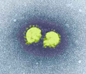
Background imageTransmission Electron Micrograph Collection: SARS virus particles, TEM

SARS virus particles, TEM
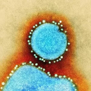
SARS virus particles. Coloured transmission electron micrograph (TEM) of two SARS virus particles (green). Severe acute respiratory syndrome (SARS)

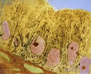
Background imageTransmission Electron Micrograph Collection: Uterus cells, TEM

Uterus cells, TEM
Uterus cells. Coloured transmission electron micrograph (TEM) of endometrial cells (yellow) from the uterus. These cells form the endometrium, the epithelial tissue lining inside the uterus

Background imageTransmission Electron Micrograph Collection: Liver cells, TEM

Liver cells, TEM
Liver cells. Coloured transmission electron micrograph (TEM) of cells in a liver, showing their tessellating boundaries (red) and nuclei (beige). Magnification: x9000 when printed 10 centimetres wide


For sale as Licensed Images

Choose your image, Select your licence and Download the media

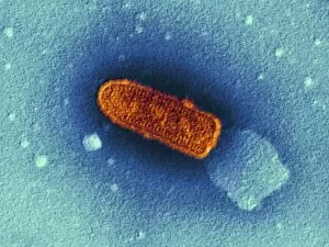
"Exploring the Microscopic World: Unveiling Intricate Structures through Transmission Electron Micrograph" In the realm of science, transmission electron micrographs (TEM) have revolutionized our understanding of various biological entities. These powerful images provide a glimpse into the hidden intricacies that make up our world at an unimaginably small scale. One captivating TEM showcases regenerating nerve cells, offering hope for those seeking to understand and treat neurological disorders. The image captures the delicate process of nerve cell regrowth, highlighting their remarkable ability to heal and restore function. Another fascinating TEM reveals fat cells in all their glory - spherical structures filled with lipid droplets that play crucial roles in energy storage and insulation within our bodies. This microscopic view sheds light on how these adipocytes contribute to overall health and metabolism. Moving on, we encounter an E. coli bacterium captured by TEM, showcasing its distinctive rod-shaped structure. This notorious bacterium serves as both a model organism for research purposes and a cause of concern due to its potential pathogenicity. Delving deeper into the microscopic world, plasma cells come into focus through another mesmerizing TEM image. These specialized white blood cells produce antibodies vital for immune defense against invading pathogens - a testament to the intricate mechanisms at work within our bodies. The hauntingly beautiful transmission electron micrograph of influenza virus particles provides insight into one of humanity's greatest challenges - infectious diseases. Studying such images aids scientists in developing effective vaccines and antiviral treatments against these elusive viral foes. Hepatitis C viruses also reveal themselves under TEM scrutiny, reminding us of the ongoing battle against this chronic liver disease affecting millions worldwide. Understanding their structure helps researchers devise strategies for prevention and treatment. Shifting gears slightly from infections to anatomy, an eye muscle is showcased through TEM imagery – revealing its unique fiber arrangement responsible for precise eye movements essential for vision coordination. A Purkinje nerve cell takes center stage next; this neuron found in the cerebellum plays a crucial role in coordinating movement and balance.