Photo Mug : Eye anatomy, SEM


![]()
Home Decor from Science Photo Library
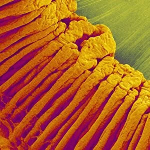
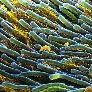
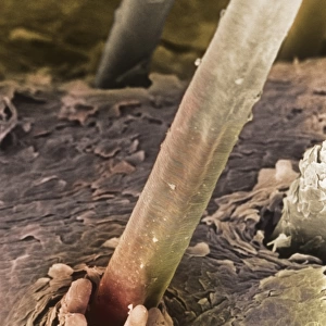
Eye anatomy, SEM
Eye anatomy. Coloured scanning electron micrograph (SEM) showing part of the ciliary body (orange) and iris (right) of an eye. The ciliary body is a ring-shaped structure that surrounds the iris and joins to ligaments that hold the lens in place behind the iris. It also contains the ciliary muscle that is contracted to alter the curvature of the lens and focus light on the retina at the back of the eye. Magnification: x20 when printed 10 centimetres wide
Science Photo Library features Science and Medical images including photos and illustrations
Media ID 6339195
© STEVE GSCHMEISSNER/SCIENCE PHOTO LIBRARY
Ciliary Body Colored False Colored Inside Internal Iris Ligament Ligaments Muscles Muscular Ocular Ophtalmological Ophthalmology Physiological Physiology Sight Tissue Vision False Coloured
Large Photo Mug (15 oz)
Elevate your coffee or tea experience with our premium white ceramic mug. Its wide, comfortable handle makes drinking easy, and you can rely on it to be both microwave and dishwasher safe. Sold in single units, preview may show both sides of the same mug so you can see how the picture wraps around.
Elevate your coffee or tea experience with our premium white ceramic mug. Its wide, comfortable handle makes drinking easy, and you can rely on it to be both microwave and dishwasher safe. Sold in single units, preview may show both sides of the same mug so you can see how the picture wraps around.
These are individually made so all sizes are approximate
MADE IN THE USA
Safe Shipping with 30 Day Money Back Guarantee
FREE PERSONALISATION*
We are proud to offer a range of customisation features including Personalised Captions, Color Filters and Picture Zoom Tools
SECURE PAYMENTS
We happily accept a wide range of payment options so you can pay for the things you need in the way that is most convenient for you
* Options may vary by product and licensing agreement. Zoomed Pictures can be adjusted in the Cart.