Photographic Print : Suboccipital muscles and nerve, artwork C014 / 5098

![]()
Photo Prints from Science Photo Library
Suboccipital muscles and nerve, artwork C014 / 5098
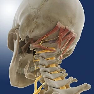
Suboccipital muscles. Computer artwork of the back of the base of the skull showing nerves (yellow) and the suboccipital muscles (pink). The two muscles at centre, which attach at the base of the skull (occipital bone) and the first vertebra of the spine (the atlas), are the rectus capitis posterior minor. Either side of those are the rectus capitis posterior major, which attach at the occipital bone and the spinous process of the second vertebra of the spine (the axis). Outside of these are the obliquus capitis superior, which attach the occipital bone to the transverse process of the atlas. These last two muscles are innervated by the occipital nerve. The horizontal muscle attached to the transverse process of the atlas and the spinous process of the axis is the obliquus capitis inferior. These muscles are responsible for extending and rotating the head. The spinal cord runs down the centre of the spine
Science Photo Library features Science and Medical images including photos and illustrations
Media ID 9215271
© SPRINGER MEDIZIN/SCIENCE PHOTO LIBRARY
Atlas Axis Back Base Bones Brachial Plexus Cervical Spine Cervical Vertebrae From Below Hyoid Bone Muscular System Musculoskeletal System Neck Nerve Nerves Oblique Occipital Bone Posterior Spinal Cord Spinal Nerve Vertebral Column Nervous System
10"x10" Photo Print
Discover the intricacies of human anatomy with our Media Storehouse Suboccipital Muscles and Nerve Print. This captivating artwork, SPRINGER MEDIZIN/SCIENCE PHOTO LIBRARY's C014 / 5098, showcases the delicate interplay of nerves (yellow) and suboccipital muscles (pink) at the base of the skull. Bring the complexity of the human body into your home or office with this stunning, high-quality photographic print. Ideal for medical professionals, students, or anyone with an interest in anatomy.
Photo prints are produced on Kodak professional photo paper resulting in timeless and breath-taking prints which are also ideal for framing. The colors produced are rich and vivid, with accurate blacks and pristine whites, resulting in prints that are truly timeless and magnificent. Whether you're looking to display your prints in your home, office, or gallery, our range of photographic prints are sure to impress. Dimensions refers to the size of the paper in inches.
Our Photo Prints are in a large range of sizes and are printed on Archival Quality Paper for excellent colour reproduction and longevity. They are ideal for framing (our Framed Prints use these) at a reasonable cost. Alternatives include cheaper Poster Prints and higher quality Fine Art Paper, the choice of which is largely dependant on your budget.
Estimated Product Size is 25.4cm x 25.4cm (10" x 10")
These are individually made so all sizes are approximate
Artwork printed orientated as per the preview above, with landscape (horizontal) or portrait (vertical) orientation to match the source image.
EDITORS COMMENTS
This print showcases the intricate details of the suboccipital muscles and nerves, offering a fascinating glimpse into the complexity of human anatomy. In this computer artwork, we are presented with a view from below, focusing on the back of the base of the skull. The pink-colored suboccipital muscles take center stage in this image. At the core, we find two rectus capitis posterior minor muscles that connect to both the occipital bone and the first vertebra of the spine. Surrounding them are larger rectus capitis posterior major muscles that attach to the occipital bone and spinous process of the second vertebra. Extending outward from these central muscles are obliquus capitis superior fibers which link together the occipital bone and transverse process of atlas. These last two muscles receive innervation from none other than our yellow-highlighted occipital nerve. Notably visible is also an obliquus capitis inferior muscle running horizontally between transverse processes of atlas and spinous process axis – responsible for extending and rotating our head. As we delve deeper into this anatomical wonderland, it becomes evident how interconnected everything truly is. The spinal cord gracefully runs down through each cervical vertebrae within our vertebral column, serving as a vital conduit for communication between body and brain. This stunning illustration not only highlights our musculoskeletal system but also sheds light on various components such as bones (including hyoid bone), nerves (like brachial plexus), and even offers insights into our nervous system's functioning. A true testament to nature's brilliance!
MADE IN THE USA
Safe Shipping with 30 Day Money Back Guarantee
FREE PERSONALISATION*
We are proud to offer a range of customisation features including Personalised Captions, Color Filters and Picture Zoom Tools
SECURE PAYMENTS
We happily accept a wide range of payment options so you can pay for the things you need in the way that is most convenient for you
* Options may vary by product and licensing agreement. Zoomed Pictures can be adjusted in the Cart.