Premium Framed Print : Goblet cell, TEM


![]()
Framed Photos from Science Photo Library
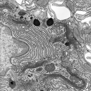
Goblet cell, TEM
Goblet cell. Transmission electron micrograph (TEM) of a section through a goblet cell in the lining (epithelium, top) of the small intestine, showing mucous being discharged (upper centre) onto the surface of the epithelium. Mucous is rich in carbohydrates and, following release from the goblet cell, becomes hydrated and expands enormously in volume forming a gel-like layer over the surface of the epithelial cells. This protects the epithelium and traps cell debris and particles that are not digested. Magnification: x4, 000 when printed 10 centimetres tall
Science Photo Library features Science and Medical images including photos and illustrations
Media ID 9241995
© MICROSCAPE/SCIENCE PHOTO LIBRARY
Black And White Bowel Bowels Cell Biology Cytological Cytology Digestive System Epithelial Epithelium Gastrointestinal Tract Goblet Cell Granule Granules Histological Histology Intestinal Intestine Intestines Lining Mucus Organelle Organelles Protective Secreting Secretory Small Intestine Transmission Electron Micrograph Transmission Electron Microscope Cells Section Sectioned
14"x16" Premium Frame
Contemporary style Premium Wooden Frame with 8"x10" Print. Complete with 2" White Mat and 1.25" thick MDF frame. Printed on 260 gsm premium paper. Glazed with shatter proof UV coated acrylic glass. Backing is paper covered backing with rubber bumpers. Supplied ready to hang with a pre-installed sawtooth/wire hanger. Care Instructions: Spot clean with a damp cloth. Securely packaged in a clear plastic bag and envelope in a reinforced cardboard shipper
FSC Real Wood Frame and Double Mounted with White Conservation Mountboard - Professionally Made and Ready to Hang
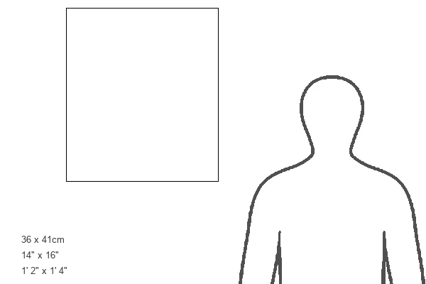
Estimated Image Size (if not cropped) is 20.3cm x 25.4cm (8" x 10")
Estimated Product Size is 35.6cm x 40.6cm (14" x 16")
These are individually made so all sizes are approximate
Artwork printed orientated as per the preview above, with portrait (vertical) orientation to match the source image.
EDITORS COMMENTS
This print from Science Photo Library showcases the intricate structure of a goblet cell in the lining of the small intestine. Taken using a transmission electron microscope (TEM), this image reveals the fascinating process of mucous discharge by the goblet cell. In the upper center of the image, we can observe mucous being released onto the surface of the epithelium. This gel-like substance is rich in carbohydrates and plays a crucial role in protecting and maintaining the health of our intestinal lining. Once discharged, it rapidly expands in volume due to hydration, forming a protective layer over the epithelial cells. The primary function of this mucus layer is to safeguard our intestines by trapping cell debris and undigested particles that could potentially harm or irritate our digestive system. By doing so, it ensures that only essential nutrients are absorbed while preventing any harmful substances from entering further into our body. With a magnification level of x4,000 when printed at 10 centimeters tall, this stunning photograph provides us with an up-close look at these remarkable organelles within our bowels. It serves as a reminder of how complex and intricately designed even microscopic structures can be within our biological systems.
MADE IN THE USA
Safe Shipping with 30 Day Money Back Guarantee
FREE PERSONALISATION*
We are proud to offer a range of customisation features including Personalised Captions, Color Filters and Picture Zoom Tools
SECURE PAYMENTS
We happily accept a wide range of payment options so you can pay for the things you need in the way that is most convenient for you
* Options may vary by product and licensing agreement. Zoomed Pictures can be adjusted in the Cart.