Premium Framed Print : Iris pigment epithelium, SEM


![]()
Framed Photos from Science Photo Library
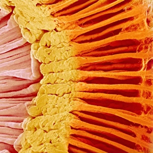
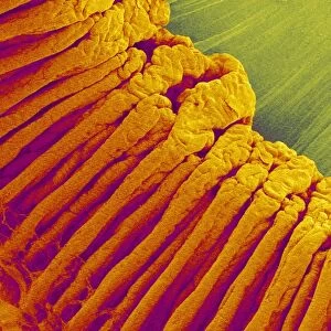
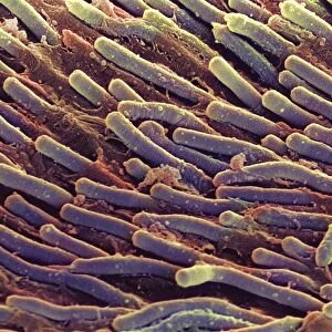
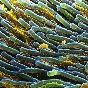
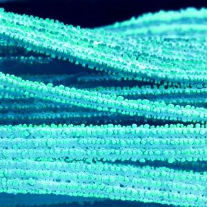
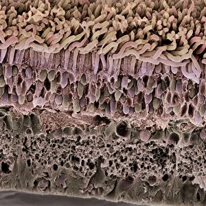
Iris pigment epithelium, SEM
Iris pigment epithelium. Coloured scanning electron micrograph (SEM) of a section through the iris of an eye, showing the iris pigment epithelium (IPE). The IPE is a layer of cuboidal cells (pink) that lies behind the iris. Each cell contains numerous large melanosomes (blue), which contain the pigment melanin. The concentration of this melanin is one of the factors that determine the colour of a persons eye. Magnification: x3, 300 when printed 10 centimetres wide
Science Photo Library features Science and Medical images including photos and illustrations
Media ID 6350495
© STEVE GSCHMEISSNER/SCIENCE PHOTO LIBRARY
Colored Colour Epithelial False Colored Inside Internal Melanin Ocular Ophtalmological Ophthalmology Physiological Physiology Sight Tissue Vision Cells False Coloured Section Sectioned
14"x16" Premium Frame
Contemporary style Premium Wooden Frame with 8"x10" Print. Complete with 2" White Mat and 1.25" thick MDF frame. Printed on 260 gsm premium paper. Glazed with shatter proof UV coated acrylic glass. Backing is paper covered backing with rubber bumpers. Supplied ready to hang with a pre-installed sawtooth/wire hanger. Care Instructions: Spot clean with a damp cloth. Securely packaged in a clear plastic bag and envelope in a reinforced cardboard shipper
FSC Real Wood Frame and Double Mounted with White Conservation Mountboard - Professionally Made and Ready to Hang
Estimated Image Size (if not cropped) is 25.4cm x 20.3cm (10" x 8")
Estimated Product Size is 40.6cm x 35.6cm (16" x 14")
These are individually made so all sizes are approximate
Artwork printed orientated as per the preview above, with landscape (horizontal) orientation to match the source image.
EDITORS COMMENTS
This print showcases the intricate beauty of the iris pigment epithelium (IPE) found in our eyes. In this coloured scanning electron micrograph, we are granted a glimpse into the hidden world of biology and tissue that contributes to our vision. The IPE, depicted here as a layer of delicate pink cuboidal cells, resides behind the iris and plays a crucial role in determining eye color. Each cell within this layer contains numerous large melanosomes, represented by their striking blue hue. These melanosomes house melanin pigment, which is responsible for giving color to our eyes. With a magnification of x3,300 when printed at 10 centimeters wide, this image allows us to appreciate the astonishing level of detail present in even the smallest components of our bodies. It serves as a reminder that there is so much more than meets the naked eye when it comes to understanding human anatomy and physiology. Photographer Steve Gschmeissner skillfully captures both artistry and scientific precision with his false-colored scanning electron micrograph. This mesmerizing composition not only appeals to those interested in ophthalmology or ocular health but also provides an opportunity for all viewers to marvel at nature's remarkable design within ourselves.
MADE IN THE USA
Safe Shipping with 30 Day Money Back Guarantee
FREE PERSONALISATION*
We are proud to offer a range of customisation features including Personalised Captions, Color Filters and Picture Zoom Tools
SECURE PAYMENTS
We happily accept a wide range of payment options so you can pay for the things you need in the way that is most convenient for you
* Options may vary by product and licensing agreement. Zoomed Pictures can be adjusted in the Cart.