
Premium Framed Print : Mint leaf surface, SEM


![]()
Framed Photos from Science Photo Library
Mint leaf surface, SEM
Mint leaf surface. Coloured scanning electron micrograph (SEM) of the surface of a mint (Menta sp.) leaf. The white structures are oil glands, modified glandular hairs that secrete oils that give this plant its aroma. The olive green structures are stomata, pores in the leaf surface that are the site of gaseous exchange in the plant. The opening and closing of the stomata is controlled by semi-circular guard cells, which swell and become turgid to close the openings
Science Photo Library features Science and Medical images including photos and illustrations
Media ID 6340203
© DAVID MCCARTHY/SCIENCE PHOTO LIBRARY
Close Colored Cuticle Edible Epidermis Gaseous Exchange Glandular Guard Cell Herb Mint Modified Hair Oil Gland Pore Secretory Stoma Stomata Surface Trichome Trichomes
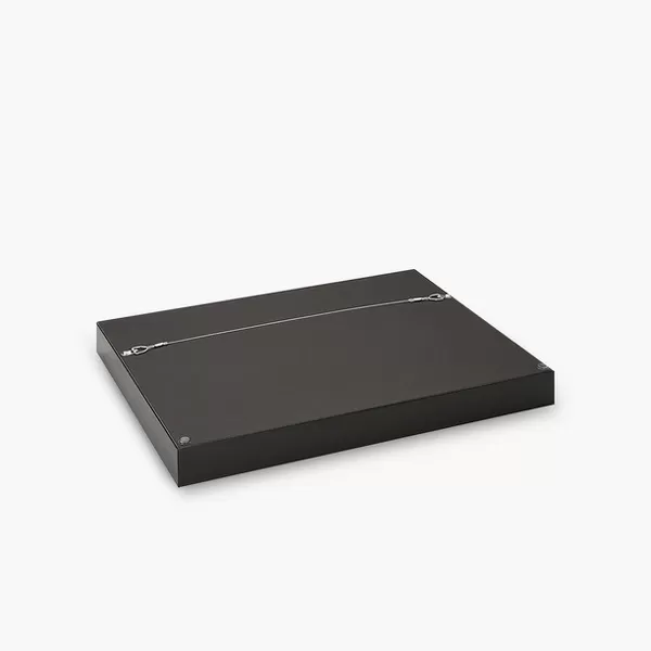
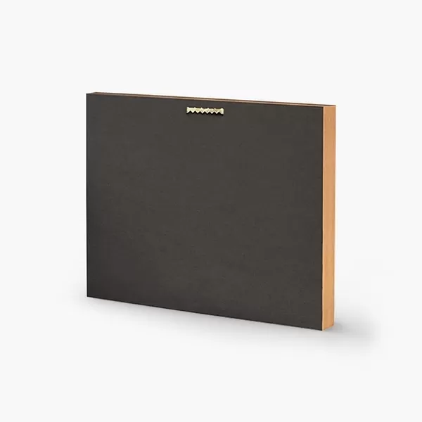
14"x16" Premium Frame
Contemporary style Premium Wooden Frame with 8"x10" Print. Complete with 2" White Mat and 1.25" thick MDF frame. Printed on 260 gsm premium paper. Glazed with shatter proof UV coated acrylic glass. Backing is paper covered backing with rubber bumpers. Supplied ready to hang with a pre-installed sawtooth/wire hanger. Care Instructions: Spot clean with a damp cloth. Securely packaged in a clear plastic bag and envelope in a reinforced cardboard shipper
FSC Real Wood Frame and Double Mounted with White Conservation Mountboard - Professionally Made and Ready to Hang
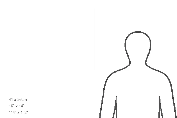
Estimated Image Size (if not cropped) is 25.4cm x 20.3cm (10" x 8")
Estimated Product Size is 40.6cm x 35.6cm (16" x 14")
These are individually made so all sizes are approximate
Artwork printed orientated as per the preview above, with landscape (horizontal) orientation to match the source image.
EDITORS COMMENTS
This print showcases the intricate beauty of a mint leaf surface, captured through a colored scanning electron microscope (SEM). The image reveals a mesmerizing world of botanical wonders. The white structures scattered across the leaf are oil glands, specialized glandular hairs that exude aromatic oils responsible for the distinct fragrance of this plant. Meanwhile, the olive green formations represent stomata - tiny pores on the leaf's surface crucial for gaseous exchange in plants. These stomata play a vital role in regulating plant respiration. The SEM image also highlights semi-circular guard cells surrounding each stoma. These remarkable cells control the opening and closing of these microscopic gates by swelling and becoming turgid when necessary to maintain optimal gas exchange within the leaf. Every detail is meticulously captured in this photograph: from the delicate epidermis to secretory cuticles and trichomes adorning the mint leaf's surface. This stunning visual representation offers an opportunity to marvel at nature's precision and complexity. Whether you have an interest in botany or simply appreciate natural beauty, this print will undoubtedly captivate your imagination with its vibrant colors and intricate details. It serves as a reminder of how even something as seemingly ordinary as a mint leaf can hold extraordinary secrets waiting to be discovered under closer examination.
MADE IN THE USA
Safe Shipping with 30 Day Money Back Guarantee
FREE PERSONALISATION*
We are proud to offer a range of customisation features including Personalised Captions, Color Filters and Picture Zoom Tools
SECURE PAYMENTS
We happily accept a wide range of payment options so you can pay for the things you need in the way that is most convenient for you
* Options may vary by product and licensing agreement. Zoomed Pictures can be adjusted in the Cart.
