Chromatin structure, diagram
![]()

Wall Art and Photo Gifts from Science Photo Library
Chromatin structure, diagram
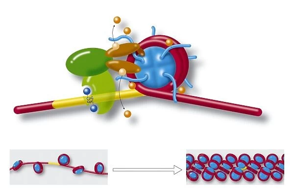
Chromatin structure, diagram. The main artwork shows various molecules and a strand of DNA (deoxyribonucleic acid, red) looping round a cylindrical histone core (blue), forming a structure known as a nucleosome. The yellow region is a section of cytosine-guanine (labelled CpG-GpC) di-nucleotides that play a role in chromatin formation. Also shown here is acetylation, deacetylation, methylation, and demethylation. Strings of nucleosomes (lower left) form the structure called beads on a string. Further compacting (lower right) condenses the chromatin even more to fit inside cell nuclei
Science Photo Library features Science and Medical images including photos and illustrations
Media ID 6326543
© ART FOR SCIENCE/SCIENCE PHOTO LIBRARY
Chromatin Coil Coiling Compact Compacting Condensing Diagram Euchromatin Fibre Genetic Heterochromatin Histone Histones Methylation Molecular Biology Nucleosome Promoter Proteins Structural Bio Chemistry Biochemical Condense Deoxyribonucleic Acid Genetics Molecular
EDITORS COMMENTS
This print showcases the intricate structure of chromatin, offering a glimpse into the fascinating world of molecular biology. The main artwork depicts a strand of DNA elegantly looping around a cylindrical histone core, forming what is known as a nucleosome. The vibrant red color represents deoxyribonucleic acid (DNA), which plays a crucial role in genetic information storage. Highlighted within the image is a yellow region consisting of cytosine-guanine di-nucleotides labeled CpG-GpC. These specific molecules contribute to the formation of chromatin and its structural organization. Additionally, various processes such as acetylation, deacetylation, methylation, and demethylation are visually represented here. The lower left portion reveals strings of nucleosomes arranged like beads on a string – an arrangement referred to as "beads on a string". As we move towards the lower right corner, we witness further compaction and condensation of chromatin to fit inside cell nuclei. Through this artful representation created by Science Photo Library, viewers gain insight into the complex nature of chromatin and its vital role in genetic regulation. This diagram serves as both an educational tool for those studying biochemistry or genetics and an aesthetic marvel that captures the beauty found within our cells' molecular machinery.
MADE IN THE USA
Safe Shipping with 30 Day Money Back Guarantee
FREE PERSONALISATION*
We are proud to offer a range of customisation features including Personalised Captions, Color Filters and Picture Zoom Tools
SECURE PAYMENTS
We happily accept a wide range of payment options so you can pay for the things you need in the way that is most convenient for you
* Options may vary by product and licensing agreement. Zoomed Pictures can be adjusted in the Cart.

