
Chromatin Collection
Chromatin, the intricate web of genetic material within our cells, holds the key to understanding life's blueprint


For sale as Licensed Images
Choose your image, Select your licence and Download the media
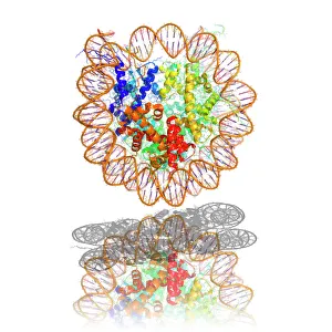
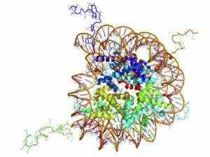
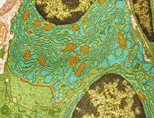
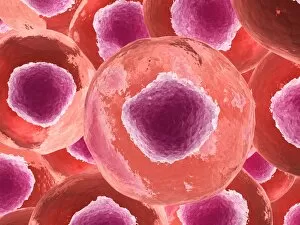
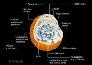
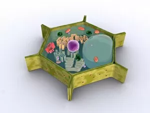
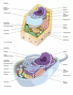
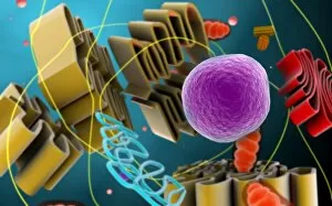
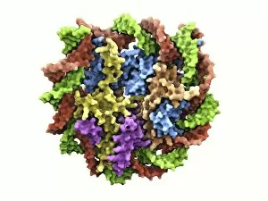
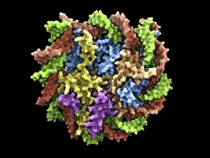
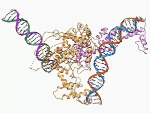
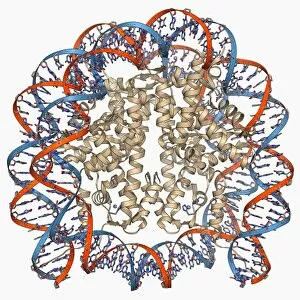
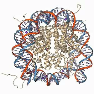
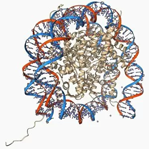
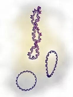
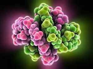
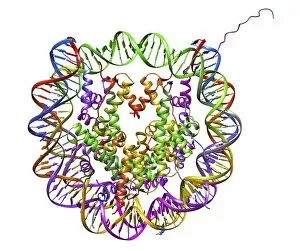
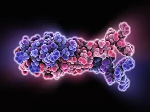
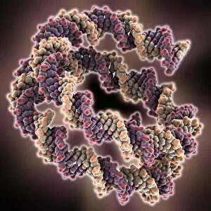
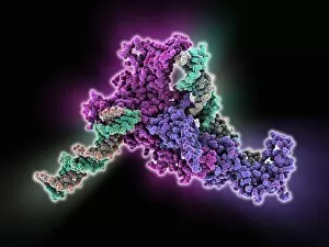
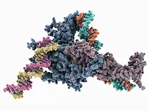
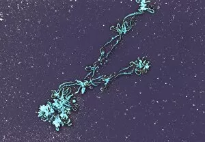
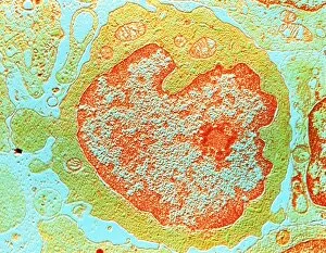
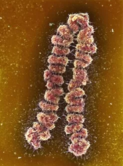
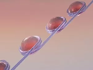
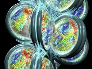
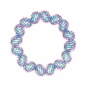
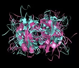
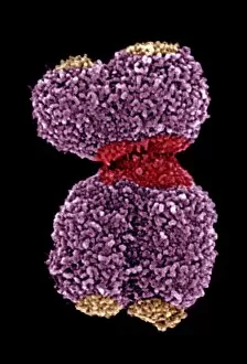
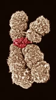
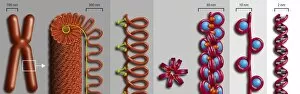
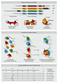
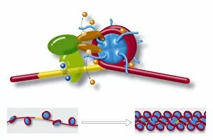
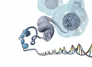
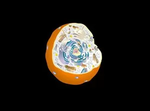
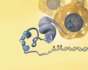
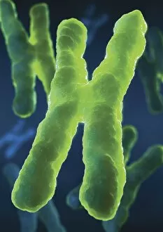
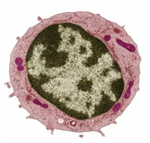
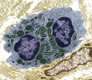
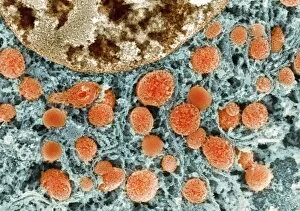
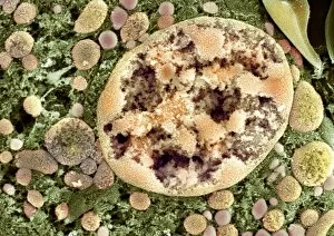
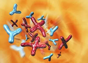
Chromatin, the intricate web of genetic material within our cells, holds the key to understanding life's blueprint. At its core lies the nucleosome molecule, a fundamental building block consisting of DNA wrapped around proteins. In a stunning molecular model, we witness the elegant dance between these two entities. Peering through an electron microscope, we uncover plasma cells with their chromatin in full view. The TEM image reveals the intricate organization and compactness of this vital substance within these specialized immune system warriors. Moving on to human chromosomes captured by SEM, we are mesmerized by their striking beauty. These thread-like structures hold our entire genetic code and play a crucial role in inheritance and development. Shifting gears to plant cells, a conceptual image showcases not only chromatin but also other essential components that make up these green powerhouses, and is here where photosynthesis takes place and life flourishes against all odds. Delving deeper into animal cell territory under microscopic scrutiny once again unveils chromatin's presence. Its delicate strands intertwine amidst various organelles like mitochondria and ribosomes – orchestrating cellular functions critical for survival. Comparing plant and animal cell anatomy side by side brings forth fascinating insights into evolutionary divergence. With labels highlighting distinct features such as chloroplasts or centrioles, it becomes evident how nature has tailored each organism's chromatin architecture to suit its unique needs. In multiple microscopic views of animal cells, we witness firsthand the sheer complexity hidden beneath our skin's surface. Chromatin intricately woven alongside countless other cellular components reminds us that life is indeed an extraordinary tapestry waiting to be unraveled. As scientists continue unraveling chromatin's mysteries, one thing remains certain: this enigmatic entity holds profound significance in shaping who we are as living beings. From nucleosomes to chromosomes across diverse organisms' cells – every glimpse deepens our appreciation for this remarkable phenomenon known as chromatin.