Fine Art Print > Popular Themes > DNA
Fine Art Print : Rough endoplasmic reticulum, TEM

![]()
Fine Art Prints from Science Photo Library
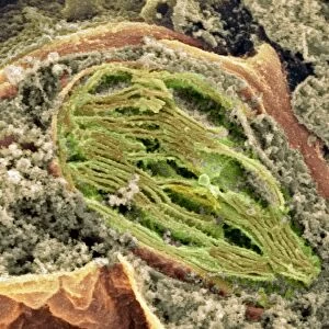
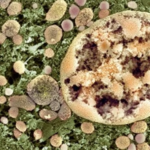
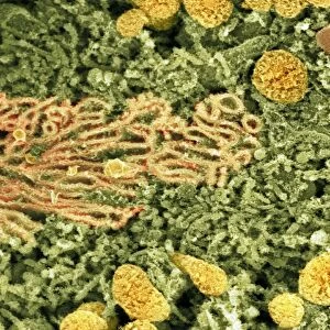
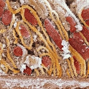
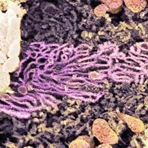
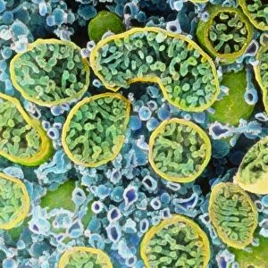
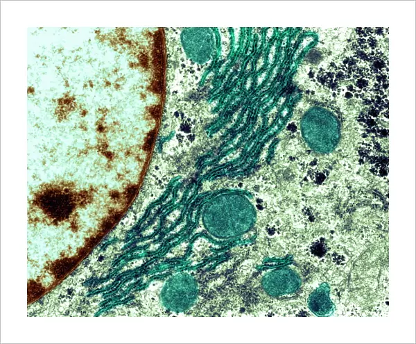
Rough endoplasmic reticulum, TEM
Rough endoplasmic reticulum, coloured transmission electron micrograph (TEM). This section shows the rough endoplasmic reticulum (ER, folds, centre), a membranous structure that occurs in cells. The cell nucleus is partially seen at left. Rough ER has ribosomes on its surface where protein synthesis occurs. The instructions for the protein synthesis come from the DNA (deoxyribonucleic acid) in the nucleus. The rounded structures may be vesicles that have broken off the rough ER to transport the proteins elsewhere in the cell. That would mean that this is the Golgi apparatus. Magnification: x20, 000 when printed 10cm wide
Science Photo Library features Science and Medical images including photos and illustrations
Media ID 6403199
© BIOMEDICAL IMAGING UNIT, SOUTHAMPTON GENERAL HOSPITAL/SCIENCE PHOTO LIBRARY
Cellular Cross Section Cytological Cytology Endoplasmic Reticulum False Colour Golgi Apparatus Histological Histology Membrane Nucleus Organelle Organelles Physiological Physiology Protein Synthesis Ribosome Ribosomes Rough Transmission Electron Microscope Vesicle Vesicles False Coloured Section Sectioned
20"x16" (+3" Border) Fine Art Print
Discover the intricacies of cellular structures with our Fine Art Prints from Media Storehouse. This captivating image showcases the Rough Endoplasmic Reticulum, as captured in stunning detail through Transmission Electron Microscopy (TEM) by Science Photo Library. Witness the mesmerizing folds of this membranous structure, a vital component of cellular function, and bring the beauty of science into your home or office space.
20x16 image printed on 26x22 Fine Art Rag Paper with 3" (76mm) white border. Our Fine Art Prints are printed on 300gsm 100% acid free, PH neutral paper with archival properties. This printing method is used by museums and art collections to exhibit photographs and art reproductions.
Our fine art prints are high-quality prints made using a paper called Photo Rag. This 100% cotton rag fibre paper is known for its exceptional image sharpness, rich colors, and high level of detail, making it a popular choice for professional photographers and artists. Photo rag paper is our clear recommendation for a fine art paper print. If you can afford to spend more on a higher quality paper, then Photo Rag is our clear recommendation for a fine art paper print.
Estimated Image Size (if not cropped) is 50.8cm x 40.6cm (20" x 16")
Estimated Product Size is 66cm x 55.9cm (26" x 22")
These are individually made so all sizes are approximate
Artwork printed orientated as per the preview above, with landscape (horizontal) orientation to match the source image.
EDITORS COMMENTS
This print showcases the intricate structure of the rough endoplasmic reticulum (ER) in a cell. In this coloured transmission electron micrograph, we can observe the folds and membranes that make up this vital organelle. The rough ER is distinguished by the presence of ribosomes on its surface, where protein synthesis takes place. The DNA within the cell nucleus provides instructions for protein synthesis, which are then carried out by the ribosomes on the rough ER. As proteins are synthesized, they may be transported to other parts of the cell through vesicles that have broken off from the rough ER. These rounded structures visible in the image could potentially be these transport vesicles destined for another crucial cellular component –the Golgi apparatus. With a magnification of x20,000 when printed at 10cm wide, this photograph offers an extraordinary glimpse into cellular biology and physiology. It highlights not only how cells function but also emphasizes their complexity and organization at a microscopic level. This stunning image was captured using a transmission electron microscope (TEM), allowing us to explore cross-sections of cells with incredible detail. Provided by Science Photo Library, it serves as a valuable resource for researchers and enthusiasts alike who seek to delve deeper into cytology, histology, and biological sciences as a whole.
MADE IN THE USA
Safe Shipping with 30 Day Money Back Guarantee
FREE PERSONALISATION*
We are proud to offer a range of customisation features including Personalised Captions, Color Filters and Picture Zoom Tools
SECURE PAYMENTS
We happily accept a wide range of payment options so you can pay for the things you need in the way that is most convenient for you
* Options may vary by product and licensing agreement. Zoomed Pictures can be adjusted in the Cart.