
Ribosomes Collection
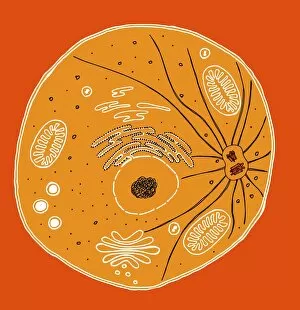
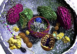
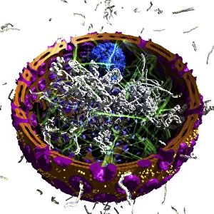
Ribosomes: The Tiny Workhorses of the Cell In the intricate world of cellular biology, ribosomes stand as the unsung heroes

For sale as Licensed Images
Choose your image, Select your licence and Download the media
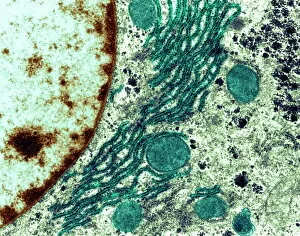
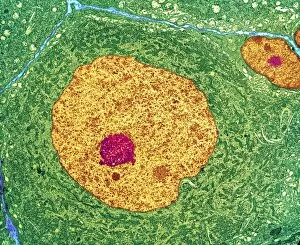
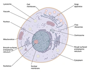
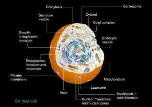
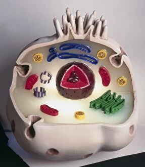
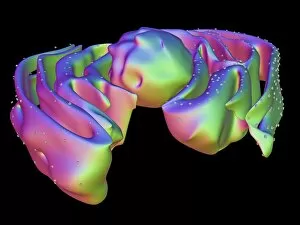
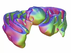
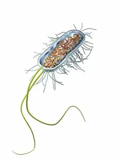
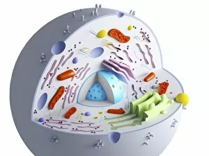
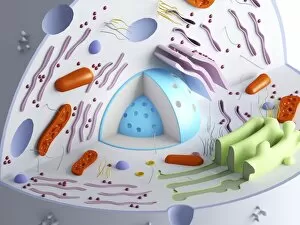
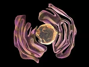
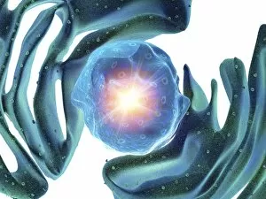
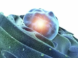
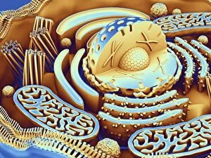
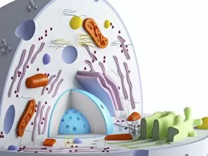
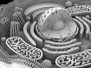
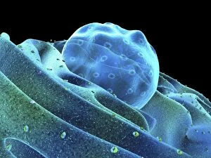
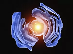
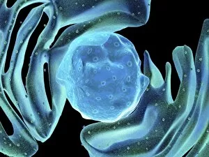
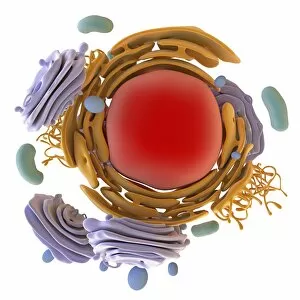
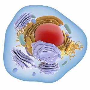
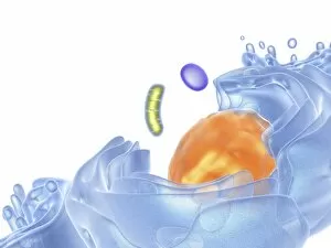
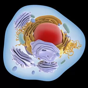
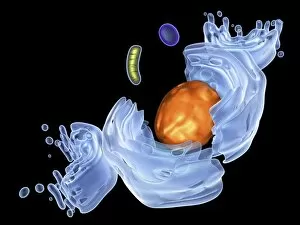
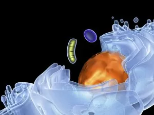
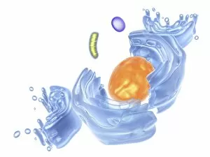
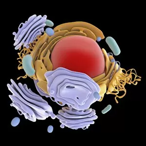
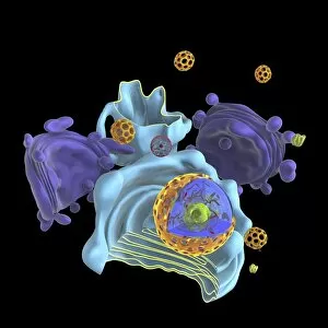
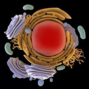
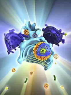
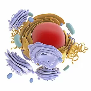
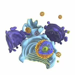
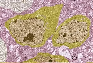
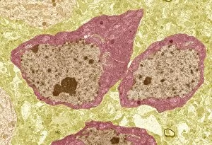
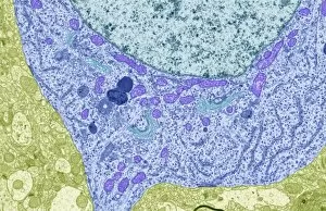
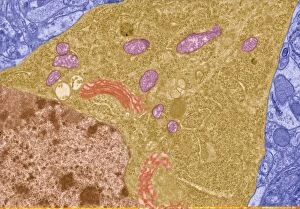
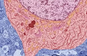
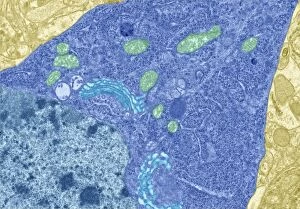
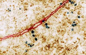
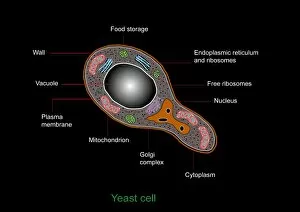
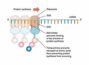
Ribosomes: The Tiny Workhorses of the Cell In the intricate world of cellular biology, ribosomes stand as the unsung heroes, tirelessly carrying out vital tasks within our cells. These tiny structures play a crucial role in protein synthesis, ensuring that life-sustaining molecules are produced efficiently. Underneath the electron microscope's lens, we can observe ribosomes at work within the rough endoplasmic reticulum (ER). This network of interconnected membranes serves as a manufacturing hub for proteins destined to be exported or integrated into cell membranes. With their characteristic appearance, these ER-bound ribosomes resemble small dots scattered throughout its surface. Zooming further into nerve cells through transmission electron microscopy (TEM), we witness an astonishing sight. Ribosomes dotting along dendrites and axons reveal their presence even in remote corners of our nervous system. Their abundance signifies their importance in maintaining neuronal function and facilitating communication between brain cells. Stepping back to admire animal cell structure as a whole, we encounter an intricately detailed model showcasing various organelles working harmoniously together. Among them stands the mighty ribosome – a fundamental component responsible for translating genetic information stored within DNA into functional proteins. Delving deeper into specific images captured by TEM and artwork illustrations, we gain insight into how these remarkable structures operate. Whether it is observing ribosomes embedded on rough ER surfaces or witnessing their presence within prokaryotic cells or animal cells' organelles - each image reveals another piece of this captivating puzzle. The nucleus and endoplasmic reticulum images highlight how closely intertwined these two components are with ribosomal activity. It emphasizes that while nucleoli produce essential RNA molecules needed for protein synthesis, it is ultimately carried out by ribosomes attached to ER membranes. As our understanding grows about these microscopic marvels called ribosomes, so does our appreciation for their significance in sustaining life itself.