Framed Print : Liver cells, SEM


![]()
Framed Photos from Science Photo Library
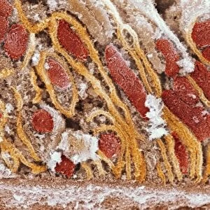
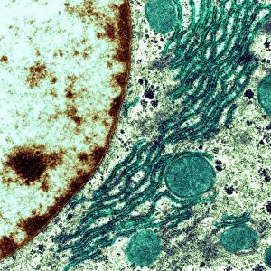
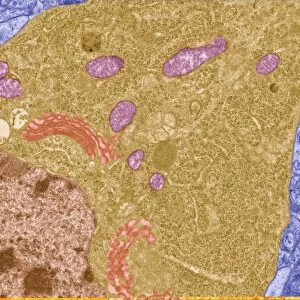
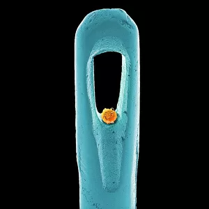
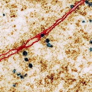
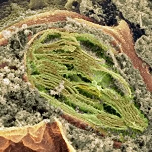
Liver cells, SEM
Liver cells. Scanning electron micrograph (SEM) of liver cells or hepatocytes. Several of the cells (for example, at upper left) are undergoing cell division. Hepatocytes are specialised epithelial cells and the most abundant cells in the liver. They are arranged in units known as lobules, each lobule surrounding a vein (not seen). Hepatocytes are involved with the various functions of the liver, including metabolism, detoxification of the blood and bile production. Magnification: x3900 at 6.5x8.5 inch size
Science Photo Library features Science and Medical images including photos and illustrations
Media ID 6450675
© DAVID MCCARTHY/SCIENCE PHOTO LIBRARY
Cytokinesis Cytology Detoxification Dividing Division Hepatic Hepatocyte Hepatocytes Lobule Metabolism Mono Chrome
12"x10" Modern Frame
Discover the intricacies of life with our Media Storehouse Framed Prints featuring this stunning Scanning Electron Micrograph (SEM) image of Liver Cells by Science Photo Library. Witness the beauty of hepatocytes in action, with several cells undergoing cell division, capturing the essence of life's continuous renewal. Bring this mesmerizing scientific discovery into your home or office space, and ignite conversations about the wonders of biology. Each print is professionally framed and printed on premium quality paper for a vibrant, long-lasting display. Add this Framed Print to your collection and embellish your surroundings with the marvels of science.
10x8 Print in an MDF Wooden Frame with 180 gsm Satin Finish Paper. Glazed using shatter proof thin plexi glass. Frame thickness is 1 inch and depth 0.75 inch. Fluted cardboard backing held with clips. Supplied ready to hang with sawtooth hanger and rubber bumpers. Spot clean with a damp cloth. Packaged foam wrapped in a card.
Contemporary Framed and Mounted Prints - Professionally Made and Ready to Hang
Estimated Image Size (if not cropped) is 25.4cm x 25.4cm (10" x 10")
Estimated Product Size is 30.5cm x 25.4cm (12" x 10")
These are individually made so all sizes are approximate
Artwork printed orientated as per the preview above, with landscape (horizontal) or portrait (vertical) orientation to match the source image.
EDITORS COMMENTS
This print showcases the intricate beauty of liver cells, captured through a scanning electron micrograph (SEM). The image reveals hepatocytes, which are specialized epithelial cells and the most abundant in the liver. These remarkable cells are arranged in lobules surrounding veins, although not visible in this particular shot. The photograph offers a glimpse into the dynamic nature of these hepatocytes as some undergo cell division, exemplified by those at the upper left corner. This process known as cytokinesis plays a crucial role in maintaining healthy liver function. Hepatocytes are involved in various vital functions such as metabolism, detoxification of blood, and bile production. At an impressive magnification of x3900 and printed at 6.5x8.5 inches size, this SEM image allows us to appreciate the complexity and intricacy within our own bodies on a microscopic level. It serves as a reminder that even at this minute scale, life continues to thrive with precision and purpose. Science Photo Library has once again provided us with an awe-inspiring visual representation of human anatomy - reminding us of both its normality and extraordinary capabilities without mentioning any commercial use or affiliation with any company.
MADE IN THE USA
Safe Shipping with 30 Day Money Back Guarantee
FREE PERSONALISATION*
We are proud to offer a range of customisation features including Personalised Captions, Color Filters and Picture Zoom Tools
SECURE PAYMENTS
We happily accept a wide range of payment options so you can pay for the things you need in the way that is most convenient for you
* Options may vary by product and licensing agreement. Zoomed Pictures can be adjusted in the Cart.