Anatomie Collection (#2)

"Exploring the Intricacies of Anatomie: From Ligaments to Artwork" Delve into the fascinating world of anatomie, where the human body and art intertwine

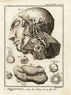
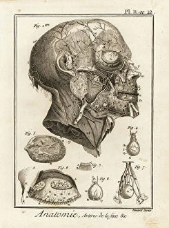
Background imageAnatomie Collection: Arteries in the face. Copperplate engraving by Robert Benard after an illustration by Albrecht von

Arteries in the face. Copperplate engraving by Robert Benard after an illustration by Albrecht von Haller from Denis
FLO4611624 Arteries in the face. Copperplate engraving by Robert Benard after an illustration by Albrecht von Haller from Denis Diderot's Encyclopedia, Pellet, Geneva

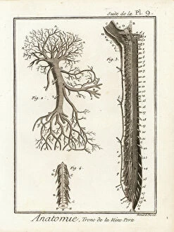
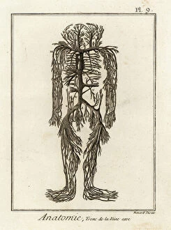
Background imageAnatomie Collection: Trunk of the hepatic portal vein. Copperplate engraving by Robert Benard from Denis Diderot's

Trunk of the hepatic portal vein. Copperplate engraving by Robert Benard from Denis Diderot's Encyclopedia, Pellet
FLO4611607 Trunk of the hepatic portal vein. Copperplate engraving by Robert Benard from Denis Diderot's Encyclopedia, Pellet, Geneva, 1779.; (add.info.: Trunk of the hepatic portal vein)

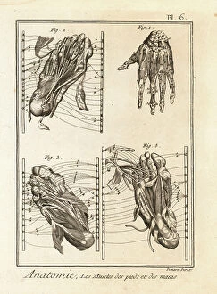
Background imageAnatomie Collection: Dissection of the muscles in the hand and foot. Copperplate engraving by Robert Benard from Denis

Dissection of the muscles in the hand and foot. Copperplate engraving by Robert Benard from Denis Diderot's
FLO4611571 Dissection of the muscles in the hand and foot. Copperplate engraving by Robert Benard from Denis Diderot's Encyclopedia, Pellet, Geneva

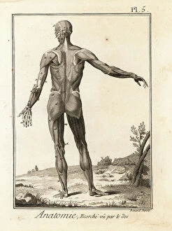
Background imageAnatomie Collection: Flayed human body revealing muscle system and tendons seen from the rear

Flayed human body revealing muscle system and tendons seen from the rear. Copperplate engraving by Robert Benard after
FLO4611562 Flayed human body revealing muscle system and tendons seen from the rear. Copperplate engraving by Robert Benard after an illustration by Bernhard Albinus from Denis Diderot's

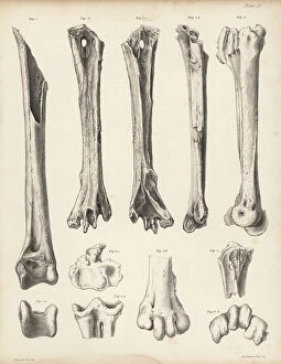
Background imageAnatomie Collection: Tibia and metatarsus of the extinct Rodrigues solitaire, Pezophaps solitaria

Tibia and metatarsus of the extinct Rodrigues solitaire, Pezophaps solitaria
FLO4612197 Tibia and metatarsus of the extinct Rodrigues solitaire, Pezophaps solitaria, in the Parisian Collection and Andersonian Collection

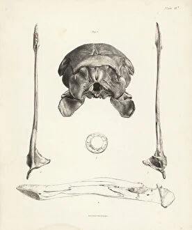
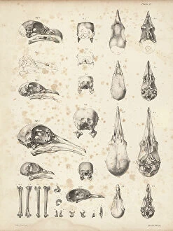
Background imageAnatomie Collection: Back view of skull, upper and lower view of lower jaw, inner view of jaw

Back view of skull, upper and lower view of lower jaw, inner view of jaw, and circle of sclerotic bones in the dodo
FLO4612158 Back view of skull, upper and lower view of lower jaw, inner view of jaw, and circle of sclerotic bones in the dodo

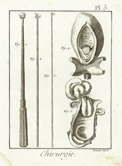
Background imageAnatomie Collection: Surgical instruments for extracting foreign bodies from the esophagus - Plate from '

Surgical instruments for extracting foreign bodies from the esophagus - Plate from " L'Encyclopedie" by
FLO4611943 Surgical instruments for extracting foreign bodies from the esophagus - Plate from " L'Encyclopedie" by Denis Diderot (1713-1784) and Jean Le Rond D'Alembert (1717-1783)

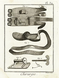
Background imageAnatomie Collection: Knee pad, slipper and mechanical mechanism - Plate from ' L'Encyclopedie' by Denis Diderot

Knee pad, slipper and mechanical mechanism - Plate from " L'Encyclopedie" by Denis Diderot (1713-1784)
FLO4611907 Knee pad, slipper and mechanical mechanism - Plate from " L'Encyclopedie" by Denis Diderot (1713-1784) and Jean Le Rond D'Alembert (1717-1783)

Background imageAnatomie Collection: The british physiologist William Harvey and the systemic circulation

The british physiologist William Harvey and the systemic circulation. Painting, France, 18th century (oil on canvas)
3434349 The british physiologist William Harvey and the systemic circulation. Painting, France, 18th century (oil on canvas); Academie des Sciences, Paris, France; Photo © CCI

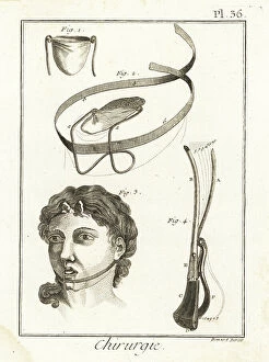
Background imageAnatomie Collection: Doctor Pibrac's tongue wound bandage and lithotoma, used to hunt the bladder - Plate taken from '

Doctor Pibrac's tongue wound bandage and lithotoma, used to hunt the bladder - Plate taken from "
FLO4611922 Doctor Pibrac's tongue wound bandage and lithotoma, used to hunt the bladder - Plate taken from " L'Encyclopedie" by Denis Diderot (1713-1784)

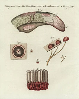
Background imageAnatomie Collection: Cow tongue under the microscope: veal tongue showing different papillae 1, foliate papilla 2

Cow tongue under the microscope: veal tongue showing different papillae 1, foliate papilla 2, filiform papillae 3
FLO4574832 Cow tongue under the microscope: veal tongue showing different papillae 1, foliate papilla 2, filiform papillae 3, and fungiform papillae 4

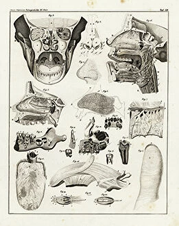
Background imageAnatomie Collection: Anatomy of the human nose, mouth, tongue, jaw and teeth, showing the senses of taste

Anatomy of the human nose, mouth, tongue, jaw and teeth, showing the senses of taste, smell and touch
FLO4684860 Anatomy of the human nose, mouth, tongue, jaw and teeth, showing the senses of taste, smell and touch. Lithograph from Lorenz Oken's Universal Natural History

Background imageAnatomie Collection: Scalp diseases, porrigo varietes and skin diseases: ulcerous pyodermatitis caused by bacteria

Scalp diseases, porrigo varietes and skin diseases: ulcerous pyodermatitis caused by bacteria - Lithography by John
FLO4618780 Scalp diseases, porrigo varietes and skin diseases: ulcerous pyodermatitis caused by bacteria - Lithography by John Pass, published in "Encyclopedia Londinensis" by John Wilkes

Background imageAnatomie Collection: Screws for compression of blood vessels by Jean Louis Petite

Screws for compression of blood vessels by Jean Louis Petite, tourniquet device and double band to adjust it
FLO4611834 Screws for compression of blood vessels by Jean Louis Petite, tourniquet device and double band to adjust it, chamois skin compress

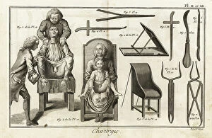
Background imageAnatomie Collection: surgical chair with patient to remove a vesical calculation through the groin - Plate taken from '

surgical chair with patient to remove a vesical calculation through the groin - Plate taken from "
FLO4611780 surgical chair with patient to remove a vesical calculation through the groin - Plate taken from " L'Encyclopedie" by Denis Diderot (1713-1784)

Background imageAnatomie Collection: Cranium 1-4 and part of sternum 5, 6 of the extinct Rodrigues solitaire

Cranium 1-4 and part of sternum 5, 6 of the extinct Rodrigues solitaire. Lithograph by Joseph Dinkel after Werner
FLO4612182 Cranium 1-4 and part of sternum 5, 6 of the extinct Rodrigues solitaire. Lithograph by Joseph Dinkel after Werner from Hugh Edwin Strickland

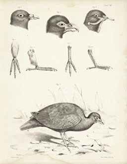
Background imageAnatomie Collection: Skulls of tooth-billed pigeon, Didunculus strigirostris, critically endangered 1, 7, 8, 9, 10 dodo

Skulls of tooth-billed pigeon, Didunculus strigirostris, critically endangered 1, 7, 8, 9, 10 dodo
FLO4612171 Skulls of tooth-billed pigeon, Didunculus strigirostris, critically endangered 1, 7, 8, 9, 10 dodo, Raphus cucullatus 2, yellow-footed green pigeon, Treron chlorigaster 3, 6

Background imageAnatomie Collection: Base of the skull, human teeth, knee cap (after Clopton Havers), mucus gland, etc

Base of the skull, human teeth, knee cap (after Clopton Havers), mucus gland, etc
FLO4611538 Base of the skull, human teeth, knee cap (after Clopton Havers), mucus gland, etc. Copperplate engraving by Robert Benard from Denis Diderot's Encyclopedia, Pellet, Geneva

Background imageAnatomie Collection: Interior of the brain and cerebellum. Copperplate engraving by Robert Benard after an illustration

Interior of the brain and cerebellum. Copperplate engraving by Robert Benard after an illustration by Albrecht von
FLO4611652 Interior of the brain and cerebellum. Copperplate engraving by Robert Benard after an illustration by Albrecht von Haller from Denis Diderot's Encyclopedia, Pellet, Geneva

Background imageAnatomie Collection: Arteries in the face. Copperplate engraving by Robert Benard after an illustration by Albrecht von

Arteries in the face. Copperplate engraving by Robert Benard after an illustration by Albrecht von Haller from Denis
FLO4611618 Arteries in the face. Copperplate engraving by Robert Benard after an illustration by Albrecht von Haller from Denis Diderot's Encyclopedia, Pellet, Geneva

Background imageAnatomie Collection: Venal system from the vena cava in the human body. Copperplate engraving by Robert Benard after an

Venal system from the vena cava in the human body. Copperplate engraving by Robert Benard after an illustration by
FLO4611599 Venal system from the vena cava in the human body. Copperplate engraving by Robert Benard after an illustration by James Drake from Denis Diderot's Encyclopedia, Pellet, Geneva

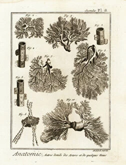
Background imageAnatomie Collection: Details of arteries, veins, lymph acorns, etc. Copperplate engraving by Robert Benard after

Details of arteries, veins, lymph acorns, etc. Copperplate engraving by Robert Benard after from Denis Diderot's
FLO4611594 Details of arteries, veins, lymph acorns, etc. Copperplate engraving by Robert Benard after from Denis Diderot's Encyclopedia, Pellet, Geneva, 1779.; (add.info.: Details of arteries)

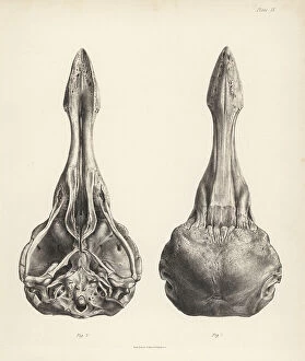
Background imageAnatomie Collection: Upper and lower views of the skull of a dodo. Lithograph from Hugh Edwin Strickland

Upper and lower views of the skull of a dodo. Lithograph from Hugh Edwin Strickland
FLO4612153 Upper and lower views of the skull of a dodo. Lithograph from Hugh Edwin Strickland and Alexander Gordon Melville's The Dodo and its Kindred, London, Reeve, Benham and Reeve

Background imageAnatomie Collection: Tooth billed pigeon, Didunculus strigirostris 1, 2 (critically endangered), Bruce's green pigeon

Tooth billed pigeon, Didunculus strigirostris 1, 2 (critically endangered), Bruce's green pigeon
FLO4612139 Tooth billed pigeon, Didunculus strigirostris 1, 2 (critically endangered), Bruce's green pigeon, Treron waalia (Treron abyssinica 3, 5) and Levaillant's dove

Background imageAnatomie Collection: Jean Louis Petite's machine to compress femoral artere during thigh amputation, bandage

Jean Louis Petite's machine to compress femoral artere during thigh amputation, bandage
FLO4611844 Jean Louis Petite's machine to compress femoral artere during thigh amputation, bandage, aneurysm needle and scissors - Plate taken from " L'Encyclopedie" by Denis

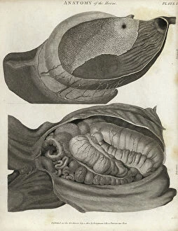
Background imageAnatomie Collection: Anatomy of the horse: stomach and intestines. Copperplate engraving by Scott from Abraham Rees

Anatomy of the horse: stomach and intestines. Copperplate engraving by Scott from Abraham Rees Cyclopedia or Universal
FLO4593968 Anatomy of the horse: stomach and intestines. Copperplate engraving by Scott from Abraham Rees Cyclopedia or Universal Dictionary of Arts, Sciences and Literature, Longman, Hurst, Rees

Background imageAnatomie Collection: Skeleton of a European perch, Perca fluviatilis, from Dr. Barclay's Museum, Edinburgh

Skeleton of a European perch, Perca fluviatilis, from Dr. Barclay's Museum, Edinburgh
FLO4565818 Skeleton of a European perch, Perca fluviatilis, from Dr. Barclay's Museum, Edinburgh. Copperplate engraving by Edward Mitchell after an anatomical illustration by Robert Kaye Greville

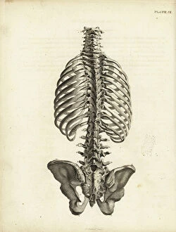
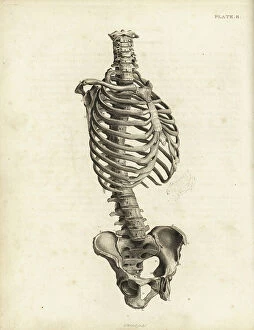
Background imageAnatomie Collection: Rear view of the human trunk skeleton including spine, ribs and pelvis

Rear view of the human trunk skeleton including spine, ribs and pelvis. Copperplate engraving by Edward Mitchell after
FLO4565792 Rear view of the human trunk skeleton including spine, ribs and pelvis. Copperplate engraving by Edward Mitchell after an anatomical illustration by Jean-Joseph Sue from John Barclay's

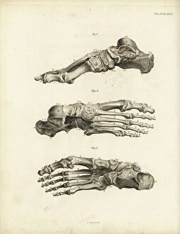
Background imageAnatomie Collection: Views of the bones in the human foot from ankle to toe. Copperplate engraving by Edward Mitchell

Views of the bones in the human foot from ankle to toe. Copperplate engraving by Edward Mitchell after an anatomical
FLO4565738 Views of the bones in the human foot from ankle to toe. Copperplate engraving by Edward Mitchell after an anatomical illustration by Jean-Joseph Sue from John Barclay's A Series of

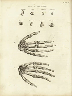
Background imageAnatomie Collection: Bones of the carpus, metacarpus and fingers in the human hand

Bones of the carpus, metacarpus and fingers in the human hand. Copperplate engraving by Edward Mitchell after an
FLO4565722 Bones of the carpus, metacarpus and fingers in the human hand. Copperplate engraving by Edward Mitchell after an anatomical illustration from John Barclay's A Series of Engravings of

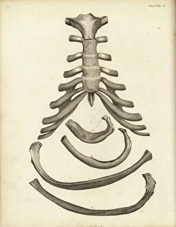
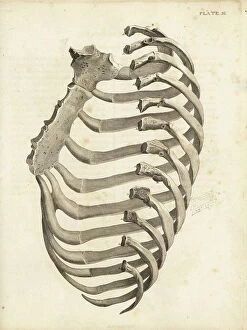
Background imageAnatomie Collection: Sternum and cartilage of the human ribcage, with several ribs

Sternum and cartilage of the human ribcage, with several ribs. Copperplate engraving by Edward Mitchell after an
FLO4565702 Sternum and cartilage of the human ribcage, with several ribs. Copperplate engraving by Edward Mitchell after an anatomical illustration by Jean-Joseph Sue from John Barclay's A Series

Background imageAnatomie Collection: Ribs and sternum in the human ribcage. Copperplate engraving by Edward Mitchell after an

Ribs and sternum in the human ribcage. Copperplate engraving by Edward Mitchell after an anatomical illustration by
FLO4565699 Ribs and sternum in the human ribcage. Copperplate engraving by Edward Mitchell after an anatomical illustration by Jean-Joseph Sue from John Barclay's A Series of Engravings of

Background imageAnatomie Collection: Front view of the human trunk skeleton including spine, ribs and pelvis

Front view of the human trunk skeleton including spine, ribs and pelvis. Copperplate engraving by Edward Mitchell after
FLO4565690 Front view of the human trunk skeleton including spine, ribs and pelvis. Copperplate engraving by Edward Mitchell after an anatomical illustration by Jean-Joseph Suefrom John Barclay's

Background imageAnatomie Collection: Prickly cockle - bucarde - Acanthocardia echinata (Cardium echinatum) 1

Prickly cockle - bucarde - Acanthocardia echinata (Cardium echinatum) 1, and slender sea pen - plume elegante
FLO4576265 Prickly cockle - bucarde - Acanthocardia echinata (Cardium echinatum) 1, and slender sea pen - plume elegante - Virgularia mirabilis (Pennatula mirabilis) 2

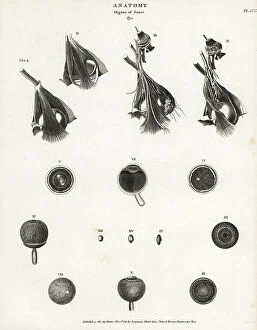
Background imageAnatomie Collection: Anatomy of the human eye, showing eyeball, optic nerve, muscle, cornea, retina, sections through eye

Anatomy of the human eye, showing eyeball, optic nerve, muscle, cornea, retina, sections through eye
FLO4591961 Anatomy of the human eye, showing eyeball, optic nerve, muscle, cornea, retina, sections through eye. Copperplate engraving by Milton from Abraham Rees Cyclopedia or Universal Dictionary

Background imageAnatomie Collection: Anatomy of human internal organs from the back showing stomach, liver, intestines, gallbladder, etc

Anatomy of human internal organs from the back showing stomach, liver, intestines, gallbladder, etc
FLO4590759 Anatomy of human internal organs from the back showing stomach, liver, intestines, gallbladder, etc. Copperplate engraving by Milton from Abraham Rees Cyclopedia or Universal Dictionary of

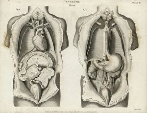
Background imageAnatomie Collection: Anatomy of human internal organs from the front showing heart, stomach, intestines

Anatomy of human internal organs from the front showing heart, stomach, intestines, kidneys and bladder
FLO4590679 Anatomy of human internal organs from the front showing heart, stomach, intestines, kidneys and bladder. Copperplate engraving by Milton from Abraham Rees Cyclopedia or Universal

Background imageAnatomie Collection: Head muscles. Lithograph of Lebrun, based on a drawing by Leveille

Head muscles. Lithograph of Lebrun, based on a drawing by Leveille, in Petity Atlas complet d'Anatomie descriptive
FLO4672607 Head muscles. Lithograph of Lebrun, based on a drawing by Leveille, in Petity Atlas complet d'Anatomie descriptive du Corps Humain, by Dr

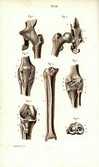
Background imageAnatomie Collection: Hip joint and thigh bone. Lithograph of Corbie, in Petity Atlas complet d'Anatomie descriptive du

Hip joint and thigh bone. Lithograph of Corbie, in Petity Atlas complet d'Anatomie descriptive du Corps Humain
FLO4672595 Hip joint and thigh bone. Lithograph of Corbie, in Petity Atlas complet d'Anatomie descriptive du Corps Humain, by Dr

Background imageAnatomie Collection: Sympathic nervous system in the thoracic, lumbar and sacral plexus. Lithograph by Davesne

Sympathic nervous system in the thoracic, lumbar and sacral plexus. Lithograph by Davesne
FLO4672529 Sympathic nervous system in the thoracic, lumbar and sacral plexus. Lithograph by Davesne, based on a drawing by Leveille

Background imageAnatomie Collection: Sympathic nervous system in the cardiac plexus. Lithograph by Davesne

Sympathic nervous system in the cardiac plexus. Lithograph by Davesne, based on a drawing by Leveille
FLO4672521 Sympathic nervous system in the cardiac plexus. Lithograph by Davesne, based on a drawing by Leveille, in Petiure Atlas complet d'Anatomie descriptive du Corps Humain, by Dr

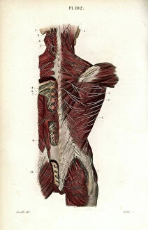
Background imageAnatomie Collection: Nervous system of the back. Lithograph of Corbie, based on a drawing by Leveille

Nervous system of the back. Lithograph of Corbie, based on a drawing by Leveille
FLO4672457 Nervous system of the back. Lithograph of Corbie, based on a drawing by Leveille, in Petity Atlas complet d'Anatomie descriptive du Corps Humain, by Dr

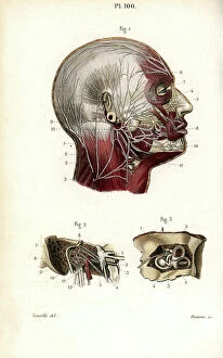
Background imageAnatomie Collection: Facial and auditory nerves. Lithograph by Davesne, based on a drawing by Leveille

Facial and auditory nerves. Lithograph by Davesne, based on a drawing by Leveille
FLO4672442 Facial and auditory nerves. Lithograph by Davesne, based on a drawing by Leveille, in Petite Atlas complet d'Anatomie descriptive du Corps Humain, by Dr

Background imageAnatomie Collection: Intercostal nerves. Lithograph of Corbie, based on a drawing by Leveille

Intercostal nerves. Lithograph of Corbie, based on a drawing by Leveille, in Petity Atlas complet d'Anatomie
FLO4672415 Intercostal nerves. Lithograph of Corbie, based on a drawing by Leveille, in Petity Atlas complet d'Anatomie descriptive du Corps Humain, by Dr

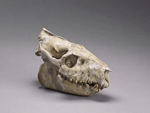
Background imageAnatomie Collection: Merycoidodon culbertsoni (skull of an Oreodon (hillock teeth)

Merycoidodon culbertsoni (skull of an Oreodon (hillock teeth), Museum d'histoire naturelle de Marseille
CSO4987831 Merycoidodon culbertsoni (skull of an Oreodon (hillock teeth), Museum d'histoire naturelle de Marseille; (add.info.: Merycoidodon culbertsoni (skull of an Oreodon (hillock teeth)

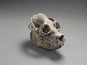
Background imageAnatomie Collection: Crane de Palaeopropithecus sp (Madagascar lemurian, extirpated species - sloth lemur of)

Crane de Palaeopropithecus sp (Madagascar lemurian, extirpated species - sloth lemur of Madagascar, extinct)
CSO4987821 Crane de Palaeopropithecus sp (Madagascar lemurian, extirpated species - sloth lemur of Madagascar, extinct), Museum d'histoire naturelle de Marseille; (add.info)

Background imageAnatomie Collection: Megaladapis sp (Skullof an Extinct Mammalia from Madagascar) Museum d'histoire naturelle de

Megaladapis sp (Skullof an Extinct Mammalia from Madagascar) Museum d'histoire naturelle de Marseille
CSO4987812 Megaladapis sp (Skullof an Extinct Mammalia from Madagascar) Museum d'histoire naturelle de Marseille; (add.info)

Background imageAnatomie Collection: Fetus head helped through the birth canal with forceps. Copperplate engraving by Andrew Bell after

Fetus head helped through the birth canal with forceps. Copperplate engraving by Andrew Bell after an illustration by
FLO4695943 Fetus head helped through the birth canal with forceps. Copperplate engraving by Andrew Bell after an illustration by Jan van Rymsdyk from William Smellie's A Set of Anatomical Tables


For sale as Licensed Images

Choose your image, Select your licence and Download the media

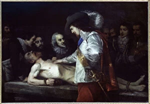
"Exploring the Intricacies of Anatomie: From Ligaments to Artwork" Delve into the fascinating world of anatomie, where the human body and art intertwine. Journey through a diverse collection of captivating images and historical references that shed light on our understanding of anatomy. Discover the delicate outer ankle ligaments in artwork C013/4452, beautifully depicted with meticulous detail. Witness how artists skillfully capture not only the physicality but also the essence of these intricate structures. Step into a different realm as dog anatomy takes center stage in another mesmerizing artwork. Uncover the intricacies of canine physiology, marveling at how their bodies are both similar and distinct from ours. Travel back in time to Uncle Tom's Cabin, as George Cruikshank's illustration brings Harriet Beecher Stowe's powerful novel to life. Explore how literature can provide insight into societal perceptions and challenges faced by individuals during that era. Max Ernst's painting "The Couple" offers an intriguing glimpse into human relationships and emotions. Delicate brushstrokes convey a depth of feeling that transcends time, inviting us to contemplate love, connection, and vulnerability. Witness Doctor Alfred Velleau conducting an autopsy amidst his attentive students in Francois Feyen Perrin's 19th-century masterpiece. This thought-provoking image captures both scientific curiosity and reverence for knowledge as medical professionals strive to unravel mysteries hidden within our bodies. Jean-Baptiste Marc Bourgery's colored engraving showcases amputations and prosthetics—a testament to humanity's resilience even in times of war-induced adversity. These artificial limbs symbolize hope for soldiers who have endured unimaginable hardships on the battlefield. "The Anatomy Lesson" series by Drs Nicolaes Tulp and Willem van der Meer transports us back centuries ago when dissection was a groundbreaking practice. These oil paintings immortalize moments where science meets artistry while unraveling secrets concealed beneath our skin.