Anatomie Collection (#5)

"Exploring the Intricacies of Anatomie: From Ligaments to Artwork" Delve into the fascinating world of anatomie, where the human body and art intertwine

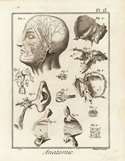
Background imageAnatomie Collection: Details of the anatomy of the human ear, showing cochlea, stapes, incus, malleus

Details of the anatomy of the human ear, showing cochlea, stapes, incus, malleus, semicircular canal, etc
FLO4611637 Details of the anatomy of the human ear, showing cochlea, stapes, incus, malleus, semicircular canal, etc. Copperplate engraving by Robert Benard after an illustration by Guichard Joseph

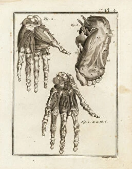
Background imageAnatomie Collection: Dissection of the human hand and foot to reveal muscle, ligament and bone

Dissection of the human hand and foot to reveal muscle, ligament and bone
FLO4611552 Dissection of the human hand and foot to reveal muscle, ligament and bone. Copperplate engraving by Robert Benard from Denis Diderot's Encyclopedia, Pellet, Geneva

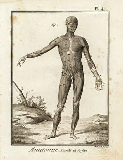
Background imageAnatomie Collection: Flayed human body revealing muscle system and tendons seen from the front

Flayed human body revealing muscle system and tendons seen from the front
FLO4611548 Flayed human body revealing muscle system and tendons seen from the front. Copperplate engraving by Robert Benard after an illustration by Bernhard Albinus from Denis Diderot's

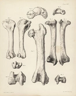
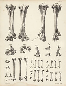
Background imageAnatomie Collection: Humerus and femur of the extinct Rodrigues solitaire, Pezophaps solitaria

Humerus and femur of the extinct Rodrigues solitaire, Pezophaps solitaria
FLO4612190 Humerus and femur of the extinct Rodrigues solitaire, Pezophaps solitaria, in the Parisian Collection and Andersonian Collection

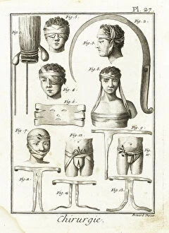
Background imageAnatomie Collection: Rectum with seistule, syringotome (syringe) for incising the anal fistula

Rectum with seistule, syringotome (syringe) for incising the anal fistula
FLO4611875 Rectum with seistule, syringotome (syringe) for incising the anal fistula, several bandages - Plate from " L'Encyclopedie" by Denis Diderot (1713-1784)

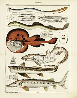
Background imageAnatomie Collection: Marine creatures: hagfish, Myxine glutinosa 1, river lamprey eel, Petromyzon fluviatilis 2

Marine creatures: hagfish, Myxine glutinosa 1, river lamprey eel, Petromyzon fluviatilis 2, torpedo ray
FLO4684894 Marine creatures: hagfish, Myxine glutinosa 1, river lamprey eel, Petromyzon fluviatilis 2, torpedo ray, Raja torpedo ocellata 3, great white shark, Squalus carcharias, vulnerable 4

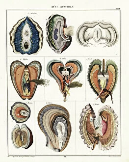
Background imageAnatomie Collection: Varieties of bivalve molluscs including clam, Tridacna, mussel, Mytilus, oyster, Ostrea

Varieties of bivalve molluscs including clam, Tridacna, mussel, Mytilus, oyster, Ostrea, pen shell, Pinna
FLO4684889 Varieties of bivalve molluscs including clam, Tridacna, mussel, Mytilus, oyster, Ostrea, pen shell, Pinna, jingle shell, Anomia, and scallop, Pecten

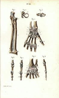
Background imageAnatomie Collection: Bone of the forearm, wrist and hand. Lithograph of Corbie, in Petity Atlas complet d'Anatomie

Bone of the forearm, wrist and hand. Lithograph of Corbie, in Petity Atlas complet d'Anatomie descriptive du Corps
FLO4672575 Bone of the forearm, wrist and hand. Lithograph of Corbie, in Petity Atlas complet d'Anatomie descriptive du Corps Humain, by Dr

Background imageAnatomie Collection: Sciatic nerve and sacred plexus. Lithograph of Corbie, based on a drawing by Leveille

Sciatic nerve and sacred plexus. Lithograph of Corbie, based on a drawing by Leveille
FLO4672464 Sciatic nerve and sacred plexus. Lithograph of Corbie, based on a drawing by Leveille, in Petity Atlas complet d'Anatomie descriptive du Corps Humain, by Dr

Background imageAnatomie Collection: Sections of the brain and pineal gland. Lithograph by Davesne, based on a drawing by Leveille

Sections of the brain and pineal gland. Lithograph by Davesne, based on a drawing by Leveille
FLO4673135 Sections of the brain and pineal gland. Lithograph by Davesne, based on a drawing by Leveille, in Petity Atlas complet d'Anatomie descriptive du Corps Humain, by Dr

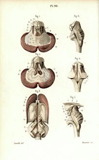
Background imageAnatomie Collection: Cerebellum section, ventricular system. Lithograph by Davesne, based on a drawing by Leveille

Cerebellum section, ventricular system. Lithograph by Davesne, based on a drawing by Leveille
FLO4673122 Cerebellum section, ventricular system. Lithograph by Davesne, based on a drawing by Leveille, in Petity Atlas complet d'Anatomie descriptive du Corps Humain, by Dr

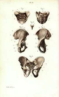
Background imageAnatomie Collection: Sacrum, coccyx and pelvic bone. Lithograph of Corbie, in Petity Atlas complet d'Anatomie

Sacrum, coccyx and pelvic bone. Lithograph of Corbie, in Petity Atlas complet d'Anatomie descriptive du Corps
FLO4673119 Sacrum, coccyx and pelvic bone. Lithograph of Corbie, in Petity Atlas complet d'Anatomie descriptive du Corps Humaine, by Dr

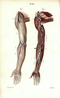
Background imageAnatomie Collection: Arm lymphatic system. Lithograph of Corbie, based on a drawing by Leveille

Arm lymphatic system. Lithograph of Corbie, based on a drawing by Leveille
FLO4673055 Arm lymphatic system. Lithograph of Corbie, based on a drawing by Leveille, in Petity Atlas complet d'Anatomie descriptive du Corps Humain, by Dr

Background imageAnatomie Collection: Lymph nodes and deep vessels of the back of the leg. Lithograph of Corbie

Lymph nodes and deep vessels of the back of the leg. Lithograph of Corbie, based on a drawing by Leveille
FLO4673037 Lymph nodes and deep vessels of the back of the leg. Lithograph of Corbie, based on a drawing by Leveille, in Petity Atlas complet d'Anatomie descriptive du Corps Humain, by Dr

Background imageAnatomie Collection: Lymphatic system of the leg and foot. Lithograph of Corbie, based on a drawing by Leveille

Lymphatic system of the leg and foot. Lithograph of Corbie, based on a drawing by Leveille
FLO4673013 Lymphatic system of the leg and foot. Lithograph of Corbie, based on a drawing by Leveille, in Petity Atlas complet d'Anatomie descriptive du Corps Humain, by Dr

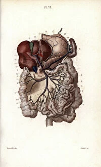
Background imageAnatomie Collection: Veins of the stomach, liver and intestines. Lithograph of Corbie, based on a drawing by Leveille

Veins of the stomach, liver and intestines. Lithograph of Corbie, based on a drawing by Leveille
FLO4672994 Veins of the stomach, liver and intestines. Lithograph of Corbie, based on a drawing by Leveille, in Petity Atlas complet d'Anatomie descriptive du Corps Humain, by Dr

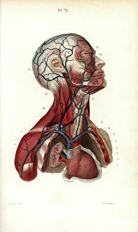
Background imageAnatomie Collection: Circulatory system of the head and torso. Lithograph of Anneshower, based on a drawing by Leveille

Circulatory system of the head and torso. Lithograph of Anneshower, based on a drawing by Leveille
FLO4672980 Circulatory system of the head and torso. Lithograph of Anneshower, based on a drawing by Leveille, in Petity Atlas complet d'Anatomie descriptive du Corps Humain, by Dr

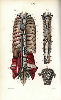
Background imageAnatomie Collection: Circulatory system of the vertebral column and uterus. Lithograph of Corbie

Circulatory system of the vertebral column and uterus. Lithograph of Corbie, based on a drawing by Leveille
FLO4672977 Circulatory system of the vertebral column and uterus. Lithograph of Corbie, based on a drawing by Leveille, in Petiure Atlas complet d'Anatomie descriptive du Corps Humain, by Dr

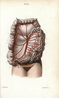
Background imageAnatomie Collection: The circulatory system of the intestines. Lithograph of Anneshower, based on a drawing by Leveille

The circulatory system of the intestines. Lithograph of Anneshower, based on a drawing by Leveille
FLO4672926 The circulatory system of the intestines. Lithograph of Anneshower, based on a drawing by Leveille, in Petity Atlas complet d'Anatomie descriptive du Corps Humain, by Dr

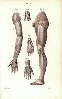
Background imageAnatomie Collection: Aponevrosis of the arm, leg and foot. Lithograph of Corbie, based on a drawing by Leveille

Aponevrosis of the arm, leg and foot. Lithograph of Corbie, based on a drawing by Leveille
FLO4672788 Aponevrosis of the arm, leg and foot. Lithograph of Corbie, based on a drawing by Leveille, in Petity Atlas complet d'Anatomie descriptive du Corps Humain, by Dr

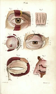
Background imageAnatomie Collection: Sections of the eye orbit and nasal cavity. Lithography of Davesne, based on a drawing by Leveille

Sections of the eye orbit and nasal cavity. Lithography of Davesne, based on a drawing by Leveille
FLO4672767 Sections of the eye orbit and nasal cavity. Lithography of Davesne, based on a drawing by Leveille, in Petity Atlas complet d'Anatomie descriptive du Corps Humain, by Dr

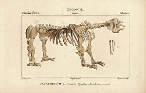
Background imageAnatomie Collection: Fossil skeleton of megatherium - Lithography, illustration by Jean Gabriel Pretre (1780-1885)

Fossil skeleton of megatherium - Lithography, illustration by Jean Gabriel Pretre (1780-1885)
FLO4651705 Fossil skeleton of megatherium - Lithography, illustration by Jean Gabriel Pretre (1780-1885) edited by Pierre Jean Francois Turpin (1775-1840)

Background imageAnatomie Collection: Anatomy of the human ear showing ear flap, anvil, stirrrup, cochlea

Anatomy of the human ear showing ear flap, anvil, stirrrup, cochlea, eustachian tube and internal parts
FLO4591705 Anatomy of the human ear showing ear flap, anvil, stirrrup, cochlea, eustachian tube and internal parts. Copperplate engraving by Milton from Abraham Rees Cyclopedia or Universal

Background imageAnatomie Collection: Anatomy of the human ear within the brain. Copperplate engraving by Milton from Abraham Rees
Anatomy of the human ear within the brain. Copperplate engraving by Milton from Abraham Rees Cyclopedia or Universal
FLO4591518 Anatomy of the human ear within the brain. Copperplate engraving by Milton from Abraham Rees Cyclopedia or Universal Dictionary of Arts, Sciences and Literature, Longman, Hurst, Rees
Background imageAnatomie Collection: Human skeleton from the back. Copperplate engraving by Milton from Abraham Rees Cyclopedia or

Human skeleton from the back. Copperplate engraving by Milton from Abraham Rees Cyclopedia or Universal Dictionary of
FLO4590971 Human skeleton from the back. Copperplate engraving by Milton from Abraham Rees Cyclopedia or Universal Dictionary of Arts, Sciences and Literature, Longman, Hurst, Rees, Orme and Brown

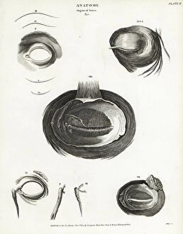
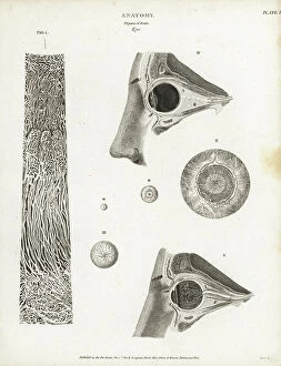
Background imageAnatomie Collection: Anatomy of the human eye, showing musculature, tear duct, etc

Anatomy of the human eye, showing musculature, tear duct, etc. Copperplate engraving by Milton from Abraham Rees
FLO4592148 Anatomy of the human eye, showing musculature, tear duct, etc. Copperplate engraving by Milton from Abraham Rees Cyclopedia or Universal Dictionary of Arts, Sciences and Literature

Background imageAnatomie Collection: Anatomy of birds: the crop or gullet. Copperplate engraving by T. Milton after a drawing by W

Anatomy of birds: the crop or gullet. Copperplate engraving by T. Milton after a drawing by W
FLO4594096 Anatomy of birds: the crop or gullet. Copperplate engraving by T. Milton after a drawing by W. Clift from Abraham Rees Cyclopedia or Universal Dictionary of Arts, Sciences and Literature

Background imageAnatomie Collection: Skeleton of a European robin, Erithacus rubecula (Motacilla rubecula) and common snipe

Skeleton of a European robin, Erithacus rubecula (Motacilla rubecula) and common snipe
FLO4565794 Skeleton of a European robin, Erithacus rubecula (Motacilla rubecula) and common snipe, Gallinago gallinago (Scollopax gallinago) from Dr. Barclay's Museum, Edinburgh

Background imageAnatomie Collection: Skeleton of an elephant with human skeleton rider, from Dr. Barclay's Museum, Edinburgh

Skeleton of an elephant with human skeleton rider, from Dr. Barclay's Museum, Edinburgh
FLO4565788 Skeleton of an elephant with human skeleton rider, from Dr. Barclay's Museum, Edinburgh. The specimen was assembled by Dr. George Ballingall, surgeon of the 33rd Regiment of Foot

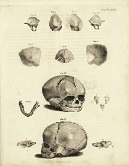
Background imageAnatomie Collection: Stages in the formation of the foetal skull. Copperplate engraving by Edward Mitchell after an

Stages in the formation of the foetal skull. Copperplate engraving by Edward Mitchell after an anatomical illustration
FLO4565769 Stages in the formation of the foetal skull. Copperplate engraving by Edward Mitchell after an anatomical illustration by Jean-Joseph Sue from John Barclay's A Series of Engravings of

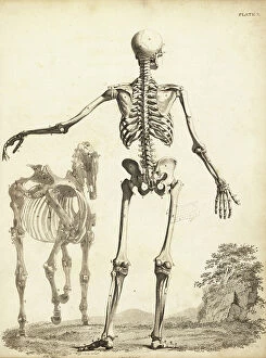
Background imageAnatomie Collection: Human skeleton from the rear, with horse skeleton by George Stubbs

Human skeleton from the rear, with horse skeleton by George Stubbs. Copperplate engraving by Edward Mitchell after an
FLO4565715 Human skeleton from the rear, with horse skeleton by George Stubbs. Copperplate engraving by Edward Mitchell after an anatomical illustration by Bernhard Siefried Albinus from John

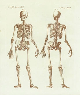
Background imageAnatomie Collection: Human skeleton from front and back. Handcoloured copperplate engraving from Bertuch's ' Bilderbuch

Human skeleton from front and back. Handcoloured copperplate engraving from Bertuch's " Bilderbuch fur
FLO4574495 Human skeleton from front and back. Handcoloured copperplate engraving from Bertuch's " Bilderbuch fur Kinder" (Picture Book for Children), Weimar, 1798

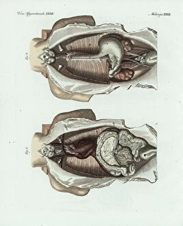
Background imageAnatomie Collection: Entrails in the human body: thyroid, stomach, esophagus, colon, pancreas, kidney, etc

Entrails in the human body: thyroid, stomach, esophagus, colon, pancreas, kidney, etc
FLO4574738 Entrails in the human body: thyroid, stomach, esophagus, colon, pancreas, kidney, etc. Handcoloured copperplate engraving from Bertuch's "Bilderbuch fur Kinder"

Background imageAnatomie Collection: Loxodonta sp (Coupe d'un crane d'elephant - Section of an elephant skull)

Loxodonta sp (Coupe d'un crane d'elephant - Section of an elephant skull)
CSO4987833 Loxodonta sp (Coupe d'un crane d'elephant - Section of an elephant skull), Museum d'histoire naturelle de Marseille; (add.info)

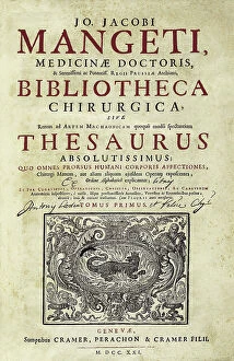
Background imageAnatomie Collection: MANGET, Jean-Jacques (1652 - 1742). Bibliotheca Surgica. 1721. MANGET, Jean-Jacques (1652 - 1742)

MANGET, Jean-Jacques (1652 - 1742). Bibliotheca Surgica. 1721. MANGET, Jean-Jacques (1652 - 1742)
CUL4875531 MANGET, Jean-Jacques (1652 - 1742). Bibliotheca Surgica. 1721. MANGET, Jean-Jacques (1652 - 1742). Swiss physician and writer. Cover of first edition, published in Geneva. Engraving

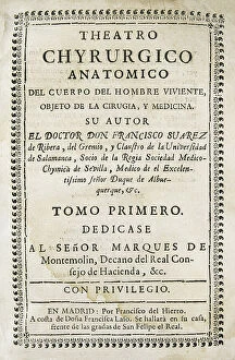
Background imageAnatomie Collection: SUAREZ de RIBERA, Francisco (S.XVIII). Spanish doctor. Illustration of ' Theatro Chyrurgico

SUAREZ de RIBERA, Francisco (S.XVIII). Spanish doctor. Illustration of " Theatro Chyrurgico anatomico del cuerpo
CUL4871750 SUAREZ de RIBERA, Francisco (S.XVIII). Spanish doctor. Illustration of " Theatro Chyrurgico anatomico del cuerpo del hombre viviente

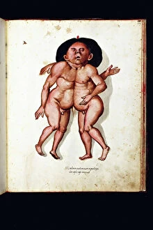
Background imageAnatomie Collection: Monster creatures. Illustrated chart of a manuscript of Natural History by Ulisse Aldrovandi

Monster creatures. Illustrated chart of a manuscript of Natural History by Ulisse Aldrovandi (1522-1605)
VEN5029583 Monster creatures. Illustrated chart of a manuscript of Natural History by Ulisse Aldrovandi (1522-1605), Ulisse Aldrovandi fonds, University Library of Bologna, ed

Background imageAnatomie Collection: Monster creatures. Illustrated chart of a manuscript of Natural History by Ulisse Aldrovandi

Monster creatures. Illustrated chart of a manuscript of Natural History by Ulisse Aldrovandi (1522-1605)
VEN5029542 Monster creatures. Illustrated chart of a manuscript of Natural History by Ulisse Aldrovandi (1522-1605), Ulisse Aldrovandi fonds, University Library of Bologna, ed

Background imageAnatomie Collection: Monster creatures. Illustrated chart of a manuscript of Natural History by Ulisse Aldrovandi

Monster creatures. Illustrated chart of a manuscript of Natural History by Ulisse Aldrovandi (1522-1605)
VEN5029514 Monster creatures. Illustrated chart of a manuscript of Natural History by Ulisse Aldrovandi (1522-1605), Ulisse Aldrovandi fonds, University Library of Bologna, ed

Background imageAnatomie Collection: Monster creature: woman with a monkey head. Illustrated chart of a manuscript of Natural History

Monster creature: woman with a monkey head. Illustrated chart of a manuscript of Natural History by Ulisse Aldrovandi
VEN5029505 Monster creature: woman with a monkey head. Illustrated chart of a manuscript of Natural History by Ulisse Aldrovandi (1522-1605), Ulisse Aldrovandi fonds, University Library of Bologna

Background imageAnatomie Collection: Anatomy of the human eye showing eyeball, cornea, lens, optic nerve

Anatomy of the human eye showing eyeball, cornea, lens, optic nerve, etc.CopperPlate engraving by Milton from Abraham
FLO4591962 Anatomy of the human eye showing eyeball, cornea, lens, optic nerve, etc.CopperPlate engraving by Milton from Abraham Rees Cyclopedia or Universal Dictionary of Arts

Background imageAnatomie Collection: Anatomy of the human eye, showing socket, surface of the eyeball, retina, etc

Anatomy of the human eye, showing socket, surface of the eyeball, retina, etc
FLO4591881 Anatomy of the human eye, showing socket, surface of the eyeball, retina, etc. Copperplate engraving by Milton from Abraham Rees Cyclopedia or Universal Dictionary of Arts

Background imageAnatomie Collection: Metatarsus bones of the dodo, Raphus cucullatus 1-10, crowned pigeon, Goura coronata 12-17

Metatarsus bones of the dodo, Raphus cucullatus 1-10, crowned pigeon, Goura coronata 12-17, tooth-billed pigeon
FLO4612222 Metatarsus bones of the dodo, Raphus cucullatus 1-10, crowned pigeon, Goura coronata 12-17, tooth-billed pigeon, Didunculus strigirostris 18, 19 and other birds

Background imageAnatomie Collection: Trocade a tracheotomy, brush for the stomach, tumour in the chest after Lorenz Heister

Trocade a tracheotomy, brush for the stomach, tumour in the chest after Lorenz Heister
FLO4611930 Trocade a tracheotomy, brush for the stomach, tumour in the chest after Lorenz Heister, scar after the elimination of the tumour

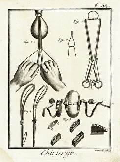
Background imageAnatomie Collection: Obstetric clamp and its use to remove a uterine polyp, tweezers

Obstetric clamp and its use to remove a uterine polyp, tweezers and speculum by Andre Levret - Plate from "
FLO4611914 Obstetric clamp and its use to remove a uterine polyp, tweezers and speculum by Andre Levret - Plate from " L'Encyclopedie" by Denis Diderot (1713-1784)

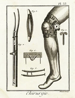
Background imageAnatomie Collection: Surgical knee pad and machine for treating disloqueous joints of Jean Louis Petite - Plate drawn

Surgical knee pad and machine for treating disloqueous joints of Jean Louis Petite - Plate drawn from "
FLO4611913 Surgical knee pad and machine for treating disloqueous joints of Jean Louis Petite - Plate drawn from " L'Encyclopedie" by Denis Diderot (1713-1784)

Background imageAnatomie Collection: Bandages, corbin beak pliers, cane spout pliers, balls removal pliers

Bandages, corbin beak pliers, cane spout pliers, balls removal pliers and various sutures - Plate taken from "
FLO4611896 Bandages, corbin beak pliers, cane spout pliers, balls removal pliers and various sutures - Plate taken from " L'Encyclopedie" by Denis Diderot (1713-1784)

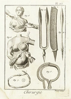
Background imageAnatomie Collection: Cruel and imperfect breast amputation, surgical pliers, razor and scalpel for mastectomy

Cruel and imperfect breast amputation, surgical pliers, razor and scalpel for mastectomy
FLO4611883 Cruel and imperfect breast amputation, surgical pliers, razor and scalpel for mastectomy, chest support instrument and its use


For sale as Licensed Images

Choose your image, Select your licence and Download the media

"Exploring the Intricacies of Anatomie: From Ligaments to Artwork" Delve into the fascinating world of anatomie, where the human body and art intertwine. Journey through a diverse collection of captivating images and historical references that shed light on our understanding of anatomy. Discover the delicate outer ankle ligaments in artwork C013/4452, beautifully depicted with meticulous detail. Witness how artists skillfully capture not only the physicality but also the essence of these intricate structures. Step into a different realm as dog anatomy takes center stage in another mesmerizing artwork. Uncover the intricacies of canine physiology, marveling at how their bodies are both similar and distinct from ours. Travel back in time to Uncle Tom's Cabin, as George Cruikshank's illustration brings Harriet Beecher Stowe's powerful novel to life. Explore how literature can provide insight into societal perceptions and challenges faced by individuals during that era. Max Ernst's painting "The Couple" offers an intriguing glimpse into human relationships and emotions. Delicate brushstrokes convey a depth of feeling that transcends time, inviting us to contemplate love, connection, and vulnerability. Witness Doctor Alfred Velleau conducting an autopsy amidst his attentive students in Francois Feyen Perrin's 19th-century masterpiece. This thought-provoking image captures both scientific curiosity and reverence for knowledge as medical professionals strive to unravel mysteries hidden within our bodies. Jean-Baptiste Marc Bourgery's colored engraving showcases amputations and prosthetics—a testament to humanity's resilience even in times of war-induced adversity. These artificial limbs symbolize hope for soldiers who have endured unimaginable hardships on the battlefield. "The Anatomy Lesson" series by Drs Nicolaes Tulp and Willem van der Meer transports us back centuries ago when dissection was a groundbreaking practice. These oil paintings immortalize moments where science meets artistry while unraveling secrets concealed beneath our skin.