Anatomie Collection (#4)

"Exploring the Intricacies of Anatomie: From Ligaments to Artwork" Delve into the fascinating world of anatomie, where the human body and art intertwine

Background imageAnatomie Collection: Side view of the skull of a dodo. Lithograph from Hugh Edwin Strickland

Side view of the skull of a dodo. Lithograph from Hugh Edwin Strickland and Alexander Gordon Melville's The Dodo
FLO4612152 Side view of the skull of a dodo. Lithograph from Hugh Edwin Strickland and Alexander Gordon Melville's The Dodo and its Kindred, London, Reeve, Benham and Reeve

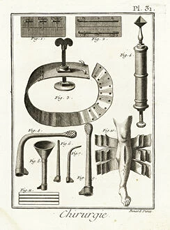
Background imageAnatomie Collection: Sutures, withers, fine-tip syringe by Dominique Anel, siphon, injection tubes

Sutures, withers, fine-tip syringe by Dominique Anel, siphon, injection tubes
FLO4611953 Sutures, withers, fine-tip syringe by Dominique Anel, siphon, injection tubes, 18-sheet bandage for complex fractures

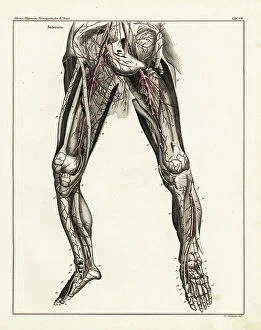
Background imageAnatomie Collection: Anatomy of the human arterial system in the legs. Handcoloured lithograph by St

Anatomy of the human arterial system in the legs. Handcoloured lithograph by St
FLO4684812 Anatomy of the human arterial system in the legs. Handcoloured lithograph by St. Schillinger from Lorenz Oken's Universal Natural History, Allgemeine Naturgeschichte fur alle Stande

Background imageAnatomie Collection: Anatomy of human intestines from the side. Lithograph from Lorenz Oken's Universal Natural History

Anatomy of human intestines from the side. Lithograph from Lorenz Oken's Universal Natural History
FLO4684807 Anatomy of human intestines from the side. Lithograph from Lorenz Oken's Universal Natural History, Allgemeine Naturgeschichte fur alle Stande, Stuttgart

Background imageAnatomie Collection: Human skeleton from the front. Engraving by Fournier after an illustration by Martin St

Human skeleton from the front. Engraving by Fournier after an illustration by Martin St
FLO4685437 Human skeleton from the front. Engraving by Fournier after an illustration by Martin St. Ange from Charles d'Orbigny's Dictionary of Natural History, Paris

Background imageAnatomie Collection: Brain sections. Lithograph by Davesne, based on a drawing by Leveille

Brain sections. Lithograph by Davesne, based on a drawing by Leveille, in Petity Atlas complet d'Anatomie
FLO4673204 Brain sections. Lithograph by Davesne, based on a drawing by Leveille, in Petity Atlas complet d'Anatomie descriptive du Corps Humain, by Dr

Background imageAnatomie Collection: Vertebral column, ribs, vertebrae and sternum. Lithograph of Corbie

Vertebral column, ribs, vertebrae and sternum. Lithograph of Corbie, in Petity Atlas complet d'Anatomie descriptive
FLO4673029 Vertebral column, ribs, vertebrae and sternum. Lithograph of Corbie, in Petity Atlas complet d'Anatomie descriptive du Corps Humain, by Dr

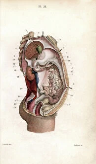
Background imageAnatomie Collection: Section through the abdomen. Lithograph of Lebrun, based on a drawing by Leveille

Section through the abdomen. Lithograph of Lebrun, based on a drawing by Leveille
FLO4672840 Section through the abdomen. Lithograph of Lebrun, based on a drawing by Leveille, in Petity Atlas complet d'Anatomie descriptive du Corps Humain, by Dr

Background imageAnatomie Collection: Aponevrosis (fibrous membrane that covers and supports the muscles) of the torso

Aponevrosis (fibrous membrane that covers and supports the muscles) of the torso, cross section and frontal view
FLO4672748 Aponevrosis (fibrous membrane that covers and supports the muscles) of the torso, cross section and frontal view

Background imageAnatomie Collection: Aponevrosis of the neck, shoulder and eye. Lithograph of Corbie, based on a drawing by Leveille

Aponevrosis of the neck, shoulder and eye. Lithograph of Corbie, based on a drawing by Leveille
FLO4672728 Aponevrosis of the neck, shoulder and eye. Lithograph of Corbie, based on a drawing by Leveille, in Petiure Atlas complet d'Anatomie descriptive du Corps Humain, by Dr

Background imageAnatomie Collection: Muscles and tendons of the leg and foot. Lithograph of Lebrun, based on a drawing by Leveille

Muscles and tendons of the leg and foot. Lithograph of Lebrun, based on a drawing by Leveille
FLO4672719 Muscles and tendons of the leg and foot. Lithograph of Lebrun, based on a drawing by Leveille, in Petity Atlas complet d'Anatomie descriptive du Corps Humain, by Dr

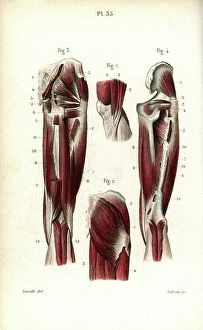
Background imageAnatomie Collection: Thigh and knee muscles and tendons. Lithograph of Lebrun, based on a drawing by Leveille

Thigh and knee muscles and tendons. Lithograph of Lebrun, based on a drawing by Leveille
FLO4672703 Thigh and knee muscles and tendons. Lithograph of Lebrun, based on a drawing by Leveille, in Petity Atlas complet d'Anatomie descriptive du Corps Humain, by Dr

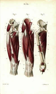
Background imageAnatomie Collection: Thigh muscles and tendons. Lithograph of Lebrun, based on a drawing by Leveille

Thigh muscles and tendons. Lithograph of Lebrun, based on a drawing by Leveille
FLO4672702 Thigh muscles and tendons. Lithograph of Lebrun, based on a drawing by Leveille, in Petity Atlas complet d'Anatomie descriptive du Corps Humain, by Dr

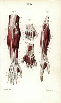
Background imageAnatomie Collection: Muscles and tendons of the forearm and wrist. Lithograph by Davesne, based on a drawing by Leveille

Muscles and tendons of the forearm and wrist. Lithograph by Davesne, based on a drawing by Leveille
FLO4672691 Muscles and tendons of the forearm and wrist. Lithograph by Davesne, based on a drawing by Leveille, in Petity Atlas complet d'Anatomie descriptive du Corps Humain, by Dr

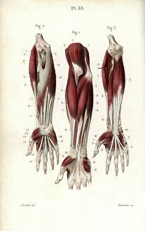
Background imageAnatomie Collection: Muscles of the forearm and wrist. Lithograph by Davesne, based on a drawing by Leveille

Muscles of the forearm and wrist. Lithograph by Davesne, based on a drawing by Leveille
FLO4672685 Muscles of the forearm and wrist. Lithograph by Davesne, based on a drawing by Leveille, in Petity Atlas complet d'Anatomie descriptive du Corps Humain, by Dr

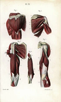
Background imageAnatomie Collection: Shoulder and arm muscles. Lithograph of Anneshower, based on a drawing by Leveille

Shoulder and arm muscles. Lithograph of Anneshower, based on a drawing by Leveille
FLO4672670 Shoulder and arm muscles. Lithograph of Anneshower, based on a drawing by Leveille, in Petiure Atlas complet d'Anatomie descriptive du Corps Humain, by Dr

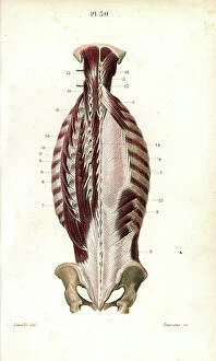
Background imageAnatomie Collection: Internal muscles of the back. Lithograph by Davesne, based on a drawing by Leveille

Internal muscles of the back. Lithograph by Davesne, based on a drawing by Leveille
FLO4672652 Internal muscles of the back. Lithograph by Davesne, based on a drawing by Leveille, in Petity Atlas complet d'Anatomie descriptive du Corps Humain, by Dr

Background imageAnatomie Collection: Torso muscles (male). Lithograph of anneshower, based on a drawing by Leveille

Torso muscles (male). Lithograph of anneshower, based on a drawing by Leveille
FLO4672627 Torso muscles (male). Lithograph of anneshower, based on a drawing by Leveille, in Petiter Atlas complet d'Anatomie descriptive du Corps Humain, by Dr

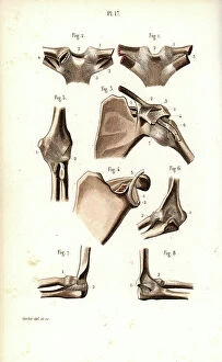
Background imageAnatomie Collection: Shoulder and elbow joints. Lithograph of Corbie, in Petity Atlas complet d'Anatomie descriptive du

Shoulder and elbow joints. Lithograph of Corbie, in Petity Atlas complet d'Anatomie descriptive du Corps Humain
FLO4672545 Shoulder and elbow joints. Lithograph of Corbie, in Petity Atlas complet d'Anatomie descriptive du Corps Humain, by Dr

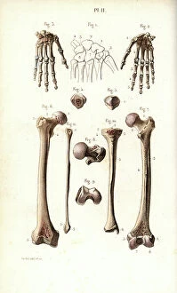
Background imageAnatomie Collection: Hand bone: carps, metacarps and phalanges and leg: femur and shin. Lithograph of Corbie
Hand bone: carps, metacarps and phalanges and leg: femur and shin. Lithograph of Corbie
FLO4672512 Hand bone: carps, metacarps and phalanges and leg: femur and shin. Lithograph of Corbie, in Petity Atlas complet d'Anatomie descriptive du Corps Humain, by Dr

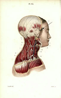
Background imageAnatomie Collection: Cervical plexus. Lithograph of Corbie, based on a drawing by Leveille

Cervical plexus. Lithograph of Corbie, based on a drawing by Leveille, in Petity Atlas complet d'Anatomie
FLO4673165 Cervical plexus. Lithograph of Corbie, based on a drawing by Leveille, in Petity Atlas complet d'Anatomie descriptive du Corps Humain, by Dr

Background imageAnatomie Collection: Veins of the legs and foot. Lithograph of Corbie, based on a drawing by Leveille

Veins of the legs and foot. Lithograph of Corbie, based on a drawing by Leveille
FLO4673005 Veins of the legs and foot. Lithograph of Corbie, based on a drawing by Leveille, in Petity Atlas complet d'Anatomie descriptive du Corps Humain, by Dr

Background imageAnatomie Collection: Axis and vertebrae. Lithograph of Corbie, in Petity Atlas complet d'Anatomie descriptive du Corps

Axis and vertebrae. Lithograph of Corbie, in Petity Atlas complet d'Anatomie descriptive du Corps Humain, by Dr
FLO4672967 Axis and vertebrae. Lithograph of Corbie, in Petity Atlas complet d'Anatomie descriptive du Corps Humain, by Dr

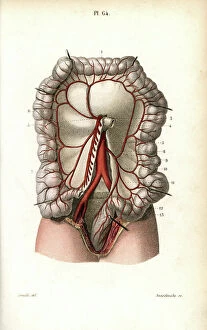
Background imageAnatomie Collection: Circulatory system of the intestines. Lithograph of Anneshower, based on a drawing by Leveille

Circulatory system of the intestines. Lithograph of Anneshower, based on a drawing by Leveille
FLO4672922 Circulatory system of the intestines. Lithograph of Anneshower, based on a drawing by Leveille, in Petity Atlas complet d'Anatomie descriptive du Corps Humain, by Dr

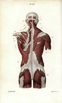
Background imageAnatomie Collection: Back and torso muscles. Lithograph by Davesne, based on a drawing by Leveille

Back and torso muscles. Lithograph by Davesne, based on a drawing by Leveille
FLO4672626 Back and torso muscles. Lithograph by Davesne, based on a drawing by Leveille, in Petity Atlas complet d'Anatomie descriptive du Corps Humain, by Dr

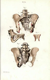
Background imageAnatomie Collection: Pelvic bone, front and back views. Lithograph of Corbie, in Petity Atlas complet d'Anatomie

Pelvic bone, front and back views. Lithograph of Corbie, in Petity Atlas complet d'Anatomie descriptive du Corps
FLO4672563 Pelvic bone, front and back views. Lithograph of Corbie, in Petity Atlas complet d'Anatomie descriptive du Corps Humain, by Dr

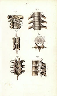
Background imageAnatomie Collection: Vertebral column, spine and neck. Lithograph of Corbie, in Petity Atlas complet d'Anatomie

Vertebral column, spine and neck. Lithograph of Corbie, in Petity Atlas complet d'Anatomie descriptive du Corps
FLO4672546 Vertebral column, spine and neck. Lithograph of Corbie, in Petity Atlas complet d'Anatomie descriptive du Corps Humain, by Dr

Background imageAnatomie Collection: Glossopharyngeal nerve. Lithograph by Davesne, based on a drawing by Leveille

Glossopharyngeal nerve. Lithograph by Davesne, based on a drawing by Leveille
FLO4672515 Glossopharyngeal nerve. Lithograph by Davesne, based on a drawing by Leveille, in Petity Atlas complet d'Anatomie descriptive du Corps Humain, by Dr

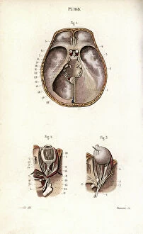
Background imageAnatomie Collection: Nerves of the skull and eye. Lithograph by Davesne, based on a drawing by Leveille

Nerves of the skull and eye. Lithograph by Davesne, based on a drawing by Leveille
FLO4672497 Nerves of the skull and eye. Lithograph by Davesne, based on a drawing by Leveille, in Petity Atlas complet d'Anatomie descriptive du Corps Humain, by Dr

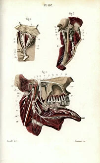
Background imageAnatomie Collection: Ophthalmic and maxillary nerves of the fifth cranial nerve. Lithograph by Davesne

Ophthalmic and maxillary nerves of the fifth cranial nerve. Lithograph by Davesne, based on a drawing by Leveille
FLO4672496 Ophthalmic and maxillary nerves of the fifth cranial nerve. Lithograph by Davesne, based on a drawing by Leveille, in Petity Atlas complet d'Anatomie descriptive du Corps Humain

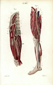
Background imageAnatomie Collection: Lumbar plexus and crural nerve. Lithograph of Corbie, based on a drawing by Leveille

Lumbar plexus and crural nerve. Lithograph of Corbie, based on a drawing by Leveille
FLO4672455 Lumbar plexus and crural nerve. Lithograph of Corbie, based on a drawing by Leveille, in Petity Atlas complet d'Anatomie descriptive du Corps Humain, by Dr

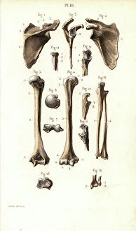
Background imageAnatomie Collection: Shoulder and arm bone: scapula, humerus, cubitus and radius. Lithograph of Corbie

Shoulder and arm bone: scapula, humerus, cubitus and radius. Lithograph of Corbie
FLO4672433 Shoulder and arm bone: scapula, humerus, cubitus and radius. Lithograph of Corbie, in Petity Atlas complet d'Anatomie descriptive du Corps Humain, by Dr

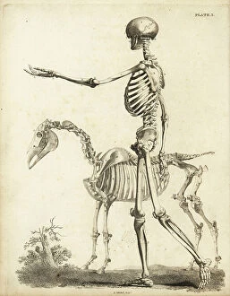
Background imageAnatomie Collection: Side view of human skeleton and horse skeleton. Copperplate engraving by Edward Mitchell after

Side view of human skeleton and horse skeleton. Copperplate engraving by Edward Mitchell after anatomical illustrations
FLO4565764 Side view of human skeleton and horse skeleton. Copperplate engraving by Edward Mitchell after anatomical illustrations by Bernhard Siegfried Albinus

Background imageAnatomie Collection: Fetal skeleton at six months with scythe 1, at three months 2 and at ten weeks 3

Fetal skeleton at six months with scythe 1, at three months 2 and at ten weeks 3
FLO4565763 Fetal skeleton at six months with scythe 1, at three months 2 and at ten weeks 3. Copperplate engraving by Edward Mitchell after an anatomical illustration by Jean-Joseph Sue from John

Background imageAnatomie Collection: Fetal skeleton at the time of birth. Copperplate engraving by Edward Mitchell after an anatomical

Fetal skeleton at the time of birth. Copperplate engraving by Edward Mitchell after an anatomical illustration by
FLO4565750 Fetal skeleton at the time of birth. Copperplate engraving by Edward Mitchell after an anatomical illustration by Jean-Joseph Sue from John Barclay's A Series of Engravings of

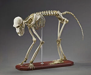
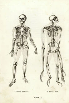
Background imageAnatomie Collection: Skeletons of a man, Homo sapiens, and endangered lar gibbon, Hylobates lar (Simia lar)

Skeletons of a man, Homo sapiens, and endangered lar gibbon, Hylobates lar (Simia lar)
FLO4720006 Skeletons of a man, Homo sapiens, and endangered lar gibbon, Hylobates lar (Simia lar). Lithograph from Georg Friedrich Treitschke's Gallery of Natural History

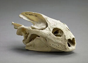
Background imageAnatomie Collection: Trionyx gangeticus (Nilssonia gangetica) (Skull of the Indian softshell turtle)

Trionyx gangeticus (Nilssonia gangetica) (Skull of the Indian softshell turtle)
CSO5006419 Trionyx gangeticus (Nilssonia gangetica) (Skull of the Indian softshell turtle), Museum d'histoire naturelle de Marseille; (add.info)

Background imageAnatomie Collection: Archaeolemur sp (Crane d'un lemurien subfossile de Madagascar - skull of an extint lemur)

Archaeolemur sp (Crane d'un lemurien subfossile de Madagascar - skull of an extint lemur from Madagascar)
CSO4987830 Archaeolemur sp (Crane d'un lemurien subfossile de Madagascar - skull of an extint lemur from Madagascar), Museum d'histoire naturelle de Marseille; (add.info)

Background imageAnatomie Collection: Papio hamadrya (Baboon hamadryas - hamadryas baboon), Museum d'histoire naturelle de Marseille

Papio hamadrya (Baboon hamadryas - hamadryas baboon), Museum d'histoire naturelle de Marseille
CSO4997023 Papio hamadrya (Baboon hamadryas - hamadryas baboon), Museum d'histoire naturelle de Marseille; (add.info.: Papio hamadrya (Baboon hamadryas - hamadryas baboon)

Background imageAnatomie Collection: Front view of a deformed female pelvis. Copperplate engraving by Andrew Bell after an illustration

Front view of a deformed female pelvis. Copperplate engraving by Andrew Bell after an illustration by Jan van Rymsdyk
FLO4696119 Front view of a deformed female pelvis. Copperplate engraving by Andrew Bell after an illustration by Jan van Rymsdyk from William Smellie's A Set of Anatomical Tables, Charles Elliot

Background imageAnatomie Collection: The abdomen of a woman in the sixth or seventh month of pregnancy, showing uterus, intestines

The abdomen of a woman in the sixth or seventh month of pregnancy, showing uterus, intestines, labia pudendi
FLO4696326 The abdomen of a woman in the sixth or seventh month of pregnancy, showing uterus, intestines, labia pudendi. Copperplate engraving by Andrew Bell after an illustration by Jan van Rymsdyk

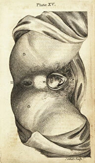
Background imageAnatomie Collection: Perineum and external parts stretched by the fetus head during late labour

Perineum and external parts stretched by the fetus head during late labour
FLO4695900 Perineum and external parts stretched by the fetus head during late labour. Copperplate engraving by Andrew Bell after an illustration by Jan van Rymsdyk from William Smellie's A Set

Background imageAnatomie Collection: Human skeleton viewed from the side. Copperplate engraving by Robert Benard after an illustration

Human skeleton viewed from the side. Copperplate engraving by Robert Benard after an illustration by Andreas Vesalius
FLO4611588 Human skeleton viewed from the side. Copperplate engraving by Robert Benard after an illustration by Andreas Vesalius from Denis Diderot's Encyclopedia, Pellet, Geneva

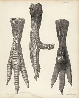
Background imageAnatomie Collection: Side, front and back of the leg of a dodo in the British Museum. Illustration drawn

Side, front and back of the leg of a dodo in the British Museum. Illustration drawn
FLO4612128 Side, front and back of the leg of a dodo in the British Museum. Illustration drawn and lithographed by Joseph Dinkel from Hugh Edwin Strickland

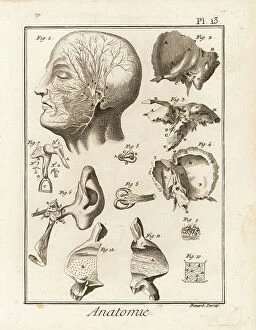
Background imageAnatomie Collection: Details of the anatomy of the human ear, showing cochlea, stapes, incus, malleus

Details of the anatomy of the human ear, showing cochlea, stapes, incus, malleus, semicircular canal, etc
FLO4611637 Details of the anatomy of the human ear, showing cochlea, stapes, incus, malleus, semicircular canal, etc. Copperplate engraving by Robert Benard after an illustration by Guichard Joseph

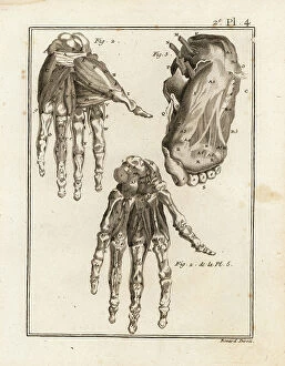
Background imageAnatomie Collection: Dissection of the human hand and foot to reveal muscle, ligament and bone

Dissection of the human hand and foot to reveal muscle, ligament and bone
FLO4611552 Dissection of the human hand and foot to reveal muscle, ligament and bone. Copperplate engraving by Robert Benard from Denis Diderot's Encyclopedia, Pellet, Geneva

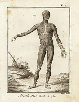
Background imageAnatomie Collection: Flayed human body revealing muscle system and tendons seen from the front

Flayed human body revealing muscle system and tendons seen from the front
FLO4611548 Flayed human body revealing muscle system and tendons seen from the front. Copperplate engraving by Robert Benard after an illustration by Bernhard Albinus from Denis Diderot's

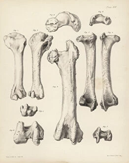
Background imageAnatomie Collection: Humerus and femur of the extinct Rodrigues solitaire, Pezophaps solitaria

Humerus and femur of the extinct Rodrigues solitaire, Pezophaps solitaria
FLO4612190 Humerus and femur of the extinct Rodrigues solitaire, Pezophaps solitaria, in the Parisian Collection and Andersonian Collection


For sale as Licensed Images

Choose your image, Select your licence and Download the media

"Exploring the Intricacies of Anatomie: From Ligaments to Artwork" Delve into the fascinating world of anatomie, where the human body and art intertwine. Journey through a diverse collection of captivating images and historical references that shed light on our understanding of anatomy. Discover the delicate outer ankle ligaments in artwork C013/4452, beautifully depicted with meticulous detail. Witness how artists skillfully capture not only the physicality but also the essence of these intricate structures. Step into a different realm as dog anatomy takes center stage in another mesmerizing artwork. Uncover the intricacies of canine physiology, marveling at how their bodies are both similar and distinct from ours. Travel back in time to Uncle Tom's Cabin, as George Cruikshank's illustration brings Harriet Beecher Stowe's powerful novel to life. Explore how literature can provide insight into societal perceptions and challenges faced by individuals during that era. Max Ernst's painting "The Couple" offers an intriguing glimpse into human relationships and emotions. Delicate brushstrokes convey a depth of feeling that transcends time, inviting us to contemplate love, connection, and vulnerability. Witness Doctor Alfred Velleau conducting an autopsy amidst his attentive students in Francois Feyen Perrin's 19th-century masterpiece. This thought-provoking image captures both scientific curiosity and reverence for knowledge as medical professionals strive to unravel mysteries hidden within our bodies. Jean-Baptiste Marc Bourgery's colored engraving showcases amputations and prosthetics—a testament to humanity's resilience even in times of war-induced adversity. These artificial limbs symbolize hope for soldiers who have endured unimaginable hardships on the battlefield. "The Anatomy Lesson" series by Drs Nicolaes Tulp and Willem van der Meer transports us back centuries ago when dissection was a groundbreaking practice. These oil paintings immortalize moments where science meets artistry while unraveling secrets concealed beneath our skin.