Anatomie Collection (#6)

"Exploring the Intricacies of Anatomie: From Ligaments to Artwork" Delve into the fascinating world of anatomie, where the human body and art intertwine

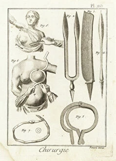
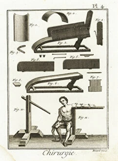
Background imageAnatomie Collection: Cruel and imperfect breast amputation, surgical pliers, razor and scalpel for mastectomy

Cruel and imperfect breast amputation, surgical pliers, razor and scalpel for mastectomy
FLO4611883 Cruel and imperfect breast amputation, surgical pliers, razor and scalpel for mastectomy, chest support instrument and its use

Background imageAnatomie Collection: Bone saw, pliers, scissors, mallet and hook for childbirth - Plate taken from ' L'Encyclopedie' by

Bone saw, pliers, scissors, mallet and hook for childbirth - Plate taken from " L'Encyclopedie" by Denis
FLO4611854 Bone saw, pliers, scissors, mallet and hook for childbirth - Plate taken from " L'Encyclopedie" by Denis Diderot (1713-1784) and Jean Le Rond D'Alembert (1717-1783)

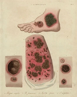
Background imageAnatomie Collection: Skin Diseases: Purple Spots, Purpura Varieties and Rose Rash - Lithography by John Pass

Skin Diseases: Purple Spots, Purpura Varieties and Rose Rash - Lithography by John Pass
FLO4618721 Skin Diseases: Purple Spots, Purpura Varieties and Rose Rash - Lithography by John Pass, published in "Encyclopedia Londinensis" by John Wilkes, J.Adlard Edition, London

Background imageAnatomie Collection: Prurigo strophulus, infant skin dermatosis and lichen simplex

Prurigo strophulus, infant skin dermatosis and lichen simplex, skin thickening after pruritus - Lithography by John
FLO4618795 Prurigo strophulus, infant skin dermatosis and lichen simplex, skin thickening after pruritus - Lithography by John Pass, published in "Encyclopedia Londinensis" by John Wilkes

Background imageAnatomie Collection: Sycosis of the beard and scalp, chronic inflammation of the hair follicles - Lithography by John

Sycosis of the beard and scalp, chronic inflammation of the hair follicles - Lithography by John Pass
FLO4618744 Sycosis of the beard and scalp, chronic inflammation of the hair follicles - Lithography by John Pass, published in "Encyclopedia Londinensis" by John Wilkes, J.Adlard Edition

Background imageAnatomie Collection: Contagious skin diseases: impetigo and rhypia. Lithograph of John Pass, published in 'Encyclopedia

Contagious skin diseases: impetigo and rhypia. Lithograph of John Pass, published in "Encyclopedia
FLO4618719 Contagious skin diseases: impetigo and rhypia. Lithograph of John Pass, published in "Encyclopedia Londinensis" by John Wilkes, edition J.Adlard, London

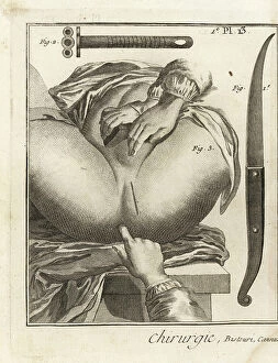
Background imageAnatomie Collection: Foubert surgical operation to remove a vesical stone - Plate taken from ' L'Encyclopedie' by Denis

Foubert surgical operation to remove a vesical stone - Plate taken from " L'Encyclopedie" by Denis
FLO4611791 Foubert surgical operation to remove a vesical stone - Plate taken from " L'Encyclopedie" by Denis Diderot (1713-1784) and Jean Le Rond D'Alembert (1717-1783)

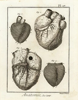
Background imageAnatomie Collection: Anatomical views of the human heart, musculature and blood circulation

Anatomical views of the human heart, musculature and blood circulation. Copperplate engraving by Robert Benard after an
FLO4611663 Anatomical views of the human heart, musculature and blood circulation. Copperplate engraving by Robert Benard after an illustration by Jean-Baptiste Senac from Denis Diderot's
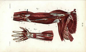
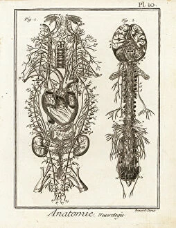
Background imageAnatomie Collection: The nervous system. Copperplate engraving by Robert Benard after illustrations by Raymond Vieussens

The nervous system. Copperplate engraving by Robert Benard after illustrations by Raymond Vieussens
FLO4611615 The nervous system. Copperplate engraving by Robert Benard after illustrations by Raymond Vieussens and Bartolomeo Eustachi from Denis Diderot's Encyclopedia, Pellet, Geneva

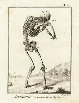
Background imageAnatomie Collection: Human skeleton viewed from the rear, in a sorrowful pose. Copperplate engraving by Robert Benard

Human skeleton viewed from the rear, in a sorrowful pose. Copperplate engraving by Robert Benard after an illustration
FLO4611543 Human skeleton viewed from the rear, in a sorrowful pose. Copperplate engraving by Robert Benard after an illustration by Andreas Vesalius from Denis Diderot's Encyclopedia, Pellet

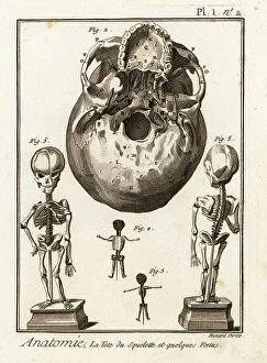
Background imageAnatomie Collection: Human skull viewed from below, with several fetal skeletons. Copperplate engraving by Robert

Human skull viewed from below, with several fetal skeletons. Copperplate engraving by Robert Benard after an
FLO4611525 Human skull viewed from below, with several fetal skeletons. Copperplate engraving by Robert Benard after an illustration by Andreas Vesalius from Denis Diderot's Encyclopedia, Pellet

Background imageAnatomie Collection: Rhesus or bandar - Rhesus macaque, Macaca mulatta (Simia rhesus)

Rhesus or bandar - Rhesus macaque, Macaca mulatta (Simia rhesus), and detail of its distinctive tail - Handcoloured
FLO4715080 Rhesus or bandar - Rhesus macaque, Macaca mulatta (Simia rhesus), and detail of its distinctive tail - Handcoloured copperplate engraving by J

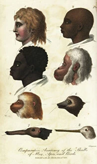
Background imageAnatomie Collection: Comparative anatomy of the heads of men, apes and birds - Handcoloured copperplate engraving by J

Comparative anatomy of the heads of men, apes and birds - Handcoloured copperplate engraving by J
FLO4714977 Comparative anatomy of the heads of men, apes and birds - Handcoloured copperplate engraving by J. Chapman from Ebenezer Sibly's Universal System of Natural History, London

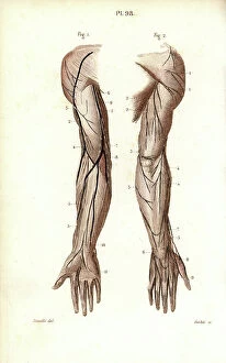
Background imageAnatomie Collection: Nerves of the arm and hand. Lithograph of Corbie, based on a drawing by Leveille

Nerves of the arm and hand. Lithograph of Corbie, based on a drawing by Leveille
FLO4673174 Nerves of the arm and hand. Lithograph of Corbie, based on a drawing by Leveille, in Petity Atlas complet d'Anatomie descriptive du Corps Humain, by Dr

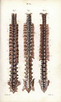
Background imageAnatomie Collection: Veins of the vertebral column. Lithograph of Corbie, based on a drawing by Leveille

Veins of the vertebral column. Lithograph of Corbie, based on a drawing by Leveille
FLO4672992 Veins of the vertebral column. Lithograph of Corbie, based on a drawing by Leveille, in Petiure Atlas complet d'Anatomie descriptive du Corps Humain, by Dr

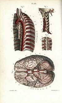
Background imageAnatomie Collection: The circulatory system of the brain has the vertebral column

The circulatory system of the brain has the vertebral column. Lithograph of Anneshower, based on a drawing by Leveille
FLO4672906 The circulatory system of the brain has the vertebral column. Lithograph of Anneshower, based on a drawing by Leveille, in Petiure Atlas complet d'Anatomie descriptive du Corps Humain

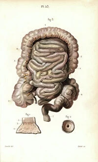
Background imageAnatomie Collection: The intestines. Lithograph of Corbie, based on a drawing by Leveille

The intestines. Lithograph of Corbie, based on a drawing by Leveille, in Petity Atlas complet d'Anatomie
FLO4672838 The intestines. Lithograph of Corbie, based on a drawing by Leveille, in Petity Atlas complet d'Anatomie descriptive du Corps Humain, by Dr

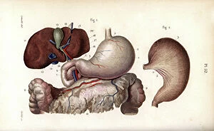
Background imageAnatomie Collection: Stomach, duodenum and liver. Lithograph of Corbie, based on a drawing by Leveille

Stomach, duodenum and liver. Lithograph of Corbie, based on a drawing by Leveille
FLO4672837 Stomach, duodenum and liver. Lithograph of Corbie, based on a drawing by Leveille, in Petity Atlas complet d'Anatomie descriptive du Corps Humain, by Dr

Background imageAnatomie Collection: Sections of the skull: nasal cavity, jaw and teeth. Lithograph of Corbie

Sections of the skull: nasal cavity, jaw and teeth. Lithograph of Corbie, in Petity Atlas complet d'Anatomie
FLO4672833 Sections of the skull: nasal cavity, jaw and teeth. Lithograph of Corbie, in Petity Atlas complet d'Anatomie descriptive du Corps Humain, by Dr

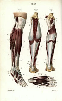
Background imageAnatomie Collection: Muscles and tendons of the leg and foot. Lithograph of Lebrun, based on a drawing by Leveille

Muscles and tendons of the leg and foot. Lithograph of Lebrun, based on a drawing by Leveille
FLO4672683 Muscles and tendons of the leg and foot. Lithograph of Lebrun, based on a drawing by Leveille, in Petity Atlas complet d'Anatomie descriptive du Corps Humain, by Dr

Background imageAnatomie Collection: Muscles of the diaphragm, odds and pelvic muscles. Lithograph by Davesne

Muscles of the diaphragm, odds and pelvic muscles. Lithograph by Davesne, based on a drawing by Leveille
FLO4672648 Muscles of the diaphragm, odds and pelvic muscles. Lithograph by Davesne, based on a drawing by Leveille, in Petity Atlas complet d'Anatomie descriptive du Corps Humain, by Dr

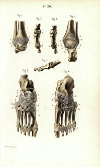
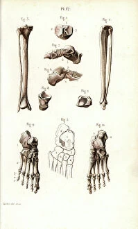
Background imageAnatomie Collection: Ankle and foot bone. Lithograph of Corbie, in Petity Atlas complet d'Anatomie descriptive du Corps

Ankle and foot bone. Lithograph of Corbie, in Petity Atlas complet d'Anatomie descriptive du Corps Humain, by Dr
FLO4672592 Ankle and foot bone. Lithograph of Corbie, in Petity Atlas complet d'Anatomie descriptive du Corps Humain, by Dr

Background imageAnatomie Collection: Spinal bone, ribs and sternum. Lithograph of Corbie, in Petity Atlas complet d'Anatomie

Spinal bone, ribs and sternum. Lithograph of Corbie, in Petity Atlas complet d'Anatomie descriptive du Corps
FLO4672534 Spinal bone, ribs and sternum. Lithograph of Corbie, in Petity Atlas complet d'Anatomie descriptive du Corps Humain, by Dr

Background imageAnatomie Collection: The thoracic canal. Lithograph of Corbie, based on a drawing by Leveille

The thoracic canal. Lithograph of Corbie, based on a drawing by Leveille, in Petity Atlas complet d'Anatomie
FLO4673048 The thoracic canal. Lithograph of Corbie, based on a drawing by Leveille, in Petity Atlas complet d'Anatomie descriptive du Corps Humain, by Dr

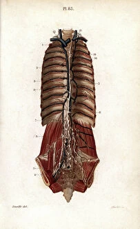
Background imageAnatomie Collection: Lymphatic system of the thorax and abdomen. Lithograph of Corbie, based on a drawing by Leveille

Lymphatic system of the thorax and abdomen. Lithograph of Corbie, based on a drawing by Leveille
FLO4673042 Lymphatic system of the thorax and abdomen. Lithograph of Corbie, based on a drawing by Leveille, in Petity Atlas complet d'Anatomie descriptive du Corps Humain, by Dr

Background imageAnatomie Collection: The circulatory system from the heart to the head. Lithograph of Choubard

The circulatory system from the heart to the head. Lithograph of Choubard, based on a drawing by Leveille
FLO4672885 The circulatory system from the heart to the head. Lithograph of Choubard, based on a drawing by Leveille, in Petity Atlas complet d'Anatomie descriptive du Corps Humain, by Dr

Background imageAnatomie Collection: Sections of the pharynx and larynx. Lithograph of Anneshower, based on a drawing by Leveille

Sections of the pharynx and larynx. Lithograph of Anneshower, based on a drawing by Leveille
FLO4672792 Sections of the pharynx and larynx. Lithograph of Anneshower, based on a drawing by Leveille, in Petity Atlas complet d'Anatomie descriptive du Corps Humain, by Dr

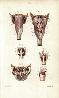
Background imageAnatomie Collection: Muscles of the pharynx and larynx. Lithograph by Davesne, based on a drawing by Leveille

Muscles of the pharynx and larynx. Lithograph by Davesne, based on a drawing by Leveille
FLO4672628 Muscles of the pharynx and larynx. Lithograph by Davesne, based on a drawing by Leveille, in Petity Atlas complet d'Anatomie descriptive du Corps Humain, by Dr

Background imageAnatomie Collection: Bone of the jaw and neck. Lithograph of Corbie, in Petity Atlas complet d'Anatomie descriptive du

Bone of the jaw and neck. Lithograph of Corbie, in Petity Atlas complet d'Anatomie descriptive du Corps Humain
FLO4672518 Bone of the jaw and neck. Lithograph of Corbie, in Petity Atlas complet d'Anatomie descriptive du Corps Humain, by Dr

Background imageAnatomie Collection: The nerves of the brachial plexus and the finger. Lithograph of Anneshower

The nerves of the brachial plexus and the finger. Lithograph of Anneshower, based on a drawing by Leveille
FLO4672483 The nerves of the brachial plexus and the finger. Lithograph of Anneshower, based on a drawing by Leveille, in Petity Atlas complet d'Anatomie descriptive du Corps Humain, by Dr

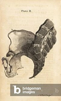
Background imageAnatomie Collection: Lateral and internal view of a healthy female pelvis. Copperplate engraving by Andrew Bell after

Lateral and internal view of a healthy female pelvis. Copperplate engraving by Andrew Bell after an illustration by Jan
FLO4695982 Lateral and internal view of a healthy female pelvis. Copperplate engraving by Andrew Bell after an illustration by Jan van Rymsdyk from William Smellie's A Set of Anatomical Tables

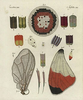
Background imageAnatomie Collection: Butterfly wing, showing structure, skeleton, scales and microscopic detail

Butterfly wing, showing structure, skeleton, scales and microscopic detail
FLO4573972 Butterfly wing, showing structure, skeleton, scales and microscopic detail. Handcoloured copperplate engraving from Bertuch's " Bilderbuch fur Kinder"

Background imageAnatomie Collection: Views of the humerus and ulna bones in the human arm. Copperplate engraving by Edward Mitchell

Views of the humerus and ulna bones in the human arm. Copperplate engraving by Edward Mitchell after an anatomical
FLO4565716 Views of the humerus and ulna bones in the human arm. Copperplate engraving by Edward Mitchell after an anatomical illustration from John Barclay's A Series of Engravings of the Human

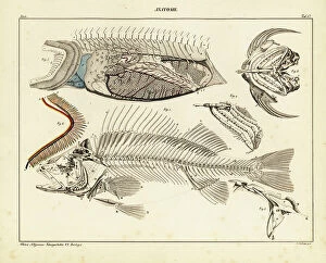
Background imageAnatomie Collection: Anatomy of a fish, showing skeleton, internal organs and gills. Lithograph by St

Anatomy of a fish, showing skeleton, internal organs and gills. Lithograph by St
FLO4684921 Anatomy of a fish, showing skeleton, internal organs and gills. Lithograph by St. Schillinger from Lorenz Oken's Universal Natural History, Allgemeine Naturgeschichte fur alle Stande

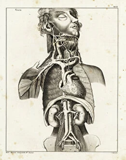
Background imageAnatomie Collection: Anatomy of the human venal system in the upper torso. Lithograph by C

Anatomy of the human venal system in the upper torso. Lithograph by C. Mayer from Lorenz Oken's Universal Natural
FLO4684833 Anatomy of the human venal system in the upper torso. Lithograph by C. Mayer from Lorenz Oken's Universal Natural History, Allgemeine Naturgeschichte fur alle Stande, Stuttgart

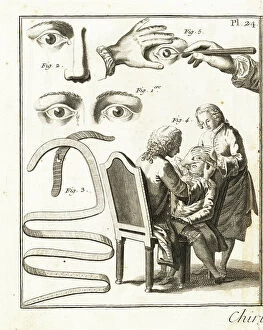
Background imageAnatomie Collection: Description of the lacrimal gland, bandage for the swelling of the lacrimal glands

Description of the lacrimal gland, bandage for the swelling of the lacrimal glands
FLO4611843 Description of the lacrimal gland, bandage for the swelling of the lacrimal glands and cataract operation - Plate taken from " L'Encyclopedie" by Denis Diderot (1713-1784)

Background imageAnatomie Collection: Monachus monachus (Skeleton of a Mediterranean monk seal), Museum of Natural History of Marseille

Monachus monachus (Skeleton of a Mediterranean monk seal), Museum of Natural History of Marseille
CSO5006631 Monachus monachus (Skeleton of a Mediterranean monk seal), Museum of Natural History of Marseille; (add.info.: Monachus monachus (Skeleton of a Mediterranean monk seal)

Background imageAnatomie Collection: Arm and hand circulatory system. Lithograph of Corbie, based on a drawing by Leveille

Arm and hand circulatory system. Lithograph of Corbie, based on a drawing by Leveille
FLO4672918 Arm and hand circulatory system. Lithograph of Corbie, based on a drawing by Leveille, in Petity Atlas complet d'Anatomie descriptive du Corps Humain, by Dr

Background imageAnatomie Collection: Sections of the eyeball and its membranes. Lithograph by Davesne, based on a drawing by Leveille

Sections of the eyeball and its membranes. Lithograph by Davesne, based on a drawing by Leveille
FLO4672801 Sections of the eyeball and its membranes. Lithograph by Davesne, based on a drawing by Leveille, in Petity Atlas complet d'Anatomie descriptive du Corps Humain, by Dr

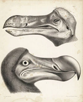
Background imageAnatomie Collection: Side view of the head of a dodo in the Ashmolean Museum and restored head in the British Museum

Side view of the head of a dodo in the Ashmolean Museum and restored head in the British Museum
FLO4612112 Side view of the head of a dodo in the Ashmolean Museum and restored head in the British Museum. Illustration drawn and lithographed by Joseph Dinkel from Hugh Edwin Strickland

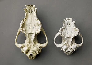
Background imageAnatomie Collection: Dog and wolf: cranes ventrals - Dog and woolf, ventral skulls)

Dog and wolf: cranes ventrals - Dog and woolf, ventral skulls), Museum d'histoire naturelle de Marseille
CSO5006416 Dog and wolf: cranes ventrals - Dog and woolf, ventral skulls), Museum d'histoire naturelle de Marseille; (add.info.: Dog and wolf: cranes ventrals - Dog and woolf, ventral skulls)

Background imageAnatomie Collection: Skin ulcer, purulent scabies, and scabies caused by a scabiei sarcoptic mite - Lithograph by John

Skin ulcer, purulent scabies, and scabies caused by a scabiei sarcoptic mite - Lithograph by John Pass
FLO4618801 Skin ulcer, purulent scabies, and scabies caused by a scabiei sarcoptic mite - Lithograph by John Pass, published in "Encyclopedia Londinensis" by John Wilkes, J.Adlard edition

Background imageAnatomie Collection: A surgeon performing trepanation and various surgical instruments, lever by Jean louis Petite

A surgeon performing trepanation and various surgical instruments, lever by Jean louis Petite, easel, trestle clamps
FLO4611817 A surgeon performing trepanation and various surgical instruments, lever by Jean louis Petite, easel, trestle clamps, cauterizer and round and triangular plates

Background imageAnatomie Collection: Chirugical equipment with a machine to reduce complex fractures

Chirugical equipment with a machine to reduce complex fractures and a machine for disloque arms - Plate taken
FLO4611937 Chirugical equipment with a machine to reduce complex fractures and a machine for disloque arms - Plate taken from " L'Encyclopedie" by Denis Diderot (1713-1784)

Background imageAnatomie Collection: Leg, ankle and foot bones. Lithograph of Corbie, in Petity Atlas complet d'Anatomie descriptive du

Leg, ankle and foot bones. Lithograph of Corbie, in Petity Atlas complet d'Anatomie descriptive du Corps Humain
FLO4672532 Leg, ankle and foot bones. Lithograph of Corbie, in Petity Atlas complet d'Anatomie descriptive du Corps Humain, by Dr

Background imageAnatomie Collection: Anatomy of the human heart. Copperplate engraving by Milton from Abraham Rees Cyclopedia or

Anatomy of the human heart. Copperplate engraving by Milton from Abraham Rees Cyclopedia or Universal Dictionary of
FLO4594159 Anatomy of the human heart. Copperplate engraving by Milton from Abraham Rees Cyclopedia or Universal Dictionary of Arts, Sciences and Literature, Longman, Hurst, Rees, Orme and Brown

Background imageAnatomie Collection: Arterial system in the human body. Copperplate engraving by Robert Benard after an illustration by

Arterial system in the human body. Copperplate engraving by Robert Benard after an illustration by James Drake
FLO4611586 Arterial system in the human body. Copperplate engraving by Robert Benard after an illustration by James Drake from Denis Diderot's Encyclopedia, Pellet, Geneva

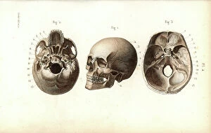
Background imageAnatomie Collection: Different views of the skull. Lithograph of Corbie, in Petity Atlas complet d'Anatomie descriptive

Different views of the skull. Lithograph of Corbie, in Petity Atlas complet d'Anatomie descriptive du Corps Humain
FLO4672756 Different views of the skull. Lithograph of Corbie, in Petity Atlas complet d'Anatomie descriptive du Corps Humain, by Dr


For sale as Licensed Images

Choose your image, Select your licence and Download the media

"Exploring the Intricacies of Anatomie: From Ligaments to Artwork" Delve into the fascinating world of anatomie, where the human body and art intertwine. Journey through a diverse collection of captivating images and historical references that shed light on our understanding of anatomy. Discover the delicate outer ankle ligaments in artwork C013/4452, beautifully depicted with meticulous detail. Witness how artists skillfully capture not only the physicality but also the essence of these intricate structures. Step into a different realm as dog anatomy takes center stage in another mesmerizing artwork. Uncover the intricacies of canine physiology, marveling at how their bodies are both similar and distinct from ours. Travel back in time to Uncle Tom's Cabin, as George Cruikshank's illustration brings Harriet Beecher Stowe's powerful novel to life. Explore how literature can provide insight into societal perceptions and challenges faced by individuals during that era. Max Ernst's painting "The Couple" offers an intriguing glimpse into human relationships and emotions. Delicate brushstrokes convey a depth of feeling that transcends time, inviting us to contemplate love, connection, and vulnerability. Witness Doctor Alfred Velleau conducting an autopsy amidst his attentive students in Francois Feyen Perrin's 19th-century masterpiece. This thought-provoking image captures both scientific curiosity and reverence for knowledge as medical professionals strive to unravel mysteries hidden within our bodies. Jean-Baptiste Marc Bourgery's colored engraving showcases amputations and prosthetics—a testament to humanity's resilience even in times of war-induced adversity. These artificial limbs symbolize hope for soldiers who have endured unimaginable hardships on the battlefield. "The Anatomy Lesson" series by Drs Nicolaes Tulp and Willem van der Meer transports us back centuries ago when dissection was a groundbreaking practice. These oil paintings immortalize moments where science meets artistry while unraveling secrets concealed beneath our skin.