
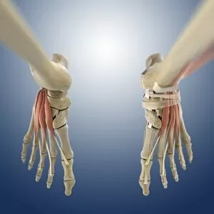
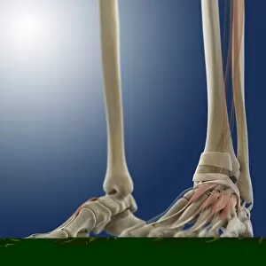
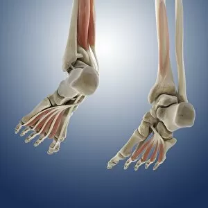
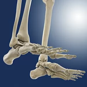
Ankle Collection (#6)
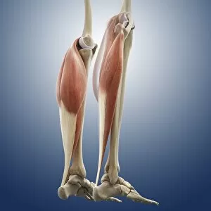
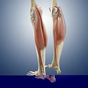
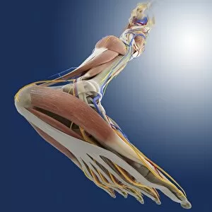
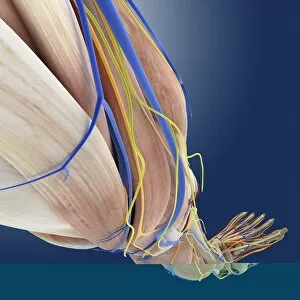
"Exploring the Ankle: From Toilets to Hairdressers, Unveiling its Secrets


For sale as Licensed Images
Choose your image, Select your licence and Download the media
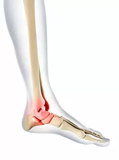
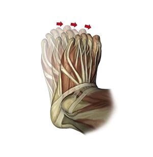
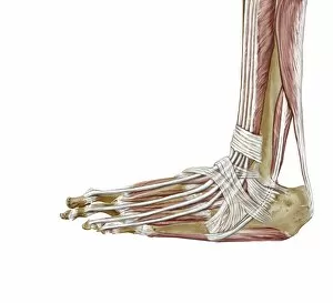
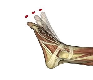
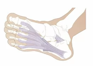
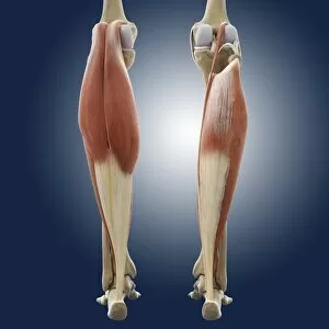
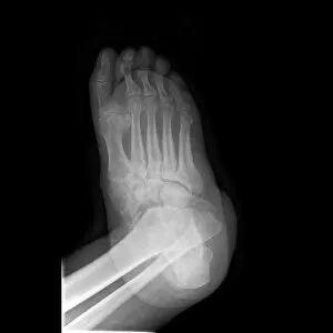
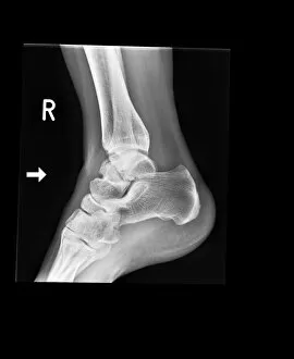
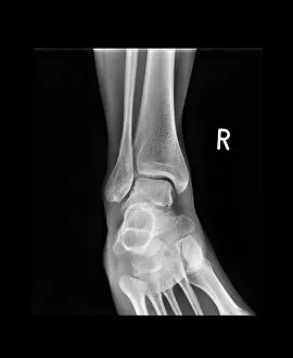
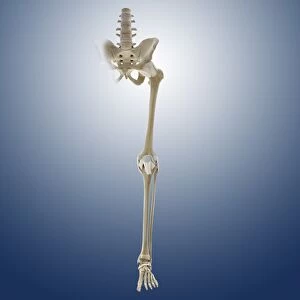
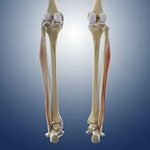
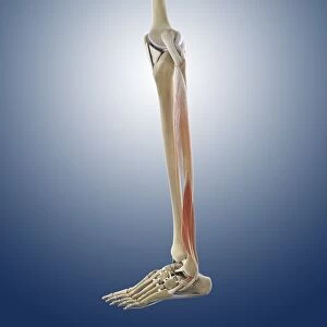
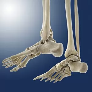
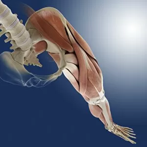
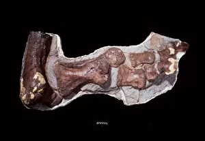
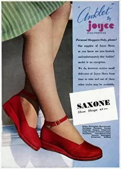
"Exploring the Ankle: From Toilets to Hairdressers, Unveiling its Secrets. " "Did you know that your ankle joint is as important as a toilet seat? Discover its fascinating role in supporting your body's weight and movement. " "Hair today, gone tomorrow. Just like hair growth, our they are constantly adapting to support our every step. " "Step into the world anatomy with this captivating image showcasing the intricate network of bones, muscles, and tendons - just like a skilled hairdresser creating a masterpiece. " "Running injuries can be painful but also inspiring. Dive into this conceptual artwork depicting the resilience and determination required to overcome ankle-related setbacks. " "A fractured ankle may seem daunting on an X-ray, but it's also a reminder of our body's incredible ability to heal and rebuild itself. " "Unleash your inner athlete with this stunning artwork capturing the power and strength of leg muscles during running - truly a sight to behold. " "Body pain got you down? Take solace in this thought-provoking artwork that explores how discomfort can lead us towards self-discovery and personal growth. " "Delve deep into anatomical wonders with a detailed diagram showcasing the bones of the right leg and hip - an educational journey for curious minds. " "Take a nostalgic trip back in time with this vintage-inspired 1950s pinup girl flaunting her flawless ankles - timeless beauty at its finest. " "Peek beneath the surface as we unveil the hidden intricacies of human foot anatomy; from skin to veins, arteries, muscles, and bones – it’s all there waiting to be discovered. " "Discovering what lies beneath: A mesmerizing MRI scan reveals secrets behind ruptured Achilles tendon – reminding us of both vulnerability and resilience within ourselves. " "Travel back millions of years as we explore the ancient anatomy of a Tyrannosaurus foot.