
Cornea Collection

The cornea, a remarkable part of the eye anatomy, is often depicted in stunning artwork

Background imageCornea Collection: Eye anatomy, artwork

Eye anatomy, artwork
Eye anatomy, computer artwork. At the front of the eye is the cornea, a transparent coating. Behind this is the lens, which is partly covered by the iris

Background imageCornea Collection: Laser eye surgery, artwork

Laser eye surgery, artwork
Laser eye surgery. Artwork showing how laser eye surgery is performed. An incision is made into the cornea (light blue), the clear dome-shaped surface that covers the front of the eye

Background imageCornea Collection: Digital illustration of dilated pupil of human eye with contraction of outer radial muscle fibres

Digital illustration of dilated pupil of human eye with contraction of outer radial muscle fibres

Background imageCornea Collection: Eye surgery, conceptual artwork

Eye surgery, conceptual artwork
Eye surgery. Conceptual artwork of a human eye with a windscreen wiper being used to produce clear vision. This represents the correction of conditions such as a cataract (cloudiness of the eyes lens)

Background imageCornea Collection: Human eye, illustration

Human eye, illustration
Eye anatomy. Cutaway illustration passing through a human eye, showing its internal anatomy and structure. The front of the eye is at right, and the structures here include the cornea

Background imageCornea Collection: External anatomy of the human eye (with labels)

External anatomy of the human eye (with labels)

Background imageCornea Collection: Common fly, Musca domestica 1, head of fly 2, magnified 3, proboscis 4,5, cornea 6, magnified 7

Common fly, Musca domestica 1, head of fly 2, magnified 3, proboscis 4,5, cornea 6, magnified 7
FLO4574014 Common fly, Musca domestica 1, head of fly 2, magnified 3, proboscis 4,5, cornea 6, magnified 7. Handcoloured copperplate engraving from Bertuch's " Bilderbuch fur Kinder"

Background imageCornea Collection: Lost In Your Eye Aquatic

Lost In Your Eye Aquatic
Alirıza ÇAKIR

Background imageCornea Collection: Lost In Your Eye Cosmic

Lost In Your Eye Cosmic
Alirıza ÇAKIR

Background imageCornea Collection: Digital illustration of constricted pupil of human eye

Digital illustration of constricted pupil of human eye
Anatomy, Biomedical Illustration, Circle, Closed, Cornea, Digitally Generated, Directly Above, Healthcare and Medicine, Human Eye, Human Muscle, Illustrative Technique, Iris, Muscle Fibre, No People

Background imageCornea Collection: Digital illustration of human eye transforming images made from light into electrical impulses

Digital illustration of human eye transforming images made from light into electrical impulses
arrow sign, biomedical illustration, blue background, connection, cornea, cross section, digitally generated, direction, eyesight, full frame, horizontal, human eye, iris, lens, no people

Background imageCornea Collection: Normal anatomy of the eye in cross section

Normal anatomy of the eye in cross section
Horizontal, Vision, Detail, Anatomy, Diagram, Muscle, Illustration, Lens, Iris, Medical, Eyeball, Biology, Lateral, Cut Out, White Background, Eye, Artwork, Cross Section, Color Image, Close Up

Background imageCornea Collection: Romania: Cornea, Peasant woman (b / w photo)

Romania: Cornea, Peasant woman (b / w photo)
6017301 Romania: Cornea, Peasant woman (b/w photo) by Hielscher, Kurt (1881-1948); Private Collection; (add.info.: Cornea, Peasant woman)

Background imageCornea Collection: Operation on a eye, plate from Traite Complet de l Anatomie de l Homme

Operation on a eye, plate from Traite Complet de l Anatomie de l Homme
CHT236314 Operation on a eye, plate from Traite Complet de l Anatomie de l Homme by Jean-Baptiste Marc Bourgery (1797-1849) 1866-67 (coloured engraving) by Jacob

Background imageCornea Collection: Close Up Of Eye

Close Up Of Eye

Background imageCornea Collection: Cross section biomedical illustration of fluid flow in chronic glaucoma with blocked trabecular

Cross section biomedical illustration of fluid flow in chronic glaucoma with blocked trabecular meshwork

Background imageCornea Collection: Retina with macular degeneration

Retina with macular degeneration

Background imageCornea Collection: Illustration of arcus senilis condition of the eye

Illustration of arcus senilis condition of the eye
Arcus senilis. Illustration of a close-up of an eye showing the condition arcus senilis, in which a grey-white circle in the periphery of the cornea occurs around the edge of the iris

Background imageCornea Collection: Retinal implant, artwork

Retinal implant, artwork
Retinal implant. Computer artwork of a biomedical chip (square, centre right) implanted just under the surface of the retina in a human eye

Background imageCornea Collection: Anatomy of the human eye

Anatomy of the human eye.. Anatomy of the human eye, showing eyeball, optic nerve, muscle, cornea, retina, sections through eye

Background imageCornea Collection: Common fly, Musca domestica, head, proboscis and cornea

Common fly, Musca domestica, head, proboscis and cornea.. Common fly, Musca domestica 1, head of fly 2, magnified 3, proboscis 4, 5, cornea 6, magnified 7

Background imageCornea Collection: Human eye, published in 1898

Human eye, published in 1898
Antique illustration of a Human eye, published in 1898

Background imageCornea Collection: Cross-section diagram of human eye

Cross-section diagram of human eye

Background imageCornea Collection: Illustration of vision of the human eye

Illustration of vision of the human eye

Background imageCornea Collection: Cross section biomedical illustration of cataract surgery

Cross section biomedical illustration of cataract surgery

Background imageCornea Collection: Cross section biomedical illustration of key anatomy of the eye

Cross section biomedical illustration of key anatomy of the eye

Background imageCornea Collection: Cross section biomedical illustration of normal eye

Cross section biomedical illustration of normal eye

Background imageCornea Collection: Cross section biomedical illustration of keratoconus or conical cornea

Cross section biomedical illustration of keratoconus or conical cornea

Background imageCornea Collection: Cross section biomedical illustration of myopia

Cross section biomedical illustration of myopia

Background imageCornea Collection: Cross section biomedical illustration of lens to correct hypermetropia

Cross section biomedical illustration of lens to correct hypermetropia

Background imageCornea Collection: Cross section biomedical illustration of anatomy of human eye

Cross section biomedical illustration of anatomy of human eye

Background imageCornea Collection: Cross section biomedical illustration of eye focusing on near object

Cross section biomedical illustration of eye focusing on near object

Background imageCornea Collection: Close-up detail of parrotfish eye, Solomon Islands

Close-up detail of parrotfish eye, Solomon Islands

Background imageCornea Collection: Conceptual image of human eye and skull

Conceptual image of human eye and skull

Background imageCornea Collection: Anatomy of human eye, perspective

Anatomy of human eye, perspective

Background imageCornea Collection: Orbital cut showing abducent nerve with ciliary ganglion and oculomotor nerve

Orbital cut showing abducent nerve with ciliary ganglion and oculomotor nerve

Background imageCornea Collection: Retina of a normal eye

Retina of a normal eye

Background imageCornea Collection: Retina with advanced diabetic retinopathy

Retina with advanced diabetic retinopathy, showing increased scar tissue, hemorrhaging and diffuse vitreous blood (blurred appearance)

Background imageCornea Collection: Astigmatism and correction with corrective lens

Astigmatism and correction with corrective lens

Background imageCornea Collection: Conceptual image of human eye cross section

Conceptual image of human eye cross section

Background imageCornea Collection: Anatomy of human eye

Anatomy of human eye

Background imageCornea Collection: Diagram of a retinal detachment

Diagram of a retinal detachment

Background imageCornea Collection: Retina of eye with glaucoma

Retina of eye with glaucoma

Background imageCornea Collection: Retina with neovascularization

Retina with neovascularization showing retinal hemorrhaging

Background imageCornea Collection: Anatomy of human eye showing focal points

Anatomy of human eye showing focal points

Background imageCornea Collection: Human eye with muscles and circulatory system

Human eye with muscles and circulatory system

Background imageCornea Collection: Conceptual image of human eye anatomy

Conceptual image of human eye anatomy

Background imageCornea Collection: Cross section of human eye

Cross section of human eye


For sale as Licensed Images

Choose your image, Select your licence and Download the media

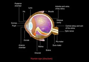
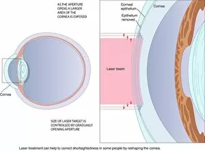
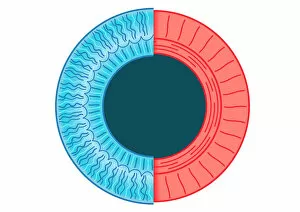
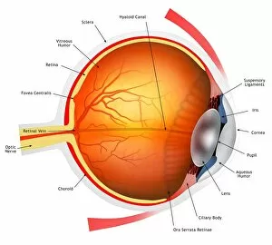
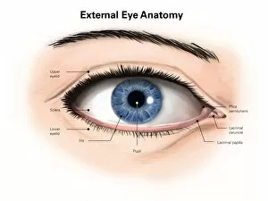
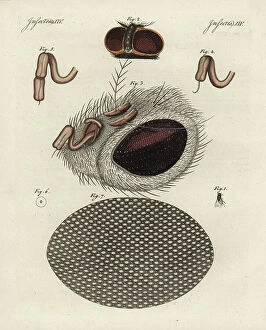
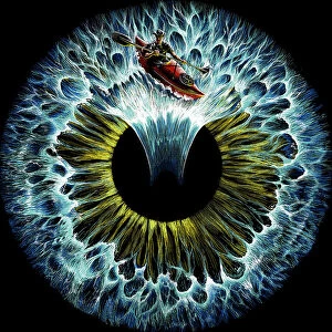
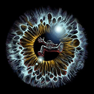
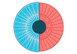
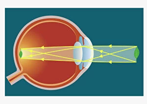
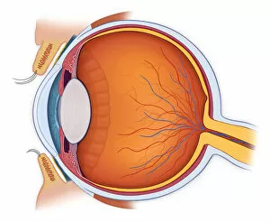
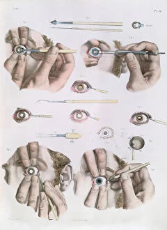
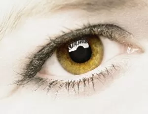
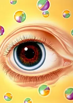
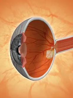
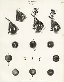
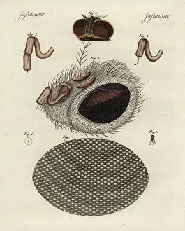
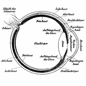
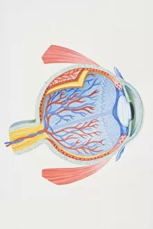
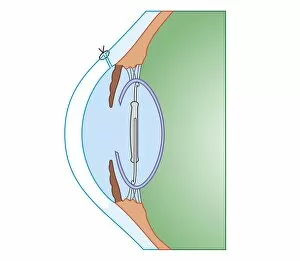
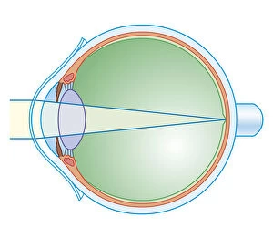
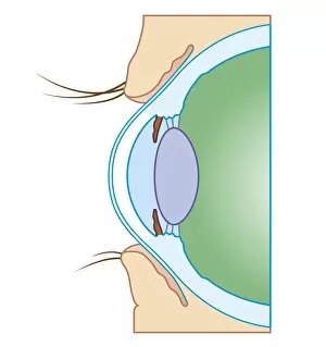
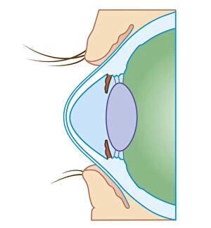
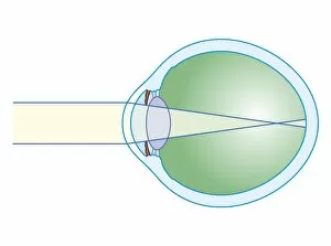
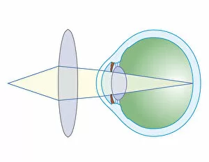
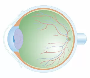
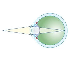
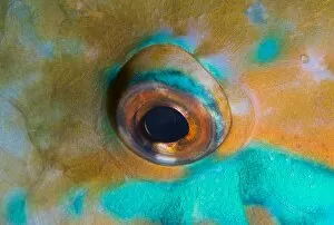
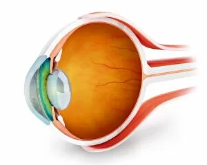
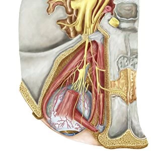
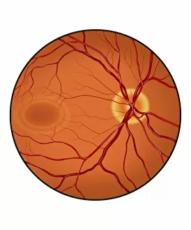
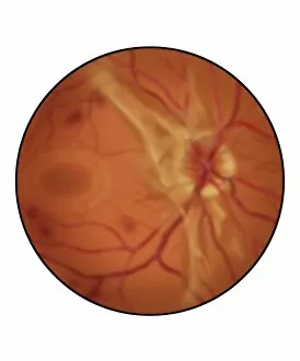
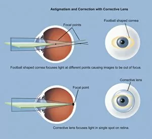
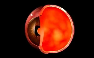
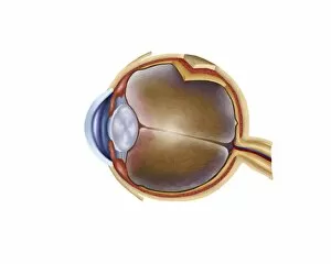
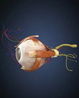
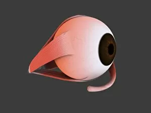
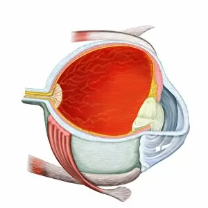
The cornea, a remarkable part of the eye anatomy, is often depicted in stunning artwork. From digital illustrations showcasing the dilation and contraction of outer radial muscle fibers to conceptual artworks representing laser eye surgery, this vital component never fails to captivate our imagination. In one captivating image, a human eye is beautifully illustrated with intricate details highlighting its external anatomy. Labels guide us through each structure, allowing us to appreciate the complexity and precision involved in vision. Another illustration takes us deeper into the normal anatomy of the eye through a cross-section view. This visual representation unveils the intricacies hidden beneath our eyelids and reminds us of the delicate balance required for optimal vision. A black-and-white photograph from Romania showcases an enchanting moment captured between a peasant woman and her cornea. The depth conveyed in this image speaks volumes about how crucial this small yet powerful organ is for connecting individuals with their surroundings. An ancient plate from Traite Complet de l'Anatomie de l'Homme offers insight into historical perspectives on eye surgery. This conceptual artwork transports us back in time, reminding us of humanity's relentless pursuit to understand and improve upon our own biology. Even nature itself provides inspiration when examining corneas; an engraving from The Pictorial Museum of Animated Nature features a common fly's head with its proboscis delicately resting against its transparent cornea. Such images remind us that even creatures so different from ourselves share similar anatomical wonders. Whether it be through scientific diagrams or artistic interpretations, exploring the beauty and complexity of corneas allows us to marvel at one of nature's most incredible creations – our eyesight itself.