Cross Section Collection (#82)

"Unveiling the Hidden World: Exploring Cross Sections in Nature and Architecture" Delving into the intricate world of honey bees

Background imageCross Section Collection: Nettle leaf gall midge larva

Nettle leaf gall midge larva, artwork. Cross- section through a stinging nettle leaf (Urtica dioica) and leaf gall. The gall is inhabited by the parasitic larva of the nettle gall midge

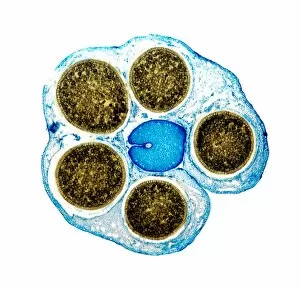
Background imageCross Section Collection: Liverwort spore cases, light micrograph

Liverwort spore cases, light micrograph
Liverwort spore cases. Light micrograph of a cross-section through five spore cases (sporophytes) from the liverwort Conocephalum conicum (Marchantiales)

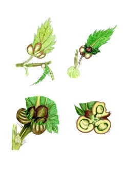
Background imageCross Section Collection: Nettle leaf galls

Nettle leaf galls, artwork. Four views of galls at the base of a stinging nettle leaf (Urtica dioica). These abnormal outgrowths are caused by the parasitic larvae of the nettle gall midge

Background imageCross Section Collection: Damaged stinging nettle

Damaged stinging nettle, artwork. Various views of a stinging nettle damaged by the weevil (Ceuthorhynchus pollinaris). At left are several enlarged views of sections of damaged stems and petioles

Background imageCross Section Collection: Maple stem, light micrograph

Maple stem, light micrograph
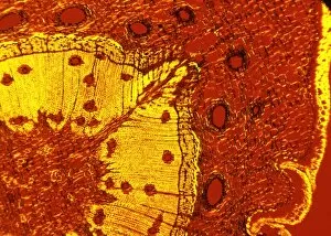
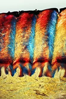
Maple stem. Polarised light micrograph of a cross-section through the stem of a field maple (Acer campestre). This shows the thick layer of cork (phellem)

Background imageCross Section Collection: Growth rings of a tree

Growth rings of a tree. Cross-section through a tree trunk showing annual growth rings. Growth rings are visible in a cross-section of a tree trunk after the tree has been felled

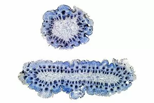
Background imageCross Section Collection: Abnormal thistle stem, light micrograph

Abnormal thistle stem, light micrograph
Abnormal thistle stem. Light micrograph of a normal (top) and abnormal (bottom) stem of the perennial sow thistle (Sonchus arvensis). The abnormal stem has grown in a flattened, or fasciated, manner

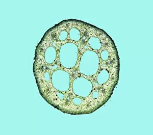
Background imageCross Section Collection: Water-lily leaf stem, light micrograph

Water-lily leaf stem, light micrograph
Water-lily leaf stem. Light micrograph of a cross- section through the leaf stem (petiole) of a white water-lily (Nymphaea alba)

Background imageCross Section Collection: Dutchmans pipe plant stem

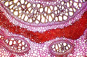
Dutchmans pipe plant stem. Light micrograph of a section through the stem of a dutchmans pipe plant (Aristolochia sipho). The inner blue circle is a layer of sclerenchyma fibres

Background imageCross Section Collection: Stem section, SEM

Stem section, SEM
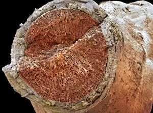
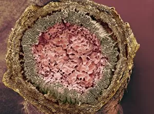
Stem section. Coloured scanning electron micrograph (SEM) of a section through a stem of a woody dicotyledon plant. This stem consists mainly of secondary xylem tissue (orange, centre)

Background imageCross Section Collection: Walnut bark

Walnut bark. Cross section through the trunk of a walnut tree (Juglans regia) revealing the pattern in its bark. This pattern is known as a rhytidome

Background imageCross Section Collection: Grass stem, SEM

Grass stem, SEM
Grass stem. Coloured scanning electron micrograph (SEM) of a section through a grass stem (family Graminaceae). At upper centre is the vascular bundle

Background imageCross Section Collection: Common horsetail stem, light micrograph

Common horsetail stem, light micrograph
Common horsetail stem. Light micrograph of a cross-section through the tip of the condensed stem of the common horsetail (Equisetum arvense), showing the central stem sourrounded by stem leaves

Background imageCross Section Collection: Bamboo stem, SEM

Bamboo stem, SEM
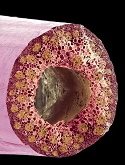
Bamboo stem. Coloured scanning electron micrograph (SEM) of a section through a bamboo stem (Phyllostachys sp.). Vascular bundles can be seen (bright yellow) containing xylem (larger openings)

Background imageCross Section Collection: Woody stem section, SEM

Woody stem section, SEM
Woody stem section. Coloured scanning electron micrograph (SEM) of a section through a stem of a woody plant. At upper centre is the pith (orange), which consists of soft, spongy parenchyma cells

Background imageCross Section Collection: Bracken rhizome, light micrograph

Bracken rhizome, light micrograph
Bracken rhizome. Light micrograph of part of a cross-section through a rhizome from the bracken Pteridium aquilinum. The centre of the rhizome is off top, while the outer surface is off bottom

Background imageCross Section Collection: Bogbean stem, light micrograph

Bogbean stem, light micrograph
Bogbean stem. Light micrograph of a cross-section through the stem of a bogbean plant (Menyanthes trifoliata), showing its internal structure

Background imageCross Section Collection: Scots pine tree stem, light micrograph

Scots pine tree stem, light micrograph
Scots pine tree stem. Polarised light micrograph of a cross-section through the stem of a Scots pine tree (Pinus sylvestris), showing resin canals

Background imageCross Section Collection: Legume root nodules, light micrograph

Legume root nodules, light micrograph
Legume root nodules. Light micrograph of sections through two root nodules from a broad bean plant (Vicia faber). These root nodules are full of nitrogen-fixing bacteria

Background imageCross Section Collection: Mistletoe vascular bundle, LM

Mistletoe vascular bundle, LM
Mistletoe vascular bundle. Light micrograph (LM) of a section of mistletoe (Viscum album) stem in cross-section, showing a vascular bundle

Background imageCross Section Collection: Liana stem

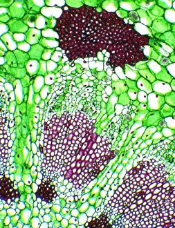
Liana stem. Light micrograph of a vascular bundle in a transverse section through a Brazilian liana stem showing the xylem and phloem

Background imageCross Section Collection: Buttercup stem, light micrograph

Buttercup stem, light micrograph
Buttercup stem. Coloured light micrograph of a section through a buttercup stem (Ranunculus sp.) showing the circular-shaped vascular bundle (right)

Background imageCross Section Collection: Root section, SEM

Root section, SEM
Root section. Coloured scanning electron micrograph (SEM) of a section through a root of a woody dicotyledon plant. This root consists of secondary xylem tissue (yellow, centre)

Background imageCross Section Collection: Laburnum tree trunk growth rings

Laburnum tree trunk growth rings
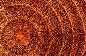
Laburnum tree trunk. Cross-section through a laburnum (Laburnum anaguroides) tree trunk showing the concentric growth rings and medullary arrays radiating from the centre

Background imageCross Section Collection: Cucumber stem, light micrograph

Cucumber stem, light micrograph
Cucumber stem. Light micrograph of a cross-section through part of a vascular bundle from a cucumber (Cucurbita sp.). This vascular bundle is from the stem of the cucumber

Background imageCross Section Collection: Mistletoe stem, LM

Mistletoe stem, LM
Mistletoe stem. Light micrograph (LM) of a mistletoe (Viscum album) stem in cross-section, showing nine vascular bundles radiating out from the centre of the stem

Background imageCross Section Collection: Leaf tissue structure, SEM

Leaf tissue structure, SEM
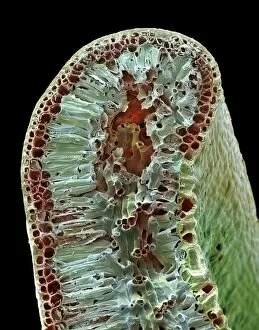
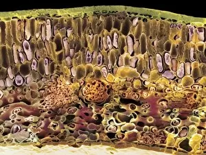
Leaf tissue structure. Coloured scanning electron micrograph (SEM) of a section through the edge of a leaf from the Common Box (Buxus sempervirens)

Background imageCross Section Collection: Conifer needle, SEM

Conifer needle, SEM
Conifer needle. Coloured scanning electron micrograph (SEM) of a section through a needle (leaf) from a conifer tree, showing the vascular bundle (large round cluster)

Background imageCross Section Collection: Leaf midrib, light micrograph

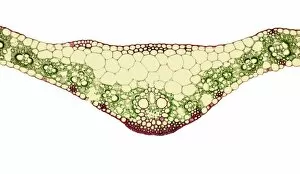
Leaf midrib, light micrograph
Leaf midrib. Light micrograph (LM) of a section through the midrib of a leaf from a monocotyledon plant. The midrib (midvein) is the continuation of a leafs stem along the centre of the leaf
Background imageCross Section Collection: Leaf bud, light micrograph

Leaf bud, light micrograph
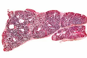
Leaf bud. Light micrograph of a cross-section through a winter leaf bud of the sycamore tree (Acer pseudoplatanus). This trees leaves are arranged in an opposite and decussate manner

Background imageCross Section Collection: Poppy ovary with developing seeds, LM

Poppy ovary with developing seeds, LM
Poppy ovary with developing seeds. Light micrograph (LM) of a cross-section through the centre of a mature poppy ovary, showing placentas bearing developing seeds (red)

Background imageCross Section Collection: Sycamore bud, light micrograph

Sycamore bud, light micrograph
Sycamore bud. Light micrograph of a cross-section through a sycamore bud (Acer pseudo-plantanus). The unexpanded leaves (red) are surrounded by protective outer scale leaves covered in cork

Background imageCross Section Collection: Sycamore leaf bud, light micrograph

Sycamore leaf bud, light micrograph
Sycamore leaf bud (Acer pseudoplatanus). Light micrograph of a cross-section through a winter leaf bud from a sycamore tree. The leaf material (red) is surrounded by a protective layer of cork (pink)

Background imageCross Section Collection: Mature poppy ovary, light micrograph

Mature poppy ovary, light micrograph
Mature poppy ovary. Light micrograph (LM) of a cross-section through a mature poppy ovary, which is incompletely divided by septa (membranes) that do not meet in the centre

Background imageCross Section Collection: Section through a pine needle, LM

Section through a pine needle, LM
Section through a pine needle. Light micrograph (LM) of a section through the needle (leaf) of a pine tree, Pinus sp.. The centre of the needle is occupied by two vascular bundles

Background imageCross Section Collection: Leaf midrib, SEM

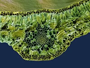
Leaf midrib, SEM
Leaf midrib. Coloured scanning electron micrograph (SEM) of a section through the midrib of a leaf from the Common Box (Buxus sempervirens)

Background imageCross Section Collection: Tulip anther, light micrograph

Tulip anther, light micrograph
Tulip anther. Light micrograph of a cross-section through the anther of a tulip bud (Tulipa sp.), showing a full anther sac with developing pollen grains (white)

Background imageCross Section Collection: Breast in pregnancy, light micrograph

Breast in pregnancy, light micrograph
Breast in pregnancy. Light micrograph of a transverse section of the human mammary gland during pregnancy. The breast is normally made up of fibro-fatty connective tissue

Background imageCross Section Collection: Scalp tissue, light micrograph

Scalp tissue, light micrograph
Scalp tissue. Polarised light micrograph of a section through the human scalp. This section shows oblique and transverse sections through hair shafts (yellow-orange)

Background imageCross Section Collection: Skin tissue, light micrograph

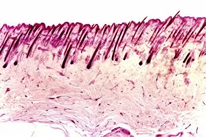
Skin tissue, light micrograph
Skin tissue. Light micrograph of a transverse section through human skin. The skin is made up of an outer epidermis (pale purple, across top) with hair follicles (deep purple)

Background imageCross Section Collection: Compact bone, light micrograph

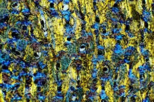
Compact bone, light micrograph
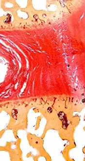
Compact bone. Polarised light micrograph of a transverse section through compact bone tissue, showing Haversian canals (circular regions)

Background imageCross Section Collection: Dogfish liver area, transverse section

Dogfish liver area, transverse section
Dogfish liver area. Light micrograph of a transverse section through the body of a lesser spotted dogfish (Scyliorhinus canicula), in the region of the liver (lower frame)

Background imageCross Section Collection: Dogfish head, transverse section

Dogfish head, transverse section
Dogfish head. Light micrograph of a transverse section through the head of a lesser spotted dogfish (Scyliorhinus canicula), in the region of the eyes (upper left and upper right)

Background imageCross Section Collection: Intervertebral disc, light micrograph

Intervertebral disc, light micrograph
Intervertebral disc. Light micrograph of a transverse section through an intervertebral disc (red) and the surrounding tissue (brown)

Background imageCross Section Collection: Heel skin tissue, light micrograph

Heel skin tissue, light micrograph
Heel skin tissue. Polarised light micrograph of a transverse section through skin from the heel of a human foot. The sole of the foot has to withstand the weight of the body

Background imageCross Section Collection: Lamprey gill region, light micrograph

Lamprey gill region, light micrograph
Lamprey gill. Light micrograph of a transverse section through the gill region of a lamprey (Petromyzon marinus), a parasitic fish. The spinal area is at upper centre

Background imageCross Section Collection: Sheeps hoof, light micrograph

Sheeps hoof, light micrograph
Sheeps hoof. Polarised light micrograph of a transverse section through the hoof of a sheep. In sheep and other hooved animals, the hoof is made of the same material as nails and claws

Background imageCross Section Collection: Sago cycad leaf, SEM

Sago cycad leaf, SEM
Sago cycad leaf. Coloured scanning electron micrograph (SEM) of a section through a sago cycad (Cycas revoluta) leaf. At lower centre and left are vascular bundles (orange clusters)


For sale as Licensed Images

Choose your image, Select your licence and Download the media

"Unveiling the Hidden World: Exploring Cross Sections in Nature and Architecture" Delving into the intricate world of honey bees, witness the mesmerizing cross section of a honeycomb revealing their remarkable life cycle. 🐝🍯 Journey deep within a Honey Bee (Apis mellifera) to discover its internal anatomy through a fascinating cross-section exploration. Marvel at the complexity of human hair follicles and skin as we unveil their hidden secrets through an enlightening cross-sectional view. Embark on a captivating expedition through the human brain, where a cross section exposes the enigmatic limbic system and primitive forebrain. As twilight descends upon Kathmandu's Boudhanath Stupa, immerse yourself in its beauty with an awe-inspiring cross-sectional perspective. Witness nature's underground marvels as we reveal a mole devouring worms beneath our feet—a captivating glimpse into their subterranean world. Step back in time to admire the architectural genius behind Florence Cathedral's dome, revealed through an intricate plan and cutaway drawing from the 16th century. Prayer flags flutter alongside Nepal's iconic Boudhanath Stupa—experience this spiritual haven from an extraordinary cross-sectional viewpoint. Encounter Australia's vibrant wildlife like never before as you explore the inner workings of a Green Tree Python through its stunning cross section. Take flight aboard Pan-American Clipper Boeing 314 with an exclusive cutaway view that unveils every detail of this magnificent aircraft from c1940. Explore Tab X’s mysterious contents by peering into its intriguing cross-section—an invitation to unravel its enigmatic nature.