Hormone Collection (#3)

"Hormone: The Intricate Symphony of the Body's Messengers" The medulla oblongata in the brain, a masterpiece of nature's artwork

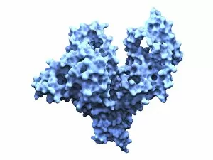
Background imageHormone Collection: Human serum albumin molecule F007 / 9905

Human serum albumin molecule F007 / 9905
Human serum albumin, molecular model. Albumin is the most abundant protein in human blood plasma. One of albumins functions is to transport fatty acids to the liver

Background imageHormone Collection: Hydrocortisone ointment F008 / 3400

Hydrocortisone ointment F008 / 3400
Hydrocortisone ointment in tube

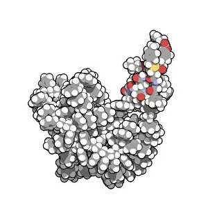
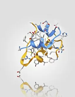
Background imageHormone Collection: Insulin A chain molecule

Insulin A chain molecule
Insulin A chain. Computer model of an A chain of human insulin that has been synthesized on a crosslinked polystyrene solid support. This is an example of solid phase peptide synthesis (SPPS)

Background imageHormone Collection: Noradrenaline norepinephrine molecule

Noradrenaline norepinephrine molecule
Noradrenaline (norepinephrine), belonging to the group of catecholamines, molecular model. Catecholamine acting also as a hormone and a neurotransmitter

Background imageHormone Collection: Adrenaline epinephrine molecule

Adrenaline epinephrine molecule
Adrenaline (epinephrine) is a hormone and a neurotransmitter. It is used to treat a number of conditions like cardiac arrest, anaphylaxis, and superficial bleeding

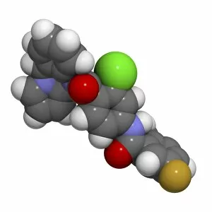
Background imageHormone Collection: Lixivaptan hyponatremia drug F007 / 0159

Lixivaptan hyponatremia drug F007 / 0159
Lixivaptan hyponatremia drug, molecular model. Lixivaptan blocks vasopressin from binding the vasopressin 2 receptor, and is used to treat heart failure in hyponatremia patients

Background imageHormone Collection: Lixivaptan hyponatremia drug F007 / 0158

Lixivaptan hyponatremia drug F007 / 0158
Lixivaptan hyponatremia drug, molecular model. Lixivaptan blocks vasopressin from binding the vasopressin 2 receptor, and is used to treat heart failure in hyponatremia patients

Background imageHormone Collection: Budesonide steroid drug F007 / 0117

Budesonide steroid drug F007 / 0117
Budesonide steroid drug, molecular model. Atoms are represented as spheres and are colour-coded: hydrogen (white), carbon (grey) and oxygen (red)

Background imageHormone Collection: Budesonide steroid drug F007 / 0118

Budesonide steroid drug F007 / 0118
Budesonide steroid drug, molecular model. Atoms are represented as spheres and are colour-coded: hydrogen (white), carbon (grey) and oxygen (red)

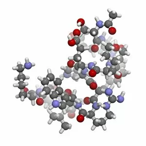
Background imageHormone Collection: Afamelanotide skin pigmentation drug F007 / 0096

Afamelanotide skin pigmentation drug F007 / 0096
Afamelanotide skin pigmentation drug, molecular model. Afamelanotide is an analogue of alpha-melanocyte stimulating hormone (a-MSH)

Background imageHormone Collection: Afamelanotide skin pigmentation drug F007 / 0095

Afamelanotide skin pigmentation drug F007 / 0095
Afamelanotide skin pigmentation drug, molecular model. Afamelanotide is an analogue of alpha-melanocyte stimulating hormone (a-MSH)

Background imageHormone Collection: Insulin molecule C014 / 2121

Insulin molecule C014 / 2121
Insulin molecule. Molecular module of insulin showing its secondary structure. Insulin is a hormone produced by the pancreas

Background imageHormone Collection: Insulin molecule F006 / 9761

Insulin molecule F006 / 9761
Insulin molecule. Molecular model of the hormone insulin from a pig. Insulin consists of two peptide chains, A and B, which are linked by disulphide bridges

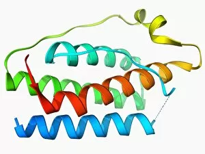
Background imageHormone Collection: Human growth hormone molecule F006 / 9683

Human growth hormone molecule F006 / 9683
Human growth hormone. Molecular model of human growth hormone (hGH, orange) bound to the extracellular domain of the human growth hormone binding protein (hGHBP)

Background imageHormone Collection: Insulin molecule F006 / 9625

Insulin molecule F006 / 9625
Insulin, molecular model. Insulin plays an important role in blood sugar regulation. It is released from the pancreas when blood sugar levels are high, for example after a meal

Background imageHormone Collection: Insulin molecule F006 / 9605

Insulin molecule F006 / 9605
Insulin molecule. Molecular model of the hormone insulin. Insulin consists of two peptide chains, A and B, which are linked by disulphide bridges

Background imageHormone Collection: Insulin molecule F006 / 9558

Insulin molecule F006 / 9558
Insulin, molecular model. Insulin plays an important role in blood sugar regulation. It is released from the pancreas when blood sugar levels are high, for example after a meal

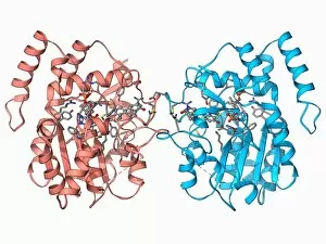
Background imageHormone Collection: Hydroxysteroid dehydrogenase molecule F006 / 9543

Hydroxysteroid dehydrogenase molecule F006 / 9543
Hydroxysteroid dehydrogenase. Molecular model of the human type 5 hydroxysteroid dehydrogenase enzyme bound to a molecule of the steroid drug androstene

Background imageHormone Collection: Neuropeptide Y neurotransmitter molecule F006 / 9493

Neuropeptide Y neurotransmitter molecule F006 / 9493
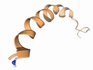
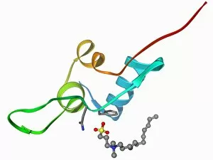
Neuropeptide Y neurotransmitter molecule. Molecular model showing the structure of the neurotransmitter neuropeptide Y (NPY). NPY is found in the brain and autonomic nervous system

Background imageHormone Collection: Androgen receptor, molecular model F006 / 9483

Androgen receptor, molecular model F006 / 9483
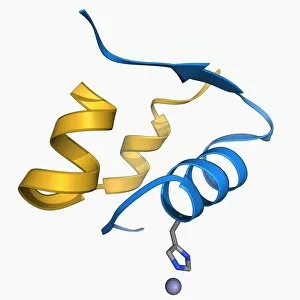
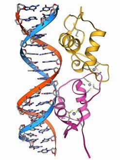
Androgen receptor. Molecular model of the DNA-binding region of an androgen receptor (pink and yellow) complexed with DNA (deoxyribonucleic acid, blue and red)

Background imageHormone Collection: Oxytocin and carrier protein F006 / 9439

Oxytocin and carrier protein F006 / 9439
Oxytocin and carrier protein. Molecular model of the hormone oxytocin bound to its carrier protein neurophysin I. Oxytocin is a neurotransmitter that plays a role in labour, sexual arousal, bonding

Background imageHormone Collection: Oestrogen receptor bound to DNA F006 / 9354

Oestrogen receptor bound to DNA F006 / 9354
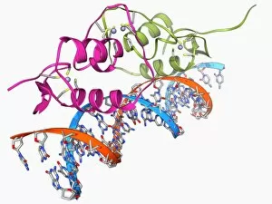
Oestrogen receptor bound to DNA. Molecular model of the DNA-binding domain (green and pink) of the oestrogen receptor bound to a molecule of DNA (deoxyribonucleic acid, blue and orange)

Background imageHormone Collection: Human growth hormone molecule F006 / 9355

Human growth hormone molecule F006 / 9355
Human growth hormone (hGH), molecular model. hGH is produced in the anterior pituitary gland of the brain. It binds to specific receptors on cells in the body to stimulate growth

Background imageHormone Collection: Insulin-like growth 1 factor molecule F006 / 9351

Insulin-like growth 1 factor molecule F006 / 9351
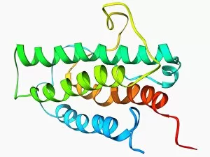
Insulin-like growth factor 1 (IGF-1), molecular model. IGFs are polypeptides that are similar in their molecular structure to insulin

Background imageHormone Collection: Erythropoietin bound to receptors F006 / 9308

Erythropoietin bound to receptors F006 / 9308
Artwork of the human erythropoietin (EPO) hormone molecule (yellow) bound to receptors (pink). Erythropoietin regulates blood oxygen levels in the body

Background imageHormone Collection: Human serum albumin molecule F006 / 9301

Human serum albumin molecule F006 / 9301
Human serum albumin, molecular model. Albumin is the most abundant protein in human blood plasma. It is seen here complexed with stearic acid, a saturated fatty acid

Background imageHormone Collection: Erythropoietin hormone molecule F006 / 9257

Erythropoietin hormone molecule F006 / 9257
Erythropoietin. Molecular model of the human erythropoietin (EPO) hormone. Erythropoietin regulates blood oxygen levels in the body

Background imageHormone Collection: Leptin molecule F006 / 9243

Leptin molecule F006 / 9243
Leptin molecule. Molecular model of the human hormone leptin. Leptin is a protein produced by adipose (fat) tissue. It interacts with receptors in the brains hypothalamus to signal when a person is

Background imageHormone Collection: Oestrogen sulfotransferase molecule F006 / 9237

Oestrogen sulfotransferase molecule F006 / 9237
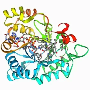
Oestrogen sulfotransferase. Molecular model of the enzyme oestrogen sulfotransferase complexed with oestradiol and ADP (adenosine diphosphate)

Background imageHormone Collection: Kidneys, anatomical artwork

Kidneys, anatomical artwork
Kidneys. Anatomical computer artwork of a mans body, showing the position of the kidneys (red, centre left and right) and adrenal glands (beige)

Background imageHormone Collection: Renal blood supply, anatomical artwork

Renal blood supply, anatomical artwork
Renal blood supply. Anatomical computer artwork of a human kidney (left) and adrenal gland (beige, upper centre), showing their main blood supply

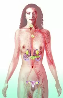
Background imageHormone Collection: Female endocrine glands, artwork C016 / 7522

Female endocrine glands, artwork C016 / 7522
Female endocrine glands. Computer artwork showing the location of the endocrine glands (yellow) in a womans body. These are (from top to bottom): thyroid, thymus, adrenal glands (x2), pancreas

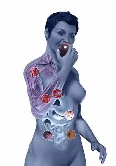
Background imageHormone Collection: Diabetic woman, artwork C016 / 9367

Diabetic woman, artwork C016 / 9367
Diabetic woman. Computer artwork showing sugar (white dots) entering the blood stream of a diabetic woman as she eats an apple. Some of the sugar is used by mitochondria (blue) to produce energy

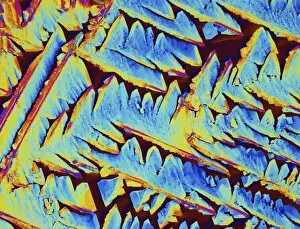
Background imageHormone Collection: Oxytocin hormone crystals, PLM C016 / 7199

Oxytocin hormone crystals, PLM C016 / 7199
Oxytocin. Polarised light micrograph (PLM) of crystals of the female hormone oxytocin. In women this hormone is secreted naturally by the pituitary gland

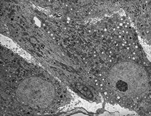
Background imageHormone Collection: Corpus luteum, TEM

Corpus luteum, TEM
Corpus luteum. Transmission electron micrograph (TEM) of a section through the corpus luteum of an ovary, showing several luteal cells


For sale as Licensed Images

Choose your image, Select your licence and Download the media

"Hormone: The Intricate Symphony of the Body's Messengers" The medulla oblongata in the brain, a masterpiece of nature's artwork, orchestrates an intricate dance of hormones. Among them, oxytocin hormone crystals shimmer like precious gems under the microscope (PLM C016 / 7196), revealing their profound role in human connection and bonding. Delving deeper into cellular realms, a colored SEM image showcases mitochondria within ovarian cells, highlighting their crucial involvement in hormone production. Meanwhile, light micrographs capture oxytocin crystals (LM C016 / 7195) and insulin crystals (C017 / 8249), unveiling their mesmerizing structures that hold vital functions within our bodies. Exploring athlete physiology through captivating artwork unveils the remarkable impact hormones have on performance and endurance. These chemical messengers drive athletes to push beyond limits and achieve greatness. Artistic depictions also shed light on the complex world of molecular biology. An intricately designed insulin molecule stands as a testament to its pivotal role in regulating blood sugar levels while offering hope for those with diabetes. The pancreas anatomy comes alive through vibrant illustrations showcasing its significance as both an endocrine and digestive organ. It secretes essential hormones such as insulin that keep our bodies functioning optimally. Testosterone takes center stage with PLM capturing crystal formations that symbolize strength and masculinity. This hormone plays a fundamental role not only in physical development but also influences mood, cognition, and overall well-being. Returning to oxytocin - known as the "love hormone" - we delve into its dual identity as both neurotransmitter and hormonal messenger. Its delicate crystalline structure reveals itself once again under microscopic scrutiny (Oxytocin hormone crystals). Lastly, an artistic portrayal of thyroid anatomy reminds us how this small gland holds immense power over metabolism regulation (artwork C013 / 4675). Hormones secreted by this butterfly-shaped organ impact our energy levels, weight management, and overall health.