Hormone Collection (#8)

"Hormone: The Intricate Symphony of the Body's Messengers" The medulla oblongata in the brain, a masterpiece of nature's artwork

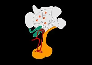
Background imageHormone Collection: Hypothalamus and hypophysis, artwork

Hypothalamus and hypophysis, artwork
Hypothalamus and hypophysis. Artwork showing the brains hypothalamus (white) and hypophysis (pituitary gland, orange), the link between the nervous system and the endocrine system

Background imageHormone Collection: Normal blood glucose level, artwork

Normal blood glucose level, artwork
Normal blood glucose (sugar) level. Artwork depicting the normal levels of blood glucose (red dots) and the hormone insulin (yellow crosses) in the human body after a meal

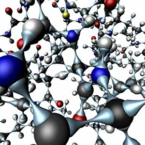
Background imageHormone Collection: Insulin molecule, close-up view

Insulin molecule, close-up view
Insulin. Computer graphic showing a part of the molecule of human insulin. Insulin is a hormone which is produced in the pancreas by cells of the Islets of Langerhans

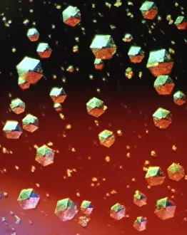
Background imageHormone Collection: Insulin crystals, light micrograph

Insulin crystals, light micrograph
Insulin. Polarised light micrograph (PLM) of crystals of the hormone insulin. The crystals appear hexagonal in shape. The insulin molecule is made up of two chains of amino acids (A & B chains)

Background imageHormone Collection: Melatonin brain function, artwork

Melatonin brain function, artwork
Melatonin brain function. Artwork showing how the hormone melatonin (blue spheres) is secreted in the brain and helps to control the bodys biological clock

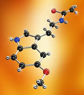
Background imageHormone Collection: Melatonin hormone molecule, artwork

Melatonin hormone molecule, artwork
Computer artwork of a ball-and-stick model of a molecule of the hormone melatonin. Atoms are colour-coded and represented as spheres; carbon (black), hydrogen (silver)

Background imageHormone Collection: Progesterone hormone, light micrograph

Progesterone hormone, light micrograph
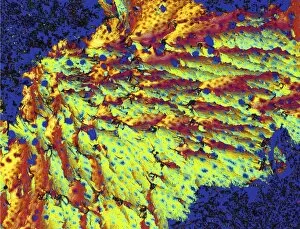
Progesterone hormone. Polarised light micrograph of crystals of female sex hormone progesterone. Progesterone is the most potent of the naturally- occurring progestogens, a class of steroid hormones

Background imageHormone Collection: Cortisone molecule

Cortisone molecule. Cortisone is a steroid hormone produced by the kidneys. It suppresses the immune system and so reduces inflammation and pain at the site of an injury

Background imageHormone Collection: Erythropoietin bound to receptors

Erythropoietin bound to receptors
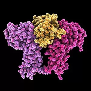
Artwork of the human erythropoietin (EPO) hormone molecule (yellow) bound to receptors (pink). Erythropoietin regulates blood oxygen levels in the body

Background imageHormone Collection: Endocrine system, artwork

Endocrine system, artwork
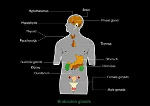
Endocrine system. Computer artwork showing the locations of hormone-producing glands (endocrine glands) in the human body

Background imageHormone Collection: Growth hormone receptor, molecular model

Growth hormone receptor, molecular model
Growth hormone receptor. Molecular model of a growth hormone receptor (orange and beige) bound to a growth hormone molecule (red)

Background imageHormone Collection: Serotonin N-acetyl transferase molecule

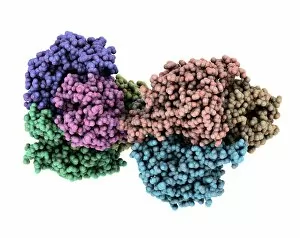
Serotonin N-acetyl transferase molecule. Computer model showing the molecular structure of the four sequence motifs (coloured) that make up the arylalkylamine N-acetyltransferase (aNAT) complex

Background imageHormone Collection: Glucagon hormone molecule

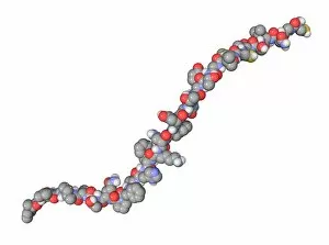
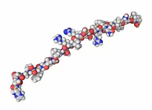
Glucagon hormone molecule. Computer model showing the structure of the human hormone glucagon. Atoms are colour-coded spheres (carbon: grey, nitrogen: blue, and oxygen: red)

Background imageHormone Collection: Gastrin hormone molecule

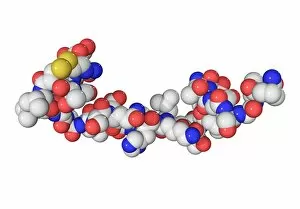
Gastrin hormone molecule. Computer model showing the structure of the human hormone gastrin. Atoms are colour-coded spheres (carbon: grey, hydrogen: light grey, nitrogen: blue, sulphur: yellow)

Background imageHormone Collection: Leptin hormone molecule

Leptin hormone molecule. Computer model showing the secondary structure of the hormone leptin. This hormone is produced by adipose (fat) tissue

Background imageHormone Collection: Aldosterone molecule

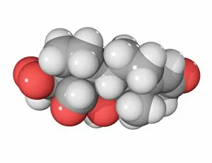
Aldosterone molecule. Molecular model showing the structure of a molecule of the steroid hormone aldosterone (C21.H28.O5). Atoms are colour-coded (carbon: grey, hydrogen: white, oxygen: red)

Background imageHormone Collection: Somatostatin hormone molecule

Somatostatin hormone molecule. Computer model showing the structure of the hormone somatostatin, also known as growth hormone-inhibiting hormone (GHIH)

Background imageHormone Collection: Atrial natriuretic peptide molecule

Atrial natriuretic peptide molecule. Computer model showing the structure of a molecule of the hormone atrial natriuretic peptide (ANP)

Background imageHormone Collection: Calcifediol molecule

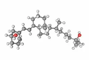
Calcifediol molecule. Computer model showing the structure of a molecule of the pre-hormone calcifediol. Atoms are colour-coded (carbon: grey, hydrogen: white)

Background imageHormone Collection: Prostacyclin molecule

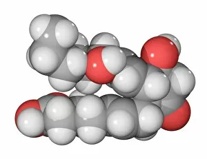
Prostacyclin molecule. Computer model showing the structure of the hormone prostacyclin, or prostaglandin I2 (PGI2). Atoms are colour-coded (carbon: dark grey, hydrogen: light grey, oxygen: red)

Background imageHormone Collection: Norepinephrine neurotransmitter molecule

Norepinephrine neurotransmitter molecule. Computer model showing the structure of the neurotransmitter and hormone norepinephrine, or noradrenaline

Background imageHormone Collection: Calcitonin hormone molecule

Calcitonin hormone molecule. Computer model showing the structure of the human hormone calcitonin. Atoms are colour-coded spheres (carbon: dark grey, hydrogen: light grey, nitrogen: blue)

Background imageHormone Collection: Adiponectin hormone molecule

Adiponectin hormone molecule. Molecular model showing the secondary structure of the human hormone adiponectin (Acrp30). Hydrogen bonding is shown by beta sheets (arrows) and random coils (wires)

Background imageHormone Collection: Prostaglandin E1 molecule

Prostaglandin E1 molecule. Computer model showing the structure of the hormone prostaglandin E1 (PGE1). Atoms are colour-coded (carbon: dark grey, hydrogen: light grey, oxygen: red)

Background imageHormone Collection: Leukotriene E4 molecule

Leukotriene E4 molecule. Computer model showing the structure of the fatty acid leukotriene E4. Atoms are colour-coded (carbon: dark grey, hydrogen: light grey, nitrogen: blue, sulphur: yellow)

Background imageHormone Collection: Secretin hormone molecule

Secretin hormone molecule. Computer model showing the structure of the hormone secretin. Atoms are colour-coded (carbon: dark grey, hydrogen: light grey, oxygen: red)

Background imageHormone Collection: Prolactin hormone molecule

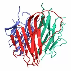
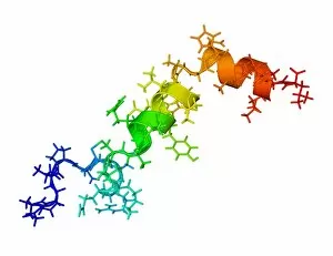
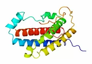
Prolactin hormone molecule. Computer model showing the secondary structure of human prolactin (hPRL), or luteotropic hormone (LTH)

Background imageHormone Collection: Oestradiol hormone molecule

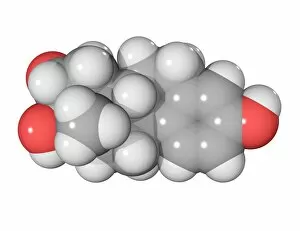
Oestradiol hormone molecule. Computer model showing the structure of the female sex hormone oestradiol. Atoms are colour-coded - carbon: dark grey, hydrogen: light grey, and oxygen: red

Background imageHormone Collection: DHEA hormone, molecular model

DHEA hormone, molecular model
DHEA hormone. Molecular model of the structure of the human hormone DHEA (dehydroepiandrosterone). Atoms are represented are colour-coded (carbon: dark grey, hydrogen: light grey, and oxygen (red)

Background imageHormone Collection: Gonadotrophin-releasing hormone molecule

Gonadotrophin-releasing hormone molecule. Computer model showing the structure of gonadotrophin-releasing hormone (GnRH). Atoms are colour-coded spheres (carbon: large grey, hydrogen: small grey)

Background imageHormone Collection: Met-enkephalin molecule

Met-enkephalin molecule. Computer model showing the structure of the neurotransmitter met-enkephalin (methionine-enkephalin)

Background imageHormone Collection: Follicle stimulating hormone molecules

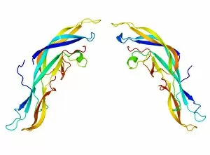
Follicle stimulating hormone molecules. Computer artwork showing the secondary structure of two strands of follicle stimulating hormone (FSH)

Background imageHormone Collection: Pancreatic hormone molecule

Pancreatic hormone molecule. Molecular model showing the secondary structure of the hormone known as pancreatic polypeptide (PP)

Background imageHormone Collection: Orexin-A hormone molecule

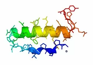
Orexin-A hormone molecule. Computer model showing the crystal structure of a molecule of the hormone orexin-A. The crystal structure consists of both the secondary structure

Background imageHormone Collection: Corticotropin-releasing factor complex

Corticotropin-releasing factor complex
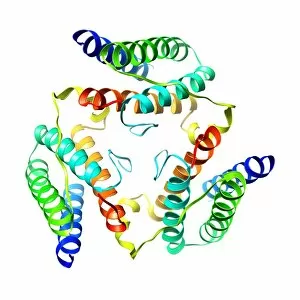
Corticotropin-releasing factor (CRF) complex. Molecular model showing the structure of the human hormone and neurotransmitter CRF in complex with its receptor

Background imageHormone Collection: Salmon calcitonin hormone molecule

Salmon calcitonin hormone molecule. Computer model showing the structure of calcitonin from salmon. Atoms are colour-coded spheres (carbon: dark grey, hydrogen: light grey, nitrogen: blue)

Background imageHormone Collection: Prostaglandin E2 molecule

Prostaglandin E2 molecule. Computer model showing the structure of the hormone prostaglandin E2 (PGE2). Atoms are colour-coded (carbon: dark grey, hydrogen: light grey, oxygen: red)

Background imageHormone Collection: Somatoliberin hormone molecule

Somatoliberin hormone molecule. Computer model showing the structure of the hormone somatoliberin, also known as growth hormone-releasing hormone (GHRH)

Background imageHormone Collection: Orexin-B hormone molecule

Orexin-B hormone molecule. Computer model showing the crystal structure of a molecule of the hormone orexin-B. The crystal structure consists of both the secondary structure

Background imageHormone Collection: Oestrone hormone molecule

Oestrone hormone molecule. Computer model showing the structure of the female sex hormone oestrone. Atoms are colour-coded - carbon: dark grey, hydrogen: light grey, and oxygen: red

Background imageHormone Collection: Latanoprost glaucoma drug molecule

Latanoprost glaucoma drug molecule
Prostacyclin molecule. Computer model showing the structure of the hormone prostacyclin, or prostaglandin I2 (PGI2). Atoms are colour-coded (carbon: dark grey, hydrogen: light grey, oxygen: red)

Background imageHormone Collection: Oestriol hormone molecule

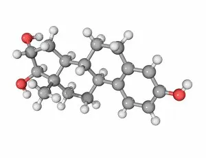
Oestriol hormone molecule. Computer model showing the structure of the female sex hormone oestriol. Atoms are colour-coded - carbon: dark grey, hydrogen: light grey, and oxygen: red

Background imageHormone Collection: Somatotrophin hormone molecule

Somatotrophin hormone molecule. Computer model showing the secondary structure of the human growth hormone somatotrophin (GH)

Background imageHormone Collection: DHT hormone, molecular model

DHT hormone, molecular model
DHT hormone. Molecular model of the structure of the male sex hormone DHT (dihydrotestosterone, or 5-alpha-dihydrotestosterone)

Background imageHormone Collection: Androstenedione hormone molecule

Androstenedione hormone molecule. Computer model showing the structure of a molecule of the steroid hormone, dietary supplement and banned drug androstenedione (C19.H26.O2)


For sale as Licensed Images

Choose your image, Select your licence and Download the media

"Hormone: The Intricate Symphony of the Body's Messengers" The medulla oblongata in the brain, a masterpiece of nature's artwork, orchestrates an intricate dance of hormones. Among them, oxytocin hormone crystals shimmer like precious gems under the microscope (PLM C016 / 7196), revealing their profound role in human connection and bonding. Delving deeper into cellular realms, a colored SEM image showcases mitochondria within ovarian cells, highlighting their crucial involvement in hormone production. Meanwhile, light micrographs capture oxytocin crystals (LM C016 / 7195) and insulin crystals (C017 / 8249), unveiling their mesmerizing structures that hold vital functions within our bodies. Exploring athlete physiology through captivating artwork unveils the remarkable impact hormones have on performance and endurance. These chemical messengers drive athletes to push beyond limits and achieve greatness. Artistic depictions also shed light on the complex world of molecular biology. An intricately designed insulin molecule stands as a testament to its pivotal role in regulating blood sugar levels while offering hope for those with diabetes. The pancreas anatomy comes alive through vibrant illustrations showcasing its significance as both an endocrine and digestive organ. It secretes essential hormones such as insulin that keep our bodies functioning optimally. Testosterone takes center stage with PLM capturing crystal formations that symbolize strength and masculinity. This hormone plays a fundamental role not only in physical development but also influences mood, cognition, and overall well-being. Returning to oxytocin - known as the "love hormone" - we delve into its dual identity as both neurotransmitter and hormonal messenger. Its delicate crystalline structure reveals itself once again under microscopic scrutiny (Oxytocin hormone crystals). Lastly, an artistic portrayal of thyroid anatomy reminds us how this small gland holds immense power over metabolism regulation (artwork C013 / 4675). Hormones secreted by this butterfly-shaped organ impact our energy levels, weight management, and overall health.