Hormone Collection (#6)

"Hormone: The Intricate Symphony of the Body's Messengers" The medulla oblongata in the brain, a masterpiece of nature's artwork

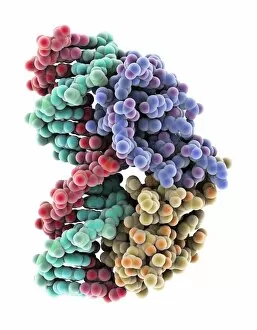
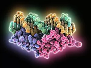
Background imageHormone Collection: Androgen receptor, molecular model C015 / 6112

Androgen receptor, molecular model C015 / 6112
Androgen receptor. Molecular model of the DNA-binding region of an androgen receptor (purple and brown) complexed with DNA (deoxyribonucleic acid, turquoise and red)
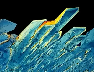
Background imageHormone Collection: Insulin crystals, light micrograph C017 / 8246

Insulin crystals, light micrograph C017 / 8246
Insulin. Polarised light micrograph (PLM) of crystals of the hormone insulin. The insulin molecule is made up of two chains of amino acids (A & B chains)

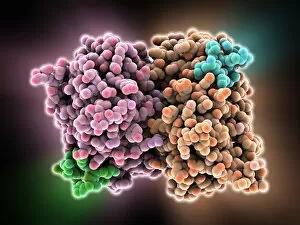
Background imageHormone Collection: Oestrogen receptor, molecular model

Oestrogen receptor, molecular model
Oestrogen receptor. Molecular model of an oestrogen receptor, bound to a glucocorticoid receptor-interacting protein. Oestrogen receptors are cytoplasmic proteins that bind oestrogens

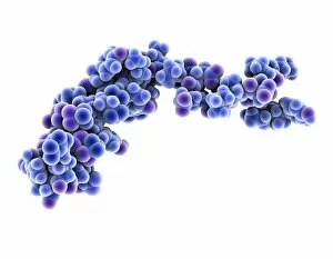
Background imageHormone Collection: Neuropeptide Y neurotransmitter molecule C015 / 6240

Neuropeptide Y neurotransmitter molecule C015 / 6240
Neuropeptide Y neurotransmitter molecule. Molecular model showing the structure of the neurotransmitter neuropeptide Y (NPY). NPY is found in the brain and autonomic nervous system

Background imageHormone Collection: Oestrogen receptor bound to DNA C015 / 5404

Oestrogen receptor bound to DNA C015 / 5404
Oestrogen receptor bound to DNA. Molecular model of the DNA-binding domain (pink and purple) of the oestrogen receptor bound to a molecule of DNA (deoxyribonucleic acid, yellow and green)

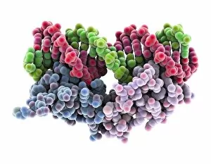
Background imageHormone Collection: Oestrogen receptor bound to DNA C015 / 5403

Oestrogen receptor bound to DNA C015 / 5403
Oestrogen receptor bound to DNA. Molecular model of the DNA-binding domain (blue and purple) of the oestrogen receptor bound to a molecule of DNA (deoxyribonucleic acid, pink and green)

Background imageHormone Collection: Pituitary gland C015 / 6420

Pituitary gland C015 / 6420
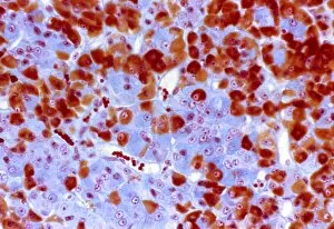
Pituitary gland. Light micrograph of a section through a healthy pituitary gland. The pituitary is a pea-sized organ found at the base of the brain

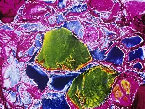
Background imageHormone Collection: Thyroid gland, SEM C015 / 6419

Thyroid gland, SEM C015 / 6419
Thyroid gland. Coloured scanning electron micrograph (SEM) of a fracture through the thyroid gland revealing several follicles (green and blue). Between the follicles is connective tissue (pink)

Background imageHormone Collection: Islet of Langerhans, TEM C015 / 6418

Islet of Langerhans, TEM C015 / 6418
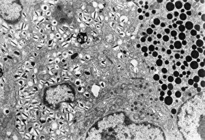
Islet of Langerhans. Transmission electron micrograph (TEM) of a section through cells clustered in an islet of Langerhans in the pancreas

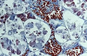
Background imageHormone Collection: Adrenal cortex, light micrograph C015 / 6417

Adrenal cortex, light micrograph C015 / 6417
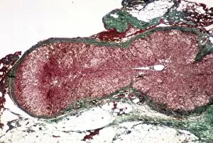
Adrenal cortex. Light micrograph of a section through a healthy adrenal cortex (red). The adrenal glands are found on top of the kidneys

Background imageHormone Collection: Erythropoietin hormone complex C016 / 4461

Erythropoietin hormone complex C016 / 4461
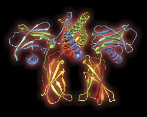
Erythropoietin hormone complex. Computer model showing the secondary structure of a molecule of the human hormone Erythropoietin (EPO), complexed with an erythropoetin receptor molecule

Background imageHormone Collection: Erythropoietin hormone complex C016 / 4458

Erythropoietin hormone complex C016 / 4458
Erythropoietin hormone complex. Computer model showing the secondary structure of a molecule of the human hormone Erythropoietin (EPO), complexed with an erythropoetin receptor molecule

Background imageHormone Collection: Erythropoietin hormone complex C016 / 4451

Erythropoietin hormone complex C016 / 4451
Erythropoietin hormone complex. Computer model showing the secondary structure of a molecule of the human hormone Erythropoietin (EPO), complexed with an erythropoetin receptor molecule

Background imageHormone Collection: Follicle-stimulating hormone complex C016 / 2794

Follicle-stimulating hormone complex C016 / 2794
Follicle-stimulating hormone (FSH) complex with receptor, molecular model. FSH helps to regulate human sexual development and reproductive processes. In females, it acts on follicles in the ovaries

Background imageHormone Collection: Follicle-stimulating hormone complex C016 / 2793

Follicle-stimulating hormone complex C016 / 2793
Follicle-stimulating hormone (FSH) complex with receptor, molecular model. FSH helps to regulate human sexual development and reproductive processes. In females, it acts on follicles in the ovaries

Background imageHormone Collection: Neuropeptide Y neurotransmitter molecule C014 / 0013

Neuropeptide Y neurotransmitter molecule C014 / 0013
Neuropeptide Y neurotransmitter molecule. Molecular model showing the structure of the neurotransmitter neuropeptide Y (NPY)

Background imageHormone Collection: Human growth hormone molecule C013 / 9006

Human growth hormone molecule C013 / 9006
Human growth hormone. Molecular model of human growth hormone (hGH, yellow) bound to the extracellular domain of the human growth hormone binding protein (hGHBP)

Background imageHormone Collection: Erythropoietin molecule C013 / 8891

Erythropoietin molecule C013 / 8891
Erythropoietin molecule (EPO), molecular model. EPO is a glycoprotein that is produced by the kidneys in response to low blood oxygen levels

Background imageHormone Collection: Erythropoietin hormone complex

Erythropoietin hormone complex. Computer model showing the secondary structure of a molecule of the human hormone Erythropoietin (EPO), complexed with an erythropoetin receptor molecule

Background imageHormone Collection: Thyroid anatomy, artwork C013 / 4674

Thyroid anatomy, artwork C013 / 4674
Thyroid anatomy. Computer artwork of the thyroid glands (pink) of the neck, shown in relation to the bodys bones and throat structures (white)

Background imageHormone Collection: Illustration of thyroid gland and microscopic view

Illustration of thyroid gland and microscopic view

Background imageHormone Collection: Computer art of thyroid gland on windpipe

Computer art of thyroid gland on windpipe
Thyroid gland. Computer graphic of the thyroid gland, seen on the blue outline of the trachea (windpipe). The thyroid gland (orange) is located in the neck on the trachea

Background imageHormone Collection: Female hormone system

Female hormone system. Computer artwork of a female figure illustrating locations of hormone producing glands (known as the endocrine system)

Background imageHormone Collection: Computer art of thyroid gland on human outline

Computer art of thyroid gland on human outline
Thyroid gland. Computer graphic of the thyroid gland, superimposed on the outline of a human figure. The thyroid gland (orange) is located in the neck

Background imageHormone Collection: Thyroid gland follicle, TEM

Thyroid gland follicle, TEM
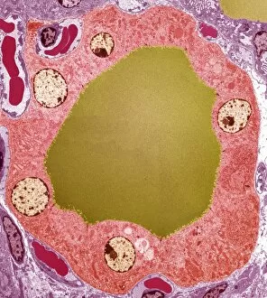
Thyroid gland follicle. Coloured transmission electron micrograph (TEM) of a follicle from the thyroid gland. The follicle consists of a layer of epithelial cells (pink)

Background imageHormone Collection: Illustration of single thyroid follicle

Illustration of single thyroid follicle
Illustration of a single human thyroid follicle, the functional unit of the thyroid gland. The follicle has been cut open to reveal the internal layer of epithelial cells surrounding a pool of

Background imageHormone Collection: Illustration of blood supply in the thyroid gland

Illustration of blood supply in the thyroid gland
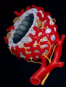
Illustration of the capillary blood supply of a follicle in the human thyroid gland. The thyroid endocrine gland consists of many closed spheroidal follicles

Background imageHormone Collection: Thyroid gland, scintigram

Thyroid gland, scintigram
Thyroid gland. Coloured scintigram of a healthy thyroid gland. This two-lobed endocrine gland is found in the neck, where it lies on top of the trachea (windpipe)

Background imageHormone Collection: Pituitary gland tissue, light micrograph

Pituitary gland tissue, light micrograph
Pituitary gland tissue. Light micrograph of a section through the pars distalis, the anterior part of the adenohypophysis in the pituitary gland

Background imageHormone Collection: Thyroid follicles

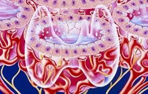
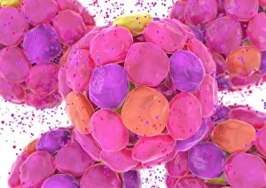
Thyroid follicles. Computer artwork of cells of thyroid follicles (pink, purple) superimposed on an artwork of the thyroid gland (red, centre)

Background imageHormone Collection: Illustration of follicles in the thyroid gland

Illustration of follicles in the thyroid gland
Thyroid gland. Illustration of the microscopic cell structure of the human thyroid gland, with an outline of the whole gland superimposed

Background imageHormone Collection: Artwork showing the thyroid gland

Artwork showing the thyroid gland

Background imageHormone Collection: Pituitary gland, TEM

Pituitary gland, TEM
Pituitary gland. Coloured transmission electron micrograph (TEM) of cells in the anterior pituitary gland, a hormone-secreting gland at the base of the brain

Background imageHormone Collection: Kideny and adrenal gland

Kideny and adrenal gland
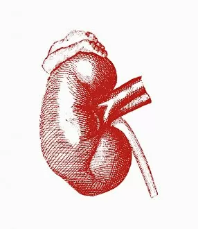
Kidney and adrenal gland, artwork. The kidney is one of a pair of organs that form urine by filtering out waste products from the blood

Background imageHormone Collection: Insulin-secreting pancreatic cells

Insulin-secreting pancreatic cells
Insulin production. Artwork of a section through beta cells secreting insulin (blue spheres) into a capillary (small blood vessel, grey tube). Beta cells are found in the pancreas

Background imageHormone Collection: Shaving foam

Shaving foam
MODEL RELEASED. Shaving foam. Nineteen year old man squeezing shaving foam into his hand in preparation for shaving. Shaving is a method of hair removal (depilation)

Background imageHormone Collection: Thyroid follicles, artwork

Thyroid follicles, artwork
Thyroid gland. Computer artwork of a thyroid gland (red, centre), superimposed on an image of cells of thyroid follicles (green, blue)

Background imageHormone Collection: Diabetes mellitus, computer artwork

Diabetes mellitus, computer artwork. Diabetes is caused by low levels of the hormone insulin and resistance to insulin by the body cells

Background imageHormone Collection: Goitre

Goitre. Artwork comparing a healthy (left) and enlarged (goitre, right) thyroid gland (lower centre, blue). An enlarged thyroid may result from hormonal imbalances

Background imageHormone Collection: Melatonin hormone molecule

Melatonin hormone molecule
Melatonin hormone, molecular model. Atoms are represented as spheres and are colour-coded: carbon (black), hydrogen (silver), nitrogen (blue) and oxygen (red)

Background imageHormone Collection: Androsterone hormone molecule

Androsterone hormone molecule
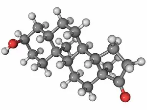
Androsterone, molecular model. This is a steroid male sex hormone. Atoms are represented as spheres and are colour-coded: carbon (grey), hydrogen (white) and oxygen (red)

Background imageHormone Collection: Dopamine, 3D molecular model

Dopamine, 3D molecular model
3D molecular model of dopamine. Dopamine is a catecholamine neurotransmitter produced in different aereas of the brain. Dopamine is also a neurohormone released by the hypothalamus

Background imageHormone Collection: Alpha-endorphin, molecular model

Alpha-endorphin, molecular model
Alpha-endorphin hormone, molecular model. Atoms are represented as spheres and are colour-coded: carbon (grey), hydrogen (white), oxygen (red), nitrogen (blue) and sulphur (yellow)

Background imageHormone Collection: Thyroid, light micrograph

Thyroid, light micrograph
Thyroid. Light micrograph of a section through a thyroid gland. The dark purple areas are follicles that are filled with a storage form form of the thyroid hormones

Background imageHormone Collection: Adrenaline, polarized light micrograph

Adrenaline, polarized light micrograph
Adrenaline. Polarized light micrograph of crystals of adrenaline (epinephrine). This is a hormone produced in the adrenal glands above the kidneys

Background imageHormone Collection: Human oestrogen-related receptor molecule

Human oestrogen-related receptor molecule
Human oestrogen-related receptor alpha, molecular model

Background imageHormone Collection: Islets of Langerhans cells, artwork

Islets of Langerhans cells, artwork
Islets of Langerhans cells, computer artwork. These cells (large, pink) of the pancreas release hormones (dots) into the blood

Background imageHormone Collection: Hypothalamus and hypophysis, diagram

Hypothalamus and hypophysis, diagram
Hypothalamus and hypophysis. Diagram showing the brains hypothalamus (white) and hypophysis (pituitary gland, orange), the link between the nervous system and the endocrine system


For sale as Licensed Images

Choose your image, Select your licence and Download the media

"Hormone: The Intricate Symphony of the Body's Messengers" The medulla oblongata in the brain, a masterpiece of nature's artwork, orchestrates an intricate dance of hormones. Among them, oxytocin hormone crystals shimmer like precious gems under the microscope (PLM C016 / 7196), revealing their profound role in human connection and bonding. Delving deeper into cellular realms, a colored SEM image showcases mitochondria within ovarian cells, highlighting their crucial involvement in hormone production. Meanwhile, light micrographs capture oxytocin crystals (LM C016 / 7195) and insulin crystals (C017 / 8249), unveiling their mesmerizing structures that hold vital functions within our bodies. Exploring athlete physiology through captivating artwork unveils the remarkable impact hormones have on performance and endurance. These chemical messengers drive athletes to push beyond limits and achieve greatness. Artistic depictions also shed light on the complex world of molecular biology. An intricately designed insulin molecule stands as a testament to its pivotal role in regulating blood sugar levels while offering hope for those with diabetes. The pancreas anatomy comes alive through vibrant illustrations showcasing its significance as both an endocrine and digestive organ. It secretes essential hormones such as insulin that keep our bodies functioning optimally. Testosterone takes center stage with PLM capturing crystal formations that symbolize strength and masculinity. This hormone plays a fundamental role not only in physical development but also influences mood, cognition, and overall well-being. Returning to oxytocin - known as the "love hormone" - we delve into its dual identity as both neurotransmitter and hormonal messenger. Its delicate crystalline structure reveals itself once again under microscopic scrutiny (Oxytocin hormone crystals). Lastly, an artistic portrayal of thyroid anatomy reminds us how this small gland holds immense power over metabolism regulation (artwork C013 / 4675). Hormones secreted by this butterfly-shaped organ impact our energy levels, weight management, and overall health.