Internal Anatomy Collection (page 4)

"Exploring the Intricacies: Unveiling Internal Anatomy through Art and Science" Delving into the hidden world of internal anatomy

Background imageInternal Anatomy Collection: Screws for compression of blood vessels by Jean Louis Petite

Screws for compression of blood vessels by Jean Louis Petite, tourniquet device and double band to adjust it
FLO4611834 Screws for compression of blood vessels by Jean Louis Petite, tourniquet device and double band to adjust it, chamois skin compress

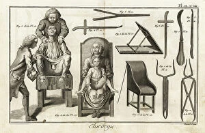
Background imageInternal Anatomy Collection: surgical chair with patient to remove a vesical calculation through the groin - Plate taken from '

surgical chair with patient to remove a vesical calculation through the groin - Plate taken from "
FLO4611780 surgical chair with patient to remove a vesical calculation through the groin - Plate taken from " L'Encyclopedie" by Denis Diderot (1713-1784)

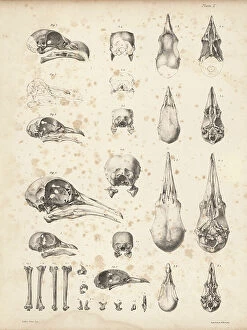
Background imageInternal Anatomy Collection: Cranium 1-4 and part of sternum 5, 6 of the extinct Rodrigues solitaire

Cranium 1-4 and part of sternum 5, 6 of the extinct Rodrigues solitaire. Lithograph by Joseph Dinkel after Werner
FLO4612182 Cranium 1-4 and part of sternum 5, 6 of the extinct Rodrigues solitaire. Lithograph by Joseph Dinkel after Werner from Hugh Edwin Strickland

Background imageInternal Anatomy Collection: Skulls of tooth-billed pigeon, Didunculus strigirostris, critically endangered 1, 7, 8, 9, 10 dodo

Skulls of tooth-billed pigeon, Didunculus strigirostris, critically endangered 1, 7, 8, 9, 10 dodo
FLO4612171 Skulls of tooth-billed pigeon, Didunculus strigirostris, critically endangered 1, 7, 8, 9, 10 dodo, Raphus cucullatus 2, yellow-footed green pigeon, Treron chlorigaster 3, 6

Background imageInternal Anatomy Collection: Base of the skull, human teeth, knee cap (after Clopton Havers), mucus gland, etc

Base of the skull, human teeth, knee cap (after Clopton Havers), mucus gland, etc
FLO4611538 Base of the skull, human teeth, knee cap (after Clopton Havers), mucus gland, etc. Copperplate engraving by Robert Benard from Denis Diderot's Encyclopedia, Pellet, Geneva

Background imageInternal Anatomy Collection: Interior of the brain and cerebellum. Copperplate engraving by Robert Benard after an illustration

Interior of the brain and cerebellum. Copperplate engraving by Robert Benard after an illustration by Albrecht von
FLO4611652 Interior of the brain and cerebellum. Copperplate engraving by Robert Benard after an illustration by Albrecht von Haller from Denis Diderot's Encyclopedia, Pellet, Geneva

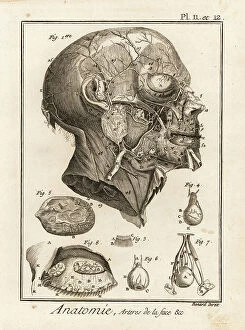
Background imageInternal Anatomy Collection: Arteries in the face. Copperplate engraving by Robert Benard after an illustration by Albrecht von

Arteries in the face. Copperplate engraving by Robert Benard after an illustration by Albrecht von Haller from Denis
FLO4611618 Arteries in the face. Copperplate engraving by Robert Benard after an illustration by Albrecht von Haller from Denis Diderot's Encyclopedia, Pellet, Geneva

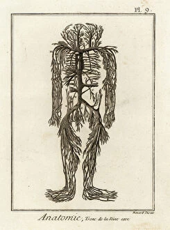
Background imageInternal Anatomy Collection: Venal system from the vena cava in the human body. Copperplate engraving by Robert Benard after an

Venal system from the vena cava in the human body. Copperplate engraving by Robert Benard after an illustration by
FLO4611599 Venal system from the vena cava in the human body. Copperplate engraving by Robert Benard after an illustration by James Drake from Denis Diderot's Encyclopedia, Pellet, Geneva

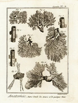
Background imageInternal Anatomy Collection: Details of arteries, veins, lymph acorns, etc. Copperplate engraving by Robert Benard after

Details of arteries, veins, lymph acorns, etc. Copperplate engraving by Robert Benard after from Denis Diderot's
FLO4611594 Details of arteries, veins, lymph acorns, etc. Copperplate engraving by Robert Benard after from Denis Diderot's Encyclopedia, Pellet, Geneva, 1779.; (add.info.: Details of arteries)

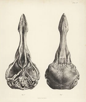
Background imageInternal Anatomy Collection: Upper and lower views of the skull of a dodo. Lithograph from Hugh Edwin Strickland

Upper and lower views of the skull of a dodo. Lithograph from Hugh Edwin Strickland
FLO4612153 Upper and lower views of the skull of a dodo. Lithograph from Hugh Edwin Strickland and Alexander Gordon Melville's The Dodo and its Kindred, London, Reeve, Benham and Reeve

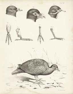
Background imageInternal Anatomy Collection: Tooth billed pigeon, Didunculus strigirostris 1, 2 (critically endangered), Bruce's green pigeon

Tooth billed pigeon, Didunculus strigirostris 1, 2 (critically endangered), Bruce's green pigeon
FLO4612139 Tooth billed pigeon, Didunculus strigirostris 1, 2 (critically endangered), Bruce's green pigeon, Treron waalia (Treron abyssinica 3, 5) and Levaillant's dove

Background imageInternal Anatomy Collection: Jean Louis Petite's machine to compress femoral artere during thigh amputation, bandage

Jean Louis Petite's machine to compress femoral artere during thigh amputation, bandage
FLO4611844 Jean Louis Petite's machine to compress femoral artere during thigh amputation, bandage, aneurysm needle and scissors - Plate taken from " L'Encyclopedie" by Denis

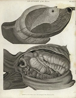
Background imageInternal Anatomy Collection: Anatomy of the horse: stomach and intestines. Copperplate engraving by Scott from Abraham Rees

Anatomy of the horse: stomach and intestines. Copperplate engraving by Scott from Abraham Rees Cyclopedia or Universal
FLO4593968 Anatomy of the horse: stomach and intestines. Copperplate engraving by Scott from Abraham Rees Cyclopedia or Universal Dictionary of Arts, Sciences and Literature, Longman, Hurst, Rees

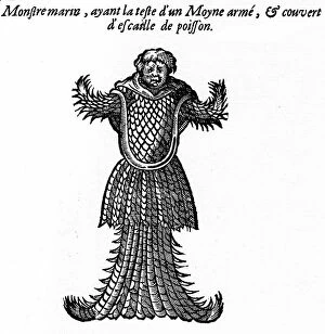
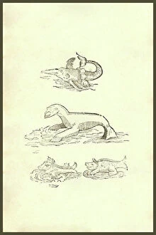
Background imageInternal Anatomy Collection: Engraving of the book of Ambroise Pare: ' Monsters and Prodiges' (first edition 1573)

Engraving of the book of Ambroise Pare: " Monsters and Prodiges" (first edition 1573)
PCT4260964 Engraving of the book of Ambroise Pare: " Monsters and Prodiges" (first edition 1573, appendix: marine monsters)

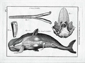
Background imageInternal Anatomy Collection: Le grand sperm whale in ' Painting Encyclopedique et Methodique des trois Regnes de la Nature' by

Le grand sperm whale in " Painting Encyclopedique et Methodique des trois Regnes de la Nature" by Pierre
FAT4299958 Le grand sperm whale in " Painting Encyclopedique et Methodique des trois Regnes de la Nature" by Pierre Joseph Bonnaterre (1762-1804), Paris

Background imageInternal Anatomy Collection: Skeleton of a European perch, Perca fluviatilis, from Dr. Barclay's Museum, Edinburgh

Skeleton of a European perch, Perca fluviatilis, from Dr. Barclay's Museum, Edinburgh
FLO4565818 Skeleton of a European perch, Perca fluviatilis, from Dr. Barclay's Museum, Edinburgh. Copperplate engraving by Edward Mitchell after an anatomical illustration by Robert Kaye Greville

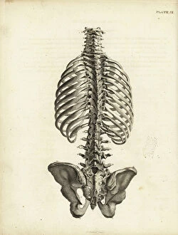
Background imageInternal Anatomy Collection: Rear view of the human trunk skeleton including spine, ribs and pelvis

Rear view of the human trunk skeleton including spine, ribs and pelvis. Copperplate engraving by Edward Mitchell after
FLO4565792 Rear view of the human trunk skeleton including spine, ribs and pelvis. Copperplate engraving by Edward Mitchell after an anatomical illustration by Jean-Joseph Sue from John Barclay's

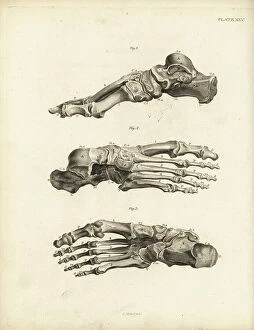
Background imageInternal Anatomy Collection: Views of the bones in the human foot from ankle to toe. Copperplate engraving by Edward Mitchell

Views of the bones in the human foot from ankle to toe. Copperplate engraving by Edward Mitchell after an anatomical
FLO4565738 Views of the bones in the human foot from ankle to toe. Copperplate engraving by Edward Mitchell after an anatomical illustration by Jean-Joseph Sue from John Barclay's A Series of

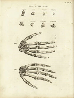
Background imageInternal Anatomy Collection: Bones of the carpus, metacarpus and fingers in the human hand

Bones of the carpus, metacarpus and fingers in the human hand. Copperplate engraving by Edward Mitchell after an
FLO4565722 Bones of the carpus, metacarpus and fingers in the human hand. Copperplate engraving by Edward Mitchell after an anatomical illustration from John Barclay's A Series of Engravings of

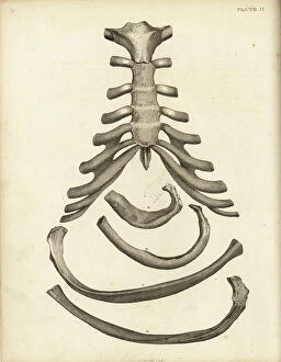
Background imageInternal Anatomy Collection: Sternum and cartilage of the human ribcage, with several ribs

Sternum and cartilage of the human ribcage, with several ribs. Copperplate engraving by Edward Mitchell after an
FLO4565702 Sternum and cartilage of the human ribcage, with several ribs. Copperplate engraving by Edward Mitchell after an anatomical illustration by Jean-Joseph Sue from John Barclay's A Series

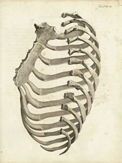
Background imageInternal Anatomy Collection: Ribs and sternum in the human ribcage. Copperplate engraving by Edward Mitchell after an

Ribs and sternum in the human ribcage. Copperplate engraving by Edward Mitchell after an anatomical illustration by
FLO4565699 Ribs and sternum in the human ribcage. Copperplate engraving by Edward Mitchell after an anatomical illustration by Jean-Joseph Sue from John Barclay's A Series of Engravings of

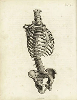
Background imageInternal Anatomy Collection: Front view of the human trunk skeleton including spine, ribs and pelvis

Front view of the human trunk skeleton including spine, ribs and pelvis. Copperplate engraving by Edward Mitchell after
FLO4565690 Front view of the human trunk skeleton including spine, ribs and pelvis. Copperplate engraving by Edward Mitchell after an anatomical illustration by Jean-Joseph Suefrom John Barclay's

Background imageInternal Anatomy Collection: Prickly cockle - bucarde - Acanthocardia echinata (Cardium echinatum) 1

Prickly cockle - bucarde - Acanthocardia echinata (Cardium echinatum) 1, and slender sea pen - plume elegante
FLO4576265 Prickly cockle - bucarde - Acanthocardia echinata (Cardium echinatum) 1, and slender sea pen - plume elegante - Virgularia mirabilis (Pennatula mirabilis) 2

Background imageInternal Anatomy Collection: Adult crane desarticule 1867

Adult crane desarticule 1867
JAB4120023 Adult crane desarticule 1867; (add.info.: Adult crane desarticule 1867); Photo ©Jaime Abecasis

Background imageInternal Anatomy Collection: Bladder anatomy 1866 illustrations showing the internal (top) and external (bottom)

Bladder anatomy 1866 illustrations showing the internal (top) and external (bottom)
JAB4119242 Bladder anatomy 1866 illustrations showing the internal (top) and external (bottom) anatomy of the male urinary bladder, showing its muscular coating (brown)

Background imageInternal Anatomy Collection: Voice box and thyroid 1866 illustration showing the anatomy of the larynx (voice box)

Voice box and thyroid 1866 illustration showing the anatomy of the larynx (voice box) and thyroid (brown)
JAB4119213 Voice box and thyroid 1866 illustration showing the anatomy of the larynx (voice box) and thyroid (brown, lower centre) Shown here are the hyoid bone (A), thyroid cartilage (B)

Background imageInternal Anatomy Collection: Chest muscles - 1866 illustration showing the structure of the muscles (brown) and tendons (white)

Chest muscles - 1866 illustration showing the structure of the muscles (brown) and tendons (white) of the human chest
JAB4119479 Chest muscles - 1866 illustration showing the structure of the muscles (brown) and tendons (white) of the human chest

Background imageInternal Anatomy Collection: Leg and foot muscles - 1866 anatomical illustration showing the muscles (brown) and tendons (white)

Leg and foot muscles - 1866 anatomical illustration showing the muscles (brown) and tendons (white)
JAB4119476 Leg and foot muscles - 1866 anatomical illustration showing the muscles (brown) and tendons (white) of the right foot (left)

Background imageInternal Anatomy Collection: Shoulder muscles - 1866 illustration of the muscles (brown) and tendons (white)

Shoulder muscles - 1866 illustration of the muscles (brown) and tendons (white)
JAB4119462 Shoulder muscles - 1866 illustration of the muscles (brown) and tendons (white) associated with the human collarbone (clavicle, top) and shoulder blade (scapula)

Background imageInternal Anatomy Collection: Chest and shoulder muscles - 1866 anatomical illustration showing the structure of the muscles

Chest and shoulder muscles - 1866 anatomical illustration showing the structure of the muscles (brown)
JAB4119433 Chest and shoulder muscles - 1866 anatomical illustration showing the structure of the muscles (brown) and tendons (white)

Background imageInternal Anatomy Collection: Hand muscles - 1866 anatomical illustration showing the muscles (brown) and tendons (white)

Hand muscles - 1866 anatomical illustration showing the muscles (brown) and tendons (white) of the human hand
JAB4119426 Hand muscles - 1866 anatomical illustration showing the muscles (brown) and tendons (white) of the human hand, with insets (left)

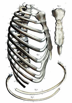
Background imageInternal Anatomy Collection: Rib cage anatomy - 1866 illustration showing the bones of the human rib cage

Rib cage anatomy - 1866 illustration showing the bones of the human rib cage, including the ribs (bottom)
JAB4119411 Rib cage anatomy - 1866 illustration showing the bones of the human rib cage, including the ribs (bottom), spine (centre-left) and sternum (breast bone, centre and upper right) Le Thorax

Background imageInternal Anatomy Collection: Pelvis anatomy - 1866 illustration showing the anatomy of the male (top) and female (bottom)

Pelvis anatomy - 1866 illustration showing the anatomy of the male (top) and female (bottom) pelvis - The pelvis
JAB4119404 Pelvis anatomy - 1866 illustration showing the anatomy of the male (top) and female (bottom) pelvis - The pelvis, anatomy, osteology - Fig 1: female pelvis Fig 2: male pelvis, Musculotion

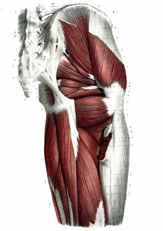
Background imageInternal Anatomy Collection: Hip muscles - 1866 illustration showing the structure of the muscles (brown) and tendons (white)

Hip muscles - 1866 illustration showing the structure of the muscles (brown) and tendons (white)
JAB4119401 Hip muscles - 1866 illustration showing the structure of the muscles (brown) and tendons (white) of the upper right leg and hip

Background imageInternal Anatomy Collection: Arm muscles - 1866 illustration showing the structure of the muscles (brown) and tendons (white)

Arm muscles - 1866 illustration showing the structure of the muscles (brown) and tendons (white)
JAB4119391 Arm muscles - 1866 illustration showing the structure of the muscles (brown) and tendons (white) of the human arm and top of the hand (bottom left and right)

Background imageInternal Anatomy Collection: Abdominal muscles - 1866 illustration showing the structure of the muscles (brown)

Abdominal muscles - 1866 illustration showing the structure of the muscles (brown) and tendons (white)
JAB4119387 Abdominal muscles - 1866 illustration showing the structure of the muscles (brown) and tendons (white) found in the rear of the human trunk - L'abdomen - Anatomy - Region anterieure de

Background imageInternal Anatomy Collection: Leg muscles - 1866 illustration showing the structure of the muscles (brown) and tendons (white)

Leg muscles - 1866 illustration showing the structure of the muscles (brown) and tendons (white)
JAB4119363 Leg muscles - 1866 illustration showing the structure of the muscles (brown) and tendons (white) of the rear of the lower leg - Anatomy du dos de la leg - Fig 1 et 2

Background imageInternal Anatomy Collection: Andreas Vesalius

Andreas Vesalius
3586025 Andreas Vesalius; (add.info.: Portrait of Andreas Vesalius (1514-1564) a Flemish/Netherlandish anatomist, physician, and author of books on human anatomy

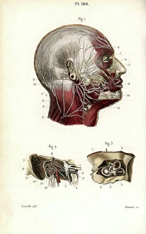
Background imageInternal Anatomy Collection: Head muscles. Lithograph of Lebrun, based on a drawing by Leveille

Head muscles. Lithograph of Lebrun, based on a drawing by Leveille, in Petity Atlas complet d'Anatomie descriptive
FLO4672607 Head muscles. Lithograph of Lebrun, based on a drawing by Leveille, in Petity Atlas complet d'Anatomie descriptive du Corps Humain, by Dr

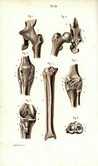
Background imageInternal Anatomy Collection: Hip joint and thigh bone. Lithograph of Corbie, in Petity Atlas complet d'Anatomie descriptive du

Hip joint and thigh bone. Lithograph of Corbie, in Petity Atlas complet d'Anatomie descriptive du Corps Humain
FLO4672595 Hip joint and thigh bone. Lithograph of Corbie, in Petity Atlas complet d'Anatomie descriptive du Corps Humain, by Dr

Background imageInternal Anatomy Collection: Sympathic nervous system in the thoracic, lumbar and sacral plexus. Lithograph by Davesne

Sympathic nervous system in the thoracic, lumbar and sacral plexus. Lithograph by Davesne
FLO4672529 Sympathic nervous system in the thoracic, lumbar and sacral plexus. Lithograph by Davesne, based on a drawing by Leveille

Background imageInternal Anatomy Collection: Sympathic nervous system in the cardiac plexus. Lithograph by Davesne

Sympathic nervous system in the cardiac plexus. Lithograph by Davesne, based on a drawing by Leveille
FLO4672521 Sympathic nervous system in the cardiac plexus. Lithograph by Davesne, based on a drawing by Leveille, in Petiure Atlas complet d'Anatomie descriptive du Corps Humain, by Dr

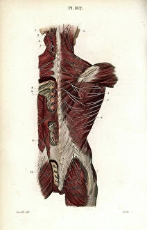
Background imageInternal Anatomy Collection: Nervous system of the back. Lithograph of Corbie, based on a drawing by Leveille

Nervous system of the back. Lithograph of Corbie, based on a drawing by Leveille
FLO4672457 Nervous system of the back. Lithograph of Corbie, based on a drawing by Leveille, in Petity Atlas complet d'Anatomie descriptive du Corps Humain, by Dr

Background imageInternal Anatomy Collection: Facial and auditory nerves. Lithograph by Davesne, based on a drawing by Leveille

Facial and auditory nerves. Lithograph by Davesne, based on a drawing by Leveille
FLO4672442 Facial and auditory nerves. Lithograph by Davesne, based on a drawing by Leveille, in Petite Atlas complet d'Anatomie descriptive du Corps Humain, by Dr

Background imageInternal Anatomy Collection: Intercostal nerves. Lithograph of Corbie, based on a drawing by Leveille

Intercostal nerves. Lithograph of Corbie, based on a drawing by Leveille, in Petity Atlas complet d'Anatomie
FLO4672415 Intercostal nerves. Lithograph of Corbie, based on a drawing by Leveille, in Petity Atlas complet d'Anatomie descriptive du Corps Humain, by Dr

Background imageInternal Anatomy Collection: Omnium Animalum : Omnium Animalum, mixed strange animal kingdom creatures

Omnium Animalum : Omnium Animalum, mixed strange animal kingdom creatures
UIG5327222 Omnium Animalum : Omnium Animalum, mixed strange animal kingdom creatures. From the 1642 book Monstrorum Historia by Ulisse Aldrovandi (Bologna, 1522-1605)

Background imageInternal Anatomy Collection: Animal Tigridi Affine : Animal Tigridi affine: an animal akin to a tiger

Animal Tigridi Affine : Animal Tigridi affine: an animal akin to a tiger. From the 1642 book Monstrorum Historia by
UIG5327220 Animal Tigridi Affine : Animal Tigridi affine: an animal akin to a tiger. From the 1642 book Monstrorum Historia by Ulisse Aldrovandi (Bologna, 1522-1605)

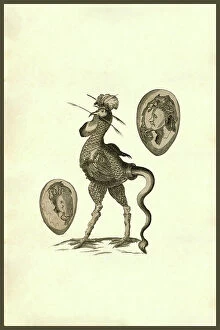
Background imageInternal Anatomy Collection: Chicken Man : Human and chicken combined creatures. From the 1642 book Monstrorum Historia by

Chicken Man : Human and chicken combined creatures. From the 1642 book Monstrorum Historia by Ulisse Aldrovandi
UIG5327206 Chicken Man : Human and chicken combined creatures. From the 1642 book Monstrorum Historia by Ulisse Aldrovandi (Bologna, 1522-1605)


For sale as Licensed Images

Choose your image, Select your licence and Download the media

"Exploring the Intricacies: Unveiling Internal Anatomy through Art and Science" Delving into the hidden world of internal anatomy, we are captivated by the wonders that lie beneath our skin. From the delicate cross-section of a Honey Bee (Apis mellifera) to the meticulous illustrations of the Channel Tunnel Project, each piece unravels a unique perspective on this captivating subject. Intriguingly, even centuries ago, artists like Giovanni Battista Bianchi skillfully captured human form in terracotta sculptures, reminding us of our intricate inner workings. Meanwhile, Andreas Vesalius revolutionized anatomical understanding with his groundbreaking studies during the Renaissance era. As we journey through time and mediums, Waldeck's original artwork takes us back to Mesoamerica in 1831. The prints from "La nature et l'homme" by Rengade depict twin fetuses in their ideal position during gemellar pregnancy – a testament to both artistry and scientific observation. The diversity within these artworks is astounding; from portraits such as Lucrezia Panciatichi's enigmatic gaze immortalized in oil on wood panel to Giulia di Alessandro de Medici holding a book – perhaps symbolizing knowledge about one's own body. Even photography joins this exploration as two male nudes perch upon a rock in an evocative black-and-white image from 1890. This photograph captures not only physicality but also invites contemplation on vulnerability and strength intertwined within our internal structures. From ancient times to modern endeavors like the Channel Tunnel Project illustration from 1906, humanity has sought to unravel its own mysteries. Through art and science alike, we continue to marvel at our internal anatomy – an ever-evolving tapestry waiting for discovery.