Internal Anatomy Collection (page 5)

"Exploring the Intricacies: Unveiling Internal Anatomy through Art and Science" Delving into the hidden world of internal anatomy

Background imageInternal Anatomy Collection: Animal Tigridi Affine : Animal Tigridi affine: an animal akin to a tiger

Animal Tigridi Affine : Animal Tigridi affine: an animal akin to a tiger. From the 1642 book Monstrorum Historia by
UIG5327220 Animal Tigridi Affine : Animal Tigridi affine: an animal akin to a tiger. From the 1642 book Monstrorum Historia by Ulisse Aldrovandi (Bologna, 1522-1605)

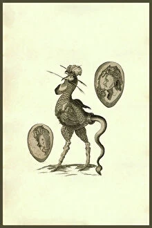
Background imageInternal Anatomy Collection: Chicken Man : Human and chicken combined creatures. From the 1642 book Monstrorum Historia by

Chicken Man : Human and chicken combined creatures. From the 1642 book Monstrorum Historia by Ulisse Aldrovandi
UIG5327206 Chicken Man : Human and chicken combined creatures. From the 1642 book Monstrorum Historia by Ulisse Aldrovandi (Bologna, 1522-1605)

Background imageInternal Anatomy Collection: Herinaceus Marinus : Herinaceus Marinus. From the 1642 book Monstrorum Historia by Ulisse

Herinaceus Marinus : Herinaceus Marinus. From the 1642 book Monstrorum Historia by Ulisse Aldrovandi (Bologna)
UIG5327205 Herinaceus Marinus : Herinaceus Marinus. From the 1642 book Monstrorum Historia by Ulisse Aldrovandi (Bologna, 1522-1605). He is considered the founder of modern Natural History

Background imageInternal Anatomy Collection: Rana piscatrix ala feu Bufo Marinus : Rana piscatrix ala feu Bufo Marinus

Rana piscatrix ala feu Bufo Marinus : Rana piscatrix ala feu Bufo Marinus
UIG5327202 Rana piscatrix ala feu Bufo Marinus : Rana piscatrix ala feu Bufo Marinus. Rana piscatrix is a type of anglerfish or monkfish today called Lophius piscatorius

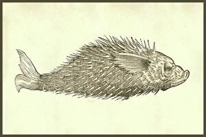
Background imageInternal Anatomy Collection: Andora Piscis aspectu fuillo : Andora Piscis aspectu fuillo. A sea cow like hairy fish monster

Andora Piscis aspectu fuillo : Andora Piscis aspectu fuillo. A sea cow like hairy fish monster
UIG5327201 Andora Piscis aspectu fuillo : Andora Piscis aspectu fuillo. A sea cow like hairy fish monster, from the 1642 book Monstrorum Historia by Ulisse Aldrovandi (Bologna, 1522-1605)

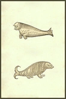
Background imageInternal Anatomy Collection: Seal & Walrus Creatures : Seal & Walrus Creatures. From the 1642 book Monstrorum Historia by

Seal & Walrus Creatures : Seal & Walrus Creatures. From the 1642 book Monstrorum Historia by Ulisse Aldrovandi
UIG5327193 Seal & Walrus Creatures : Seal & Walrus Creatures. From the 1642 book Monstrorum Historia by Ulisse Aldrovandi (Bologna, 1522-1605). He is considered the founder of modern Natural History

Background imageInternal Anatomy Collection: Human Monsters : Six people with deformities. From the 1642 book Monstrorum Historia by Ulisse

Human Monsters : Six people with deformities. From the 1642 book Monstrorum Historia by Ulisse Aldrovandi (Bologna)
UIG5327179 Human Monsters : Six people with deformities. From the 1642 book Monstrorum Historia by Ulisse Aldrovandi (Bologna, 1522-1605). He is considered the founder of modern Natural History

Background imageInternal Anatomy Collection: Monstrum biceps & septiceps : Monstrum biceps & septiceps. Multi armed men

Monstrum biceps & septiceps : Monstrum biceps & septiceps. Multi armed men
UIG5327177 Monstrum biceps & septiceps : Monstrum biceps & septiceps. Multi armed men. From the 1642 book Monstrorum Historia by Ulisse Aldrovandi (Bologna, 1522-1605)

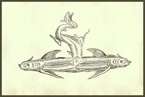
Background imageInternal Anatomy Collection: Piscis Dicephalos : Piscis Dicephalos, two headed fish. From the 1642 book Monstrorum Historia by

Piscis Dicephalos : Piscis Dicephalos, two headed fish. From the 1642 book Monstrorum Historia by Ulisse Aldrovandi
UIG5327152 Piscis Dicephalos : Piscis Dicephalos, two headed fish. From the 1642 book Monstrorum Historia by Ulisse Aldrovandi (Bologna, 1522-1605)

Background imageInternal Anatomy Collection: Capra Humana Facie : Capra Humana Facie. From the 1642 book Monstrorum Historia by Ulisse

Capra Humana Facie : Capra Humana Facie. From the 1642 book Monstrorum Historia by Ulisse Aldrovandi (Bologna)
UIG5327147 Capra Humana Facie : Capra Humana Facie. From the 1642 book Monstrorum Historia by Ulisse Aldrovandi (Bologna, 1522-1605). He is considered the founder of modern Natural History

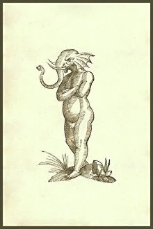
Background imageInternal Anatomy Collection: Puer capite elephantino : Puer capite elephantino, boy with an elephant's head, elephant man

Puer capite elephantino : Puer capite elephantino, boy with an elephant's head, elephant man
UIG5327138 Puer capite elephantino : Puer capite elephantino, boy with an elephant's head, elephant man. From the 1642 book Monstrorum Historia by Ulisse Aldrovandi (Bologna, 1522-1605)

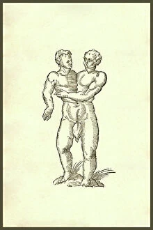
Background imageInternal Anatomy Collection: Two heads on one body, 1642 (engraving)

Two heads on one body, 1642 (engraving)
UIG5327120 Two heads on one body, 1642 (engraving); (add.info.: Monstrum hermaphroditicum from the 1642 book "Monstrorum Historia" by Ulisse Aldrovandi (Bologna)

Background imageInternal Anatomy Collection: Infans biceps obscaeno membro : Infans biceps obscaeno membro, 407, two heads on one body

Infans biceps obscaeno membro : Infans biceps obscaeno membro, 407, two heads on one body, siamese twins
UIG5327115 Infans biceps obscaeno membro : Infans biceps obscaeno membro, 407, two heads on one body, siamese twins. From the 1642 book Monstrorum Historia by Ulisse Aldrovandi (Bologna, 1522-1605)

Background imageInternal Anatomy Collection: Gryphis Tertia (Griffin) : The griffin, griffon, or gryphon is a legendary creature with the body

Gryphis Tertia (Griffin) : The griffin, griffon, or gryphon is a legendary creature with the body, tail
UIG5327074 Gryphis Tertia (Griffin) : The griffin, griffon, or gryphon is a legendary creature with the body, tail, and back legs of a lion; the head and wings of an eagle;

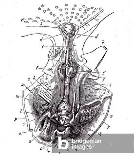
Background imageInternal Anatomy Collection: Octopus anatomy, ventral face, 1900 (engraving)

Octopus anatomy, ventral face, 1900 (engraving)
6947881 Octopus anatomy, ventral face, 1900 (engraving); Private Collection; (add.info.: Octopus anatomy, ventral face, 1900); Photo ©Jaime Abecasis

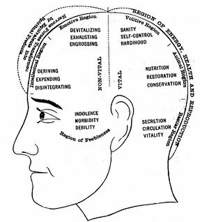
Background imageInternal Anatomy Collection: An engraving depicting a diagram showing the supposed functions of various areas of the brain

An engraving depicting a diagram showing the supposed functions of various areas of the brain, 19th century
5311364 An engraving depicting a diagram showing the supposed functions of various areas of the brain, 19th century; (add.info.: Date: 1886); Universal History Archive/UIG

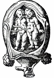
Background imageInternal Anatomy Collection: A woodcut depicting twins in the uterus. Woodcut by Jost Amman (1539-1591) a Swiss-German artist

A woodcut depicting twins in the uterus. Woodcut by Jost Amman (1539-1591) a Swiss-German artist, 16th century
5311252 A woodcut depicting twins in the uterus. Woodcut by Jost Amman (1539-1591) a Swiss-German artist, 16th century; Universal History Archive/UIG

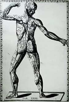
Background imageInternal Anatomy Collection: A human nervous system

A human nervous system
5308007 A human nervous system.; (add.info.: Print of a human nervous system by Bartolomeo Eustachi (d.1574) one of the founders of the science of human anatomy

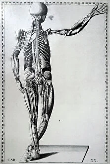
Background imageInternal Anatomy Collection: A human skeleton and nervous system

A human skeleton and nervous system
5308006 A human skeleton and nervous system.; (add.info.: Print of a human skeleton and nervous system by Bartolomeo Eustachi (d.1574) one of the founders of the science of human anatomy

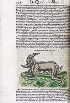
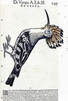
Background imageInternal Anatomy Collection: Medici Tigurini Historiae animalium

Medici Tigurini Historiae animalium
2626593 Medici Tigurini Historiae animalium; (add.info.: Illustration of a satyr or half man half beast monster, from medici Tigurini Historiae animalium Historia animalium 1564; a veterinary)

Background imageInternal Anatomy Collection: Medici Tigurini Historiae animalium

Medici Tigurini Historiae animalium
2626572 Medici Tigurini Historiae animalium; (add.info.: Illustration of a road runner bird, from medici Tigurini Historiae animalium Historia animalium 1564; a veterinary anatomical book)

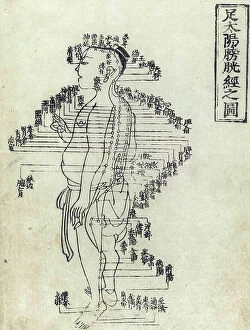
Background imageInternal Anatomy Collection: Shi si jing fa hui

Shi si jing fa hui
2626557 Shi si jing fa hui; (add.info.: Illustration from the Japanese & Chinese edition (1716) of Shi si jing fa hui, (Routes of the Fourteen Meridians and their Functions)

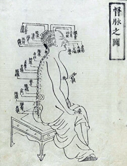
Background imageInternal Anatomy Collection: Shi si jing fa hui

Shi si jing fa hui
2626545 Shi si jing fa hui; (add.info.: Illustration from the Japanese & Chinese edition (1716) of Shi si jing fa hui, (Routes of the Fourteen Meridians and their Functions)

Background imageInternal Anatomy Collection: Fasciculus medicinae

Fasciculus medicinae
2626479 Fasciculus medicinae; (add.info.: Illustration from Fasciculus medicinae Venice; 1494 the first printed book to contain anatomical illustrations)

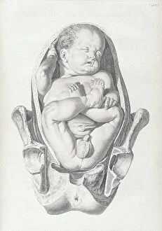
Background imageInternal Anatomy Collection: A set of anatomical tables, with explanations, and an abridgment, of the practice of midwifery

A set of anatomical tables, with explanations, and an abridgment, of the practice of midwifery, 18th (engraving)
2626227 A set of anatomical tables, with explanations, and an abridgment, of the practice of midwifery, 18th (engraving); (add.info)

Background imageInternal Anatomy Collection: A set of anatomical tables, with explanations, and an abridgment, of the practice of midwifery

A set of anatomical tables, with explanations, and an abridgment, of the practice of midwifery, 18th (engraving)
2626225 A set of anatomical tables, with explanations, and an abridgment, of the practice of midwifery, 18th (engraving); (add.info)

Background imageInternal Anatomy Collection: Fasciculus medicinae

Fasciculus medicinae
2626473 Fasciculus medicinae; (add.info.: Illustration from Fasciculus medicinae Venice; 1494 the first printed book to contain anatomical illustrations)

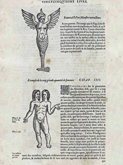
Background imageInternal Anatomy Collection: Les oeuures d'Ambroise Pare?

Les oeuures d'Ambroise Pare?
2626428 Les oeuures d'Ambroise Pare?; (add.info.: Illustration from Les oeuures d'Ambroise Pare? 1585 by Ambroise Pare?, 1510-1590 French surgeon and anatomist.); Universal History Archive/UIG

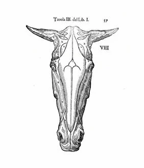
Background imageInternal Anatomy Collection: Equine anatomical Illustration

Equine anatomical Illustration
2626309 Equine anatomical Illustration; (add.info.: Equine anatomical Illustration from Anatomia del cavallo, infermita, et suoi rimedii')

Background imageInternal Anatomy Collection: Fasciculus medicinae

Fasciculus medicinae
2626475 Fasciculus medicinae; (add.info.: Illustration from Fasciculus medicinae Venice; 1494 the first printed book to contain anatomical illustrations)

Background imageInternal Anatomy Collection: A set of anatomical tables, with explanations, and an abridgment, of the practice of midwifery

A set of anatomical tables, with explanations, and an abridgment, of the practice of midwifery, 18th (engraving)
2626221 A set of anatomical tables, with explanations, and an abridgment, of the practice of midwifery, 18th (engraving); (add.info)

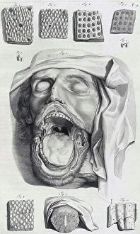
Background imageInternal Anatomy Collection: The anatomy textbook Ontleding des Menschelyken Lichaams, 1690 (engraving)

The anatomy textbook Ontleding des Menschelyken Lichaams, 1690 (engraving)
2625841 The anatomy textbook Ontleding des Menschelyken Lichaams, 1690 (engraving); (add.info.: Illustrations by Govard Bidloo, from the anatomy textbook Ontleding des menschelyken Lichaams')

Background imageInternal Anatomy Collection: The Anatomy of Human Bodies, 1698 (engraving)

The Anatomy of Human Bodies, 1698 (engraving)
2625657 The Anatomy of Human Bodies, 1698 (engraving); (add.info.: Illustration from The anatomy of humane bodies by William Cowper, (Oxford, 1698).); Universal History Archive/UIG

Background imageInternal Anatomy Collection: The Anatomy of Human Bodies, 1698 (engraving)

The Anatomy of Human Bodies, 1698 (engraving)
2625636 The Anatomy of Human Bodies, 1698 (engraving); (add.info.: Illustration from The anatomy of humane bodies by William Cowper, (Oxford, 1698).); Universal History Archive/UIG

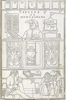
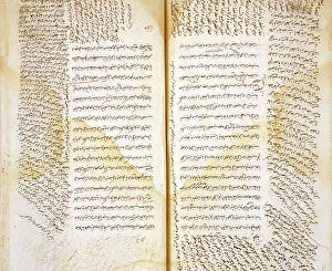
Background imageInternal Anatomy Collection: The Canon of Medicine

The Canon of Medicine
2625211 The Canon of Medicine; (add.info.: The central text consists of the anatomical sections of the Canon of Medicine by Ibn S?n? (Avicenna)

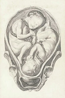
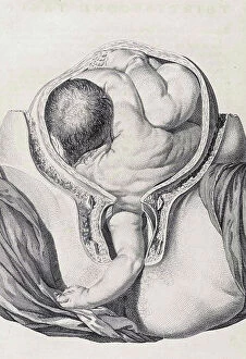
Background imageInternal Anatomy Collection: Fetus head helped through the birth canal with forceps. Copperplate engraving by Andrew Bell after

Fetus head helped through the birth canal with forceps. Copperplate engraving by Andrew Bell after an illustration by
FLO4695943 Fetus head helped through the birth canal with forceps. Copperplate engraving by Andrew Bell after an illustration by Jan van Rymsdyk from William Smellie's A Set of Anatomical Tables

Background imageInternal Anatomy Collection: BARTHOLIN, Thomas (1616-1680). Danish physician, mathematician, and theologian

BARTHOLIN, Thomas (1616-1680). Danish physician, mathematician, and theologian
CUL4875561 BARTHOLIN, Thomas (1616-1680). Danish physician, mathematician, and theologian. He is best known for his work in the discovery of the lymphatic system in humans

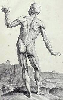
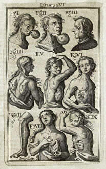
Background imageInternal Anatomy Collection: SUAREZ de RIBERA, Francisco (S.XVIII). Spanish doctor. Illustration of ' Theatro Chyrurgico

SUAREZ de RIBERA, Francisco (S.XVIII). Spanish doctor. Illustration of " Theatro Chyrurgico anatomico del cuerpo
CUL4871798 SUAREZ de RIBERA, Francisco (S.XVIII). Spanish doctor. Illustration of " Theatro Chyrurgico anatomico del cuerpo del hombre viviente

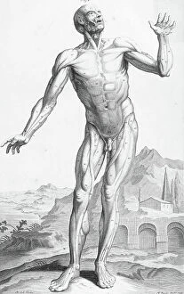
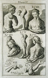
Background imageInternal Anatomy Collection: SUAREZ de RIBERA, Francisco (S.XVIII). Spanish doctor. Illustration of ' Theatro Chyrurgico

SUAREZ de RIBERA, Francisco (S.XVIII). Spanish doctor. Illustration of " Theatro Chyrurgico anatomico del cuerpo
CUL4871759 SUAREZ de RIBERA, Francisco (S.XVIII). Spanish doctor. Illustration of " Theatro Chyrurgico anatomico del cuerpo del hombre viviente

Background imageInternal Anatomy Collection: Le Coeur, extract from 'The anatomy of the human body' 1797, by Charles Bell (1774-1824)

Le Coeur, extract from "The anatomy of the human body" 1797, by Charles Bell (1774-1824)
XEE5028095 Le Coeur, extract from "The anatomy of the human body" 1797, by Charles Bell (1774-1824) - Biblioteca nazionale braidense, Milano; (add.info.: Le Coeur)

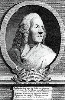
Background imageInternal Anatomy Collection: Portrait of FELIX VICQ d'AZYR (1748-1794), French physician and anatomist

Portrait of FELIX VICQ d'AZYR (1748-1794), French physician and anatomist
XEE5032021 Portrait of FELIX VICQ d'AZYR (1748-1794), French physician and anatomist; (add.info.: Portrait of FELIX VICQ d'AZYR (1748-1794), French physician and anatomist); Stefano Bianchetti

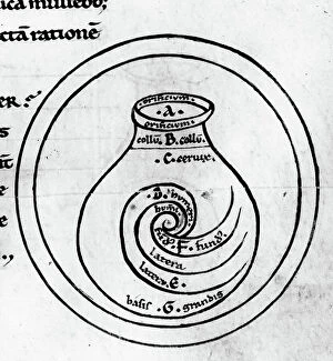
Background imageInternal Anatomy Collection: Anatomy: representation of the uterus by Garioponto (970 - 1050), Italian doctor of the Middle Ages

Anatomy: representation of the uterus by Garioponto (970 - 1050), Italian doctor of the Middle Ages
XEE5029574 Anatomy: representation of the uterus by Garioponto (970 - 1050), Italian doctor of the Middle Ages, Biblioteca Civica Bertoliana

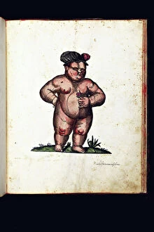
Background imageInternal Anatomy Collection: Monster creatures. Illustrated chart of a manuscript of Natural History by Ulisse Aldrovandi

Monster creatures. Illustrated chart of a manuscript of Natural History by Ulisse Aldrovandi (1522-1605)
VEN5029555 Monster creatures. Illustrated chart of a manuscript of Natural History by Ulisse Aldrovandi (1522-1605), Ulisse Aldrovandi fonds, University Library of Bologna, ed

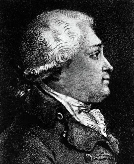
Background imageInternal Anatomy Collection: Portrait of Antoine Ferrein (1693-1769) anatomist francais

Portrait of Antoine Ferrein (1693-1769) anatomist francais
XEE5029142 Portrait of Antoine Ferrein (1693-1769) anatomist francais; (add.info.: Portrait of Antoine Ferrein (1693-1769) anatomist francais); Stefano Bianchetti

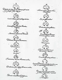
Background imageInternal Anatomy Collection: Metoposcopiae liber, The metoposcopy of 1658, by (Cardanus Hieronymus) (Jerome Cardane)

Metoposcopiae liber, The metoposcopy of 1658, by (Cardanus Hieronymus) (Jerome Cardane) Gerolamo Cardano (1501-1576)
XEE5028461 Metoposcopiae liber, The metoposcopy of 1658, by (Cardanus Hieronymus) (Jerome Cardane) Gerolamo Cardano (1501-1576) Italian doctor, mathematician

Background imageInternal Anatomy Collection: Bertillonage (Bertillon system or forensic anthropometry) a forensic technique developed by

Bertillonage (Bertillon system or forensic anthropometry) a forensic technique developed by the Frenchman Alphonse
ELD4961071 Bertillonage (Bertillon system or forensic anthropometry) a forensic technique developed by the Frenchman Alphonse Bertillon (1853-1914) in 1879

Background imageInternal Anatomy Collection: The anatomist Niels Stensen (Stenon or Steensen or Steno or stenone) (1638-1688)

The anatomist Niels Stensen (Stenon or Steensen or Steno or stenone) (1638-1688)
ELD4905480 The anatomist Niels Stensen (Stenon or Steensen or Steno or stenone) (1638-1688) crowned of Titiopolis on 19/09/1677 by Pope Innocent XI and Cardinal Barbarigo

Background imageInternal Anatomy Collection: France, Pays de la Loire, Maine-et-Loire (49), Angers: hospital of Angers, medical school

France, Pays de la Loire, Maine-et-Loire (49), Angers: hospital of Angers, medical school
LUX4771257 France, Pays de la Loire, Maine-et-Loire (49), Angers: hospital of Angers, medical school, medical students study anatomy with the help of a skeleton and the book


For sale as Licensed Images

Choose your image, Select your licence and Download the media

"Exploring the Intricacies: Unveiling Internal Anatomy through Art and Science" Delving into the hidden world of internal anatomy, we are captivated by the wonders that lie beneath our skin. From the delicate cross-section of a Honey Bee (Apis mellifera) to the meticulous illustrations of the Channel Tunnel Project, each piece unravels a unique perspective on this captivating subject. Intriguingly, even centuries ago, artists like Giovanni Battista Bianchi skillfully captured human form in terracotta sculptures, reminding us of our intricate inner workings. Meanwhile, Andreas Vesalius revolutionized anatomical understanding with his groundbreaking studies during the Renaissance era. As we journey through time and mediums, Waldeck's original artwork takes us back to Mesoamerica in 1831. The prints from "La nature et l'homme" by Rengade depict twin fetuses in their ideal position during gemellar pregnancy – a testament to both artistry and scientific observation. The diversity within these artworks is astounding; from portraits such as Lucrezia Panciatichi's enigmatic gaze immortalized in oil on wood panel to Giulia di Alessandro de Medici holding a book – perhaps symbolizing knowledge about one's own body. Even photography joins this exploration as two male nudes perch upon a rock in an evocative black-and-white image from 1890. This photograph captures not only physicality but also invites contemplation on vulnerability and strength intertwined within our internal structures. From ancient times to modern endeavors like the Channel Tunnel Project illustration from 1906, humanity has sought to unravel its own mysteries. Through art and science alike, we continue to marvel at our internal anatomy – an ever-evolving tapestry waiting for discovery.