
Internal Anatomy Collection (page 6)
"Exploring the Intricacies: Unveiling Internal Anatomy through Art and Science" Delving into the hidden world of internal anatomy


For sale as Licensed Images
Choose your image, Select your licence and Download the media
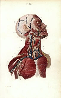
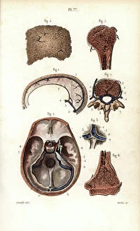
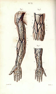
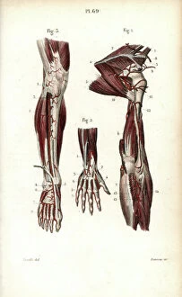
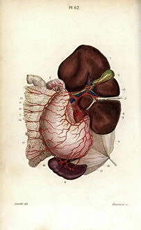
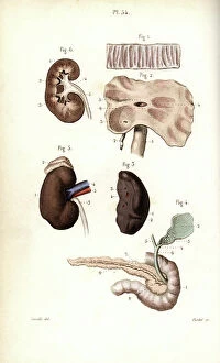
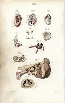
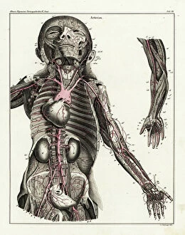
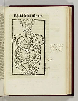
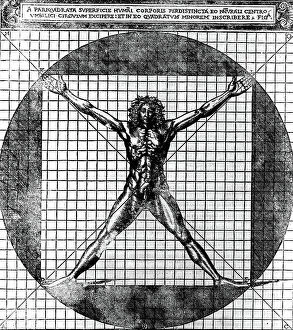
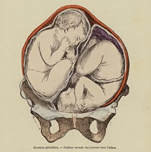
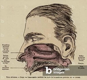
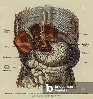
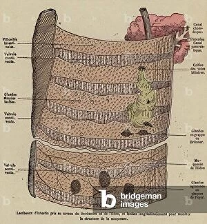
"Exploring the Intricacies: Unveiling Internal Anatomy through Art and Science" Delving into the hidden world of internal anatomy, we are captivated by the wonders that lie beneath our skin. From the delicate cross-section of a Honey Bee (Apis mellifera) to the meticulous illustrations of the Channel Tunnel Project, each piece unravels a unique perspective on this captivating subject. Intriguingly, even centuries ago, artists like Giovanni Battista Bianchi skillfully captured human form in terracotta sculptures, reminding us of our intricate inner workings. Meanwhile, Andreas Vesalius revolutionized anatomical understanding with his groundbreaking studies during the Renaissance era. As we journey through time and mediums, Waldeck's original artwork takes us back to Mesoamerica in 1831. The prints from "La nature et l'homme" by Rengade depict twin fetuses in their ideal position during gemellar pregnancy – a testament to both artistry and scientific observation. The diversity within these artworks is astounding; from portraits such as Lucrezia Panciatichi's enigmatic gaze immortalized in oil on wood panel to Giulia di Alessandro de Medici holding a book – perhaps symbolizing knowledge about one's own body. Even photography joins this exploration as two male nudes perch upon a rock in an evocative black-and-white image from 1890. This photograph captures not only physicality but also invites contemplation on vulnerability and strength intertwined within our internal structures. From ancient times to modern endeavors like the Channel Tunnel Project illustration from 1906, humanity has sought to unravel its own mysteries. Through art and science alike, we continue to marvel at our internal anatomy – an ever-evolving tapestry waiting for discovery.