Internal Anatomy Collection (page 7)

"Exploring the Intricacies: Unveiling Internal Anatomy through Art and Science" Delving into the hidden world of internal anatomy

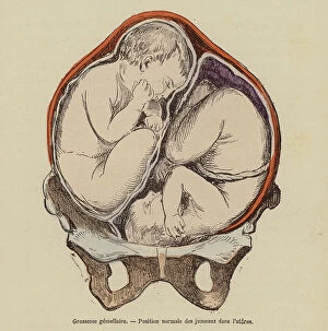
Background imageInternal Anatomy Collection: Normal position of twins in the womb during pregnancy (coloured engraving)

Normal position of twins in the womb during pregnancy (coloured engraving)
8655687 Normal position of twins in the womb during pregnancy (coloured engraving) by French School, (19th century) (after); Private Collection; (add.info)

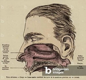
Background imageInternal Anatomy Collection: Interior of the nose (coloured engraving)

Interior of the nose (coloured engraving)
8655758 Interior of the nose (coloured engraving) by French School, (19th century) (after); Private Collection; (add.info.: Interior of the nose)

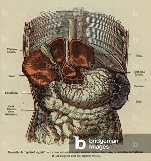
Background imageInternal Anatomy Collection: Organs of the digestive system (coloured engraving)

Organs of the digestive system (coloured engraving)
8655750 Organs of the digestive system (coloured engraving) by French School, (19th century) (after); Private Collection; (add.info.: Organs of the digestive system)

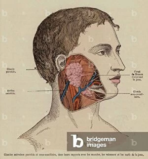
Background imageInternal Anatomy Collection: Saivary glands and muscles, nerves and blood vessels of the cheek (coloured engraving)

Saivary glands and muscles, nerves and blood vessels of the cheek (coloured engraving)
8655749 Saivary glands and muscles, nerves and blood vessels of the cheek (coloured engraving) by French School, (19th century) (after); Private Collection; (add.info.: Saivary glands and muscles)

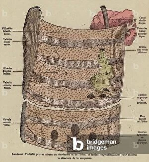
Background imageInternal Anatomy Collection: Section of the small intestine showing the structure of the mucosa (coloured engraving)

Section of the small intestine showing the structure of the mucosa (coloured engraving)
8655747 Section of the small intestine showing the structure of the mucosa (coloured engraving) by French School, (19th century) (after); Private Collection; (add.info)

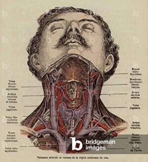
Background imageInternal Anatomy Collection: Blood vessels of the neck (coloured engraving)

Blood vessels of the neck (coloured engraving)
8655731 Blood vessels of the neck (coloured engraving) by French School, (19th century) (after); Private Collection; (add.info.: Blood vessels of the neck)

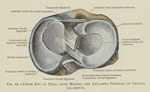
Background imageInternal Anatomy Collection: Upper End of Tibia, with Menisci and Attached Portions of Crucial Ligaments (colour litho)

Upper End of Tibia, with Menisci and Attached Portions of Crucial Ligaments (colour litho)
8667637 Upper End of Tibia, with Menisci and Attached Portions of Crucial Ligaments (colour litho) by English School, (20th century); Private Collection; (add.info.: Upper End of Tibia)

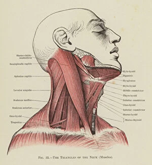
Background imageInternal Anatomy Collection: The Triangles of the Neck, Muscles (colour litho)

The Triangles of the Neck, Muscles (colour litho)
8667618 The Triangles of the Neck, Muscles (colour litho) by English School, (20th century); Private Collection; (add.info.: The Triangles of the Neck, Muscles)

Background imageInternal Anatomy Collection: Superficial Muscles on the Front of the Arm and Forearm (colour litho)

Superficial Muscles on the Front of the Arm and Forearm (colour litho)
8667617 Superficial Muscles on the Front of the Arm and Forearm (colour litho) by English School, (20th century); Private Collection; (add.info)

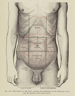
Background imageInternal Anatomy Collection: The Front of the Body (colour litho)

The Front of the Body (colour litho)
8667613 The Front of the Body (colour litho) by English School, (20th century); Private Collection; (add.info.: The Front of the Body)

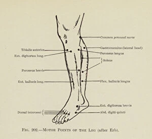
Background imageInternal Anatomy Collection: Motor Points of the Leg (litho)

Motor Points of the Leg (litho)
8667606 Motor Points of the Leg (litho) by English School, (20th century); Private Collection; (add.info.: Motor Points of the Leg)

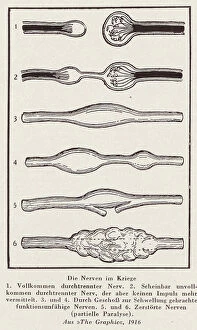
Background imageInternal Anatomy Collection: Nerves damaged by war (litho)

Nerves damaged by war (litho)
8664192 Nerves damaged by war (litho) by English School, (20th century); Private Collection; (add.info.: Nerves damaged by war)

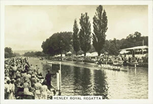
Background imageInternal Anatomy Collection: Our Countryside, 1938: Henley Royal Regatta (b/w photo)

Our Countryside, 1938: Henley Royal Regatta (b/w photo)
8671509 Our Countryside, 1938: Henley Royal Regatta (b/w photo) by English Photographer, (20th century); Private Collection; (add.info.: Henley Royal Regatta)

Background imageInternal Anatomy Collection: Traite de medecine de Mansour : Muscle et systeme nerveux humain. Manuscrit perse

Traite de medecine de Mansour : Muscle et systeme nerveux humain. Manuscrit perse
AIS5373969 Traite de medecine de Mansour : Muscle et systeme nerveux humain. Manuscrit perse. In "L'Anatomie du corps (Tachrih al-badan (al badan)" de Mansour ben Eliyas Chirazi

Background imageInternal Anatomy Collection: Normal disposition of the viscera in the human body. 'Life and health' by Dr Jules Rengade
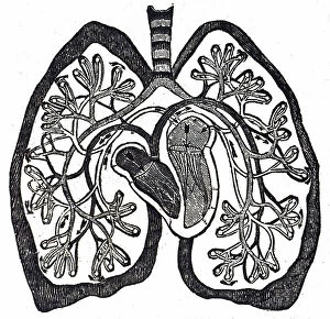
Normal disposition of the viscera in the human body. "Life and health" by Dr Jules Rengade. Paris 1881
7295202 Normal disposition of the viscera in the human body. "Life and health" by Dr Jules Rengade. Paris 1881; Private Collection; Photo ©Jaime Abecasis

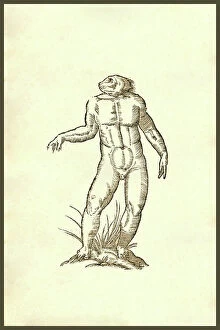
Background imageInternal Anatomy Collection: Vitulomonachus sorbini : Vitulomonachus sorbini, 371, being with a calf's body

Vitulomonachus sorbini : Vitulomonachus sorbini, 371, being with a calf's body
UIG5327101 Vitulomonachus sorbini : Vitulomonachus sorbini, 371, being with a calf's body and deformed human head walking on its hind legs. Renaissance woodcut of a monster

Background imageInternal Anatomy Collection: Vituli Marini Alia Species : Vituli marini alia species: a species of seal

Vituli Marini Alia Species : Vituli marini alia species: a species of seal
UIG5327218 Vituli Marini Alia Species : Vituli marini alia species: a species of seal. From the 1642 book Monstrorum Historia by Ulisse Aldrovandi (Bologna, 1522-1605)

Background imageInternal Anatomy Collection: Ancient Monster Men : Man like creatures. From the 1642 book Monstrorum Historia by Ulisse

Ancient Monster Men : Man like creatures. From the 1642 book Monstrorum Historia by Ulisse Aldrovandi (Bologna)
UIG5327192 Ancient Monster Men : Man like creatures. From the 1642 book Monstrorum Historia by Ulisse Aldrovandi (Bologna, 1522-1605). He is considered the founder of modern Natural History

Background imageInternal Anatomy Collection: Sea Monster Effigies : sea creatures. From the 1642 book Monstrorum Historia by Ulisse Aldrovandi

Sea Monster Effigies : sea creatures. From the 1642 book Monstrorum Historia by Ulisse Aldrovandi (Bologna, 1522-1605)
UIG5327176 Sea Monster Effigies : sea creatures. From the 1642 book Monstrorum Historia by Ulisse Aldrovandi (Bologna, 1522-1605). He is considered the founder of modern Natural History

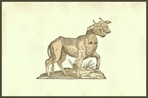
Background imageInternal Anatomy Collection: Vitulus biceps quator oculis : Vitulus biceps quator oculis, calf with one body

Vitulus biceps quator oculis : Vitulus biceps quator oculis, calf with one body and two heads with eyes on its chest
UIG5327136 Vitulus biceps quator oculis : Vitulus biceps quator oculis, calf with one body and two heads with eyes on its chest

Background imageInternal Anatomy Collection: Serpent Dicephalos : Serpent Dicephalos, two headed snake. From the 1642 book Monstrorum Historia

Serpent Dicephalos : Serpent Dicephalos, two headed snake. From the 1642 book Monstrorum Historia by Ulisse Aldrovandi
UIG5327135 Serpent Dicephalos : Serpent Dicephalos, two headed snake. From the 1642 book Monstrorum Historia by Ulisse Aldrovandi (Bologna, 1522-1605)

Background imageInternal Anatomy Collection: Pullus Dicephalos & Dirrhynchos : Pullus Dicephalos & Dirrhynchos

Pullus Dicephalos & Dirrhynchos : Pullus Dicephalos & Dirrhynchos. Chicken with two heads or deformity
UIG5327133 Pullus Dicephalos & Dirrhynchos : Pullus Dicephalos & Dirrhynchos. Chicken with two heads or deformity. From the 1642 book Monstrorum Historia by Ulisse Aldrovandi (Bologna, 1522-1605)

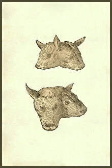
Background imageInternal Anatomy Collection: Capitis Vitulini Geminati Duplex Effigies

Capitis Vitulini Geminati Duplex Effigies
UIG5327131 Capitis Vitulini Geminati Duplex Effigies; (add.info.: Capitis Vitulini Geminati Duplex Effigies : Capitis Vitulini Geminati Duplex Effigies)

Background imageInternal Anatomy Collection: Monstrosus Marinus Daemoniforme : Renaissance woodcut of a sea demon

Monstrosus Marinus Daemoniforme : Renaissance woodcut of a sea demon. From the 1642 book Monstrorum Historia by Ulisse
UIG5327097 Monstrosus Marinus Daemoniforme : Renaissance woodcut of a sea demon. From the 1642 book Monstrorum Historia by Ulisse Aldrovandi (Bologna, 1522-1605)

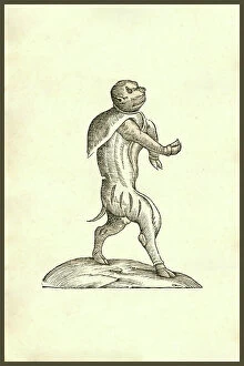
Background imageInternal Anatomy Collection: Renaissance woodcut of a squirrel monster

Renaissance woodcut of a squirrel monster
UIG5327094 Renaissance woodcut of a squirrel monster; (add.info.: Su : Renaissance woodcut of a squirrel monster. From the 1642 book Monstrorum Historia by Ulisse Aldrovandi (Bologna, 1522-1605)

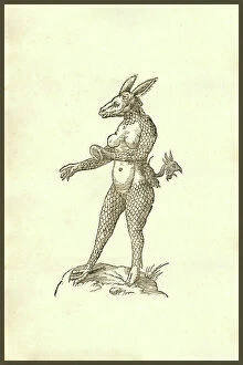
Background imageInternal Anatomy Collection: Monstrum capite asinino : Monstrum capite asinino; a scaled female monster with the head of an ass

Monstrum capite asinino : Monstrum capite asinino; a scaled female monster with the head of an ass
UIG5327092 Monstrum capite asinino : Monstrum capite asinino; a scaled female monster with the head of an ass. Renaissance woodcut of a monster

Background imageInternal Anatomy Collection: Aries Biceps Heiroglyphica : Renaissance woodcut Aries biceps, 347, two-headed ram

Aries Biceps Heiroglyphica : Renaissance woodcut Aries biceps, 347, two-headed ram
UIG5327077 Aries Biceps Heiroglyphica : Renaissance woodcut Aries biceps, 347, two-headed ram. From the 1642 book Monstrorum Historia by Ulisse Aldrovandi (Bologna, 1522-1605)

Background imageInternal Anatomy Collection: Animal Africanum Deforme : Renaissance Woodcut monsters. From the 1642 book Monstrorum Historia by

Animal Africanum Deforme : Renaissance Woodcut monsters. From the 1642 book Monstrorum Historia by Ulisse Aldrovandi
UIG5327058 Animal Africanum Deforme : Renaissance Woodcut monsters. From the 1642 book Monstrorum Historia by Ulisse Aldrovandi (Bologna, 1522-1605)

Background imageInternal Anatomy Collection: Ancient Creatures : Creatures! From the 1642 book Monstrorum Historia by Ulisse Aldrovandi

Ancient Creatures : Creatures! From the 1642 book Monstrorum Historia by Ulisse Aldrovandi (Bologna, 1522-1605)
UIG5327200 Ancient Creatures : Creatures! From the 1642 book Monstrorum Historia by Ulisse Aldrovandi (Bologna, 1522-1605). He is considered the founder of modern Natural History

Background imageInternal Anatomy Collection: Sea Monster Effigies : Triton, Siren, and Monarch sea creatures

Sea Monster Effigies : Triton, Siren, and Monarch sea creatures. From the 1642 book Monstrorum Historia by Ulisse
UIG5327182 Sea Monster Effigies : Triton, Siren, and Monarch sea creatures. From the 1642 book Monstrorum Historia by Ulisse Aldrovandi (Bologna, 1522-1605)

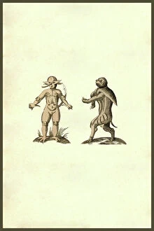
Background imageInternal Anatomy Collection: Infans capite canino : Infans capite canino, dog headed boy or man

Infans capite canino : Infans capite canino, dog headed boy or man. From the 1642 book Monstrorum Historia by Ulisse
UIG5327142 Infans capite canino : Infans capite canino, dog headed boy or man. From the 1642 book Monstrorum Historia by Ulisse Aldrovandi (Bologna, 1522-1605)

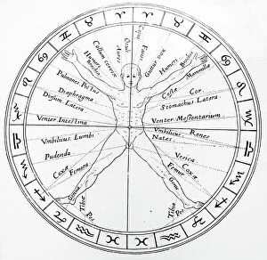
Background imageInternal Anatomy Collection: The organs of the body and the zodiacal influences

The organs of the body and the zodiacal influences
5310126 The organs of the body and the zodiacal influences; (add.info.: Woodblock engraving depicting the organs of the body and the zodiacal influences on them)

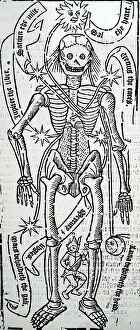
Background imageInternal Anatomy Collection: The organs of the body and the planetary influences

The organs of the body and the planetary influences
5310125 The organs of the body and the planetary influences; (add.info.: Woodblock engraving depicting the organs of the body and the planetary influences on them)

Background imageInternal Anatomy Collection: An engraving depicting Descartes drawing of the human brain, 17th century

An engraving depicting Descartes drawing of the human brain, 17th century
5311365 An engraving depicting Descartes drawing of the human brain, 17th century; Universal History Archive/UIG

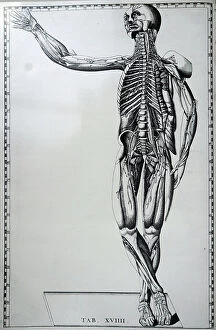
Background imageInternal Anatomy Collection: A human skeleton and nervous system, dated 18th century (engraving)

A human skeleton and nervous system, dated 18th century (engraving)
5308005 A human skeleton and nervous system, dated 18th century (engraving); (add.info.: Print of a human skeleton and nervous system by Bartolomeo Eustachi (d.1574)

Background imageInternal Anatomy Collection: A woodcut engraving depicting a blood-letting man, showing points of blood-letting connected with

A woodcut engraving depicting a blood-letting man, showing points of blood-letting connected with signs of the Zodiac
5311213 A woodcut engraving depicting a blood-letting man, showing points of blood-letting connected with signs of the Zodiac, 16th century; Universal History Archive/UIG

Background imageInternal Anatomy Collection: Diagram of the circulation of blood as understood after William Harvey's work

Diagram of the circulation of blood as understood after William Harvey's work
5309470 Diagram of the circulation of blood as understood after William Harvey's work, showing the Lesser Circulation (Pulmonary Circulation)

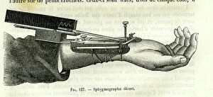
Background imageInternal Anatomy Collection: Direct sphygmograph, 1882 (engraving)

Direct sphygmograph, 1882 (engraving)
8766934 Direct sphygmograph, 1882 (engraving); Private Collection; (add.info.: Direct sphygmograph. Dr. Jaccoud-V 33 1882); Photo ©Jaime Abecasis

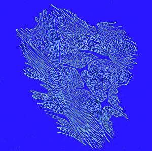
Background imageInternal Anatomy Collection: Crossing section of fibro-nucleated tumors, 1867 (engraving)

Crossing section of fibro-nucleated tumors, 1867 (engraving)
8766909 Crossing section of fibro-nucleated tumors, 1867 (engraving); Private Collection; (add.info.: Crossing section of fibro-nucleated tumors. ( Dr. Jaccoud dictionary.Vol)

Background imageInternal Anatomy Collection: Dog Skulls and Skeleton Set - Dogs skulls and skeleton, Museum d'histoire naturelle de Marseille

Dog Skulls and Skeleton Set - Dogs skulls and skeleton, Museum d'histoire naturelle de Marseille
CSO4987875 Dog Skulls and Skeleton Set - Dogs skulls and skeleton, Museum d'histoire naturelle de Marseille; (add.info.: Dog Skulls and Skeleton Set - Dogs skulls and skeleton)

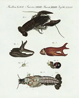
Background imageInternal Anatomy Collection: European or noble crayfish or crawfish (ecrevisse a pate rouge), Astacus astacus. Vulnerable

European or noble crayfish or crawfish (ecrevisse a pate rouge), Astacus astacus. Vulnerable
FLO4575853 European or noble crayfish or crawfish (ecrevisse a pate rouge), Astacus astacus. Vulnerable. Crayfish 1, claws 2, 3, eye 4, internal organs 5, and jaw muscle 6

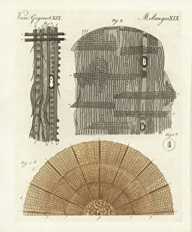
Background imageInternal Anatomy Collection: Anatomy of a tree: cross section of pine branch 1b, wood chip 1a, the same magnified 2

Anatomy of a tree: cross section of pine branch 1b, wood chip 1a, the same magnified 2, magnified further 3
FLO4574347 Anatomy of a tree: cross section of pine branch 1b, wood chip 1a, the same magnified 2, magnified further 3. Handcoloured copperplate engraving from Bertuch's " Bilderbuch fur

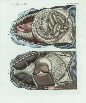
Background imageInternal Anatomy Collection: The entrails in the human body: lungs, liver, diaphragm, stomach, duodenum, colon, etc

The entrails in the human body: lungs, liver, diaphragm, stomach, duodenum, colon, etc
FLO4574696 The entrails in the human body: lungs, liver, diaphragm, stomach, duodenum, colon, etc. Handcoloured copperplate engraving from Bertuch's " Bilderbuch fur Kinder"

Background imageInternal Anatomy Collection: Human airways 1866 illustration of the voice box (larynx, top), trachea (windpipe, vertical)

Human airways 1866 illustration of the voice box (larynx, top), trachea (windpipe, vertical)
JAB4119233 Human airways 1866 illustration of the voice box (larynx, top), trachea (windpipe, vertical, centre) and bronchi (lower left and right)

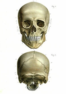
Background imageInternal Anatomy Collection: Human skull 1866 illustration showing the anatomy of the front (Fig 1) and rear (Fig 2)

Human skull 1866 illustration showing the anatomy of the front (Fig 1) and rear (Fig 2)
JAB4119205 Human skull 1866 illustration showing the anatomy of the front (Fig 1) and rear (Fig 2) of a human skull Le crane - Fig 1: Facial region: frontal bone, frontal bump, eyebrow arcade

Background imageInternal Anatomy Collection: Circulatory system. (La vie normale et la vie normale de J.Rengade - lymph nodes du cou et la)

Circulatory system. (La vie normale et la vie normale de J.Rengade - lymph nodes du cou et la aisselle.Drawing A)
JAB4120095 Circulatory system. (La vie normale et la vie normale de J.Rengade - lymph nodes du cou et la aisselle.Drawing A. Demarle 1881; (add.info.: Circulatory system)

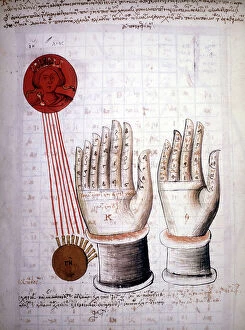
Background imageInternal Anatomy Collection: Page of a manuscript by Pedanius Dioscorides (Dioscorides) Anazarebus, Greek physician. 13th century

Page of a manuscript by Pedanius Dioscorides (Dioscorides) Anazarebus, Greek physician. 13th century
GIA5036777 Page of a manuscript by Pedanius Dioscorides (Dioscorides) Anazarebus, Greek physician. 13th century.; © Giancarlo Costa

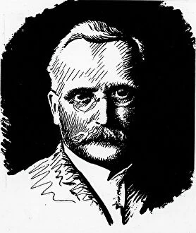
Background imageInternal Anatomy Collection: Portrait of Wilhelm Roux (1850-1924) German anatomist

Portrait of Wilhelm Roux (1850-1924) German anatomist
XEE5031769 Portrait of Wilhelm Roux (1850-1924) German anatomist; (add.info.: Portrait of Wilhelm Roux (1850-1924) German anatomist); Stefano Bianchetti


For sale as Licensed Images

Choose your image, Select your licence and Download the media

"Exploring the Intricacies: Unveiling Internal Anatomy through Art and Science" Delving into the hidden world of internal anatomy, we are captivated by the wonders that lie beneath our skin. From the delicate cross-section of a Honey Bee (Apis mellifera) to the meticulous illustrations of the Channel Tunnel Project, each piece unravels a unique perspective on this captivating subject. Intriguingly, even centuries ago, artists like Giovanni Battista Bianchi skillfully captured human form in terracotta sculptures, reminding us of our intricate inner workings. Meanwhile, Andreas Vesalius revolutionized anatomical understanding with his groundbreaking studies during the Renaissance era. As we journey through time and mediums, Waldeck's original artwork takes us back to Mesoamerica in 1831. The prints from "La nature et l'homme" by Rengade depict twin fetuses in their ideal position during gemellar pregnancy – a testament to both artistry and scientific observation. The diversity within these artworks is astounding; from portraits such as Lucrezia Panciatichi's enigmatic gaze immortalized in oil on wood panel to Giulia di Alessandro de Medici holding a book – perhaps symbolizing knowledge about one's own body. Even photography joins this exploration as two male nudes perch upon a rock in an evocative black-and-white image from 1890. This photograph captures not only physicality but also invites contemplation on vulnerability and strength intertwined within our internal structures. From ancient times to modern endeavors like the Channel Tunnel Project illustration from 1906, humanity has sought to unravel its own mysteries. Through art and science alike, we continue to marvel at our internal anatomy – an ever-evolving tapestry waiting for discovery.