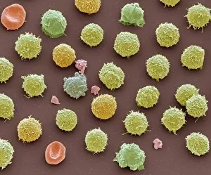
Leukocyte Collection (#4)

"Exploring the Mighty Defenders: Leukocytes and their Battle against Disease" In this captivating collection of images, we delve into the fascinating world of leukocytes

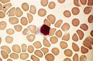
Background imageLeukocyte Collection: Malignant histiocytosis, light micrograph

Malignant histiocytosis, light micrograph
Malignant histiocytosis. Light micrograph of blood cells in a case of malignant histiocytosis. Hystiocytosis is an excess number of histiocytes

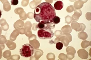
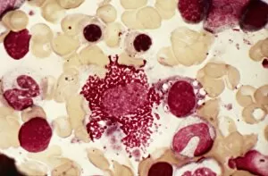
Background imageLeukocyte Collection: Chediak-Higashi syndrome, micrograph

Chediak-Higashi syndrome, micrograph
Chediak-Higashi syndrome. Light micrograph of blood cells in a case of Chediak-Higashi syndrome. The cells include neutrophil white blood cells with abnormal granules

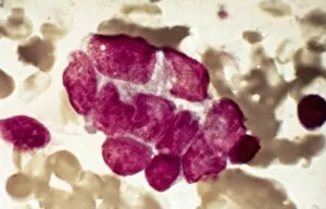
Background imageLeukocyte Collection: Metastatic breast cancer, micrograph

Metastatic breast cancer, micrograph
Metastatic breast cancer. Light micrograph of blood cells in a case of metastatic (secondary) medullary breast cancer. Medullary breast cancer is a rare form of breast cancer that is diagnosed by

Background imageLeukocyte Collection: Metastatic lung cancer, micrograph

Metastatic lung cancer, micrograph
Metastatic lung cancer. Light micrograph of blood cells in a case of metastatic (secondary) medullary lung cancer. Medullary cancer (carcinoma)

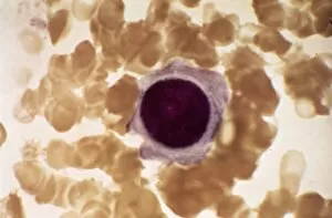
Background imageLeukocyte Collection: Alders anomaly, light micrograph

Alders anomaly, light micrograph
Alders anomaly. Light micrograph of an abnormal lymphocyte white blood cell (centre) from a blood sample in a case of Alders anomaly

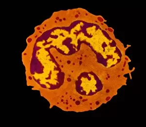
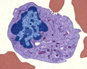
Background imageLeukocyte Collection: Granulocyte white blood cell, TEM

Granulocyte white blood cell, TEM
Granulocyte white blood cell. Coloured transmission electron micrograph (TEM) of a neutrophil granulocyte, a type of white blood cell

Background imageLeukocyte Collection: Acute leukaemia, light micrograph

Acute leukaemia, light micrograph
Acute leukaemia. Light micrograph of blood cells from bone marrow in a case of acute leukaemia. These cells include a polynuclear degranulated basophil and myeloblasts

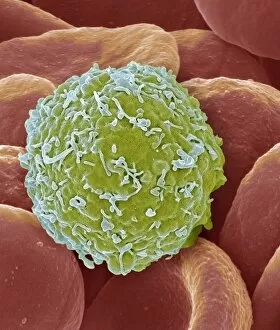
Background imageLeukocyte Collection: Leukaemia cell, SEM

Leukaemia cell, SEM
Leukaemia cell. Coloured scanning electron micrograph (SEM) of a leukaemic (cancerous) lymphocyte white blood cell (green), amongst normal red blood cells (erythrocytes, red)

Background imageLeukocyte Collection: Chronic myeloid leukaemia, micrograph

Chronic myeloid leukaemia, micrograph
Chronic myeloid leukaemia. Light micrograph of blood cells from bone marrow in a case of chronic myeloid leukaemia. Leukaemia is a cancer where certain blood cells form in excess

Background imageLeukocyte Collection: Langerhans cell histiocytosis, micrograph

Langerhans cell histiocytosis, micrograph
Langerhans cell histiocytosis. Light micrograph of blood cells in a case of Langerhans cell histiocytosis (LCH), also called X-type histiocytosis

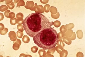
Background imageLeukocyte Collection: Hodgkins disease, light micrograph

Hodgkins disease, light micrograph
Hodgkins disease. Light micrograph of two non-dystrophic histiocyte blood cells (centre) in a case of Hodgkins disease, also called Hodgkins lymphoma

Background imageLeukocyte Collection: Antibodies and B cells, artwork C016 / 6255

Antibodies and B cells, artwork C016 / 6255
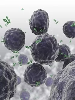
Antibodies and B cells. Computer artwork showing antibodies (green) neutralising cytokine activity in B cell (large, round) white blood cells

Background imageLeukocyte Collection: Antibodies and B cells, artwork C016 / 6254

Antibodies and B cells, artwork C016 / 6254
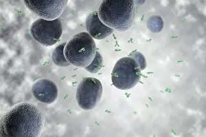
Antibodies and B cells. Computer artwork showing antibodies (green) neutralising cytokine activity in B cell (large, round) white blood cells

Background imageLeukocyte Collection: Normal and leukaemic blood, artwork C016 / 6261

Normal and leukaemic blood, artwork C016 / 6261
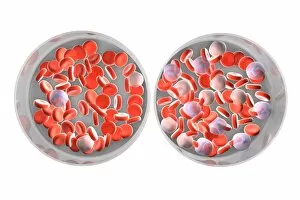
Normal and leukaemic blood. Computer artwork comparing the appearance of normal blood (left) and that affected by leukaemia (right)

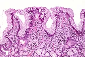
Background imageLeukocyte Collection: Gastritis, light micrograph C016 / 0508

Gastritis, light micrograph C016 / 0508
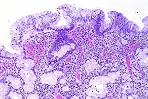
Gastritis. Light micrograph of a section through an inflamed human stomach (gastritis), showing an abnormal accumulation of white blood cells (leucocytes)

Background imageLeukocyte Collection: Gastritis, light micrograph C016 / 0507

Gastritis, light micrograph C016 / 0507
Gastritis. Light micrograph of a section through an inflamed human stomach (gastritis), showing an abnormal accumulation of white blood cells (leucocytes)

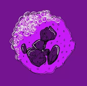
Background imageLeukocyte Collection: Neutrophil white blood cell, illustration C018 / 0741

Neutrophil white blood cell, illustration C018 / 0741
Neutrophil white blood cell, illustration. Neutrophils are the most abundant white blood cell in humans. They have a multi-lobed nucleus (dark purple)

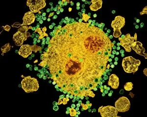
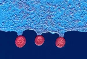
Background imageLeukocyte Collection: Budding HIV particles, TEM C018 / 0125

Budding HIV particles, TEM C018 / 0125
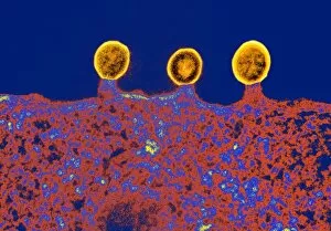
Budding HIV particles. Coloured transmision electron micrograph (TEM) of human immunodeficiency virus (HIV) particles (green) budding from the surface of a T lymphocyte (orange)

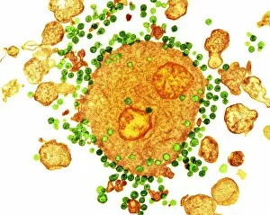
Background imageLeukocyte Collection: Budding HIV particles, TEM C018 / 0124

Budding HIV particles, TEM C018 / 0124
Budding HIV particles. Coloured transmision electron micrograph (TEM) of human immunodeficiency virus (HIV) particles (gren) budding from the surface of a T lymphocyte (orange) from the H9 cell line

Background imageLeukocyte Collection: Budding HIV particles, TEM C018 / 0123

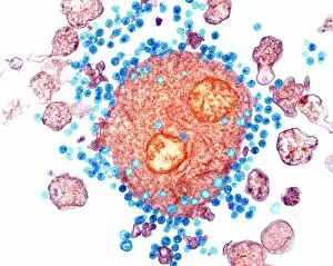
Budding HIV particles, TEM C018 / 0123
Budding HIV particles. Coloured transmision electron micrograph (TEM) of human immunodeficiency virus (HIV) particles (blue) budding from the surface of a T lymphocyte (red) from the H9 cell line

Background imageLeukocyte Collection: Budding HIV particles, TEM C018 / 0127

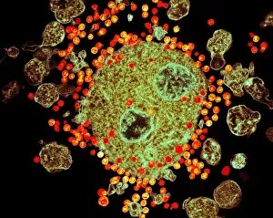
Budding HIV particles, TEM C018 / 0127
Budding HIV particles. Coloured transmision electron micrograph (TEM) of human immunodeficiency virus (HIV) particles (orange) budding from the surface of a T lymphocyte (brown)

Background imageLeukocyte Collection: Budding HIV particles, TEM C018 / 0126

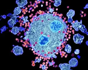
Budding HIV particles, TEM C018 / 0126
Budding HIV particles. Coloured transmision electron micrograph (TEM) of human immunodeficiency virus (HIV) particles (pink) budding from the surface of a T lymphocyte (blue) from the H9 cell line

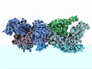
Background imageLeukocyte Collection: Human immune response molecule complex

Human immune response molecule complex. Molecular model showing a human T-cell receptor and an HLA-A leukocyte (white blood cell) antigen bound to a TAX peptide from a virus

Background imageLeukocyte Collection: Human immune response molecule complex C014 / 0871

Human immune response molecule complex C014 / 0871
Human immune response molecule complex. Molecular model showing a human T-cell receptor and an HLA-A leukocyte (white blood cell) antigen bound to a TAX peptide from a virus

Background imageLeukocyte Collection: Monocyte white blood cell, TEM

Monocyte white blood cell, TEM
Monocyte white blood cell. Transmission electron micrograph (TEM) of a section through a monocyte white blood cell, showing the cell nucleus (blue)

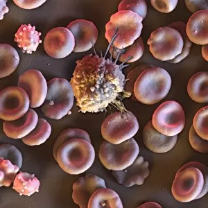
Background imageLeukocyte Collection: Blood cells, SEM C016 / 8026

Blood cells, SEM C016 / 8026
Blood cells. Coloured scanning electron micrograph (SEM) of a lymphocyte white blood cell (centre) and red blood cells (erythrocytes, circular)

Background imageLeukocyte Collection: Activated macrophage, SEM C015 / 6377

Activated macrophage, SEM C015 / 6377
Activated macrophage. Coloured scanning electron micrograph (SEM) of an active macrophage white blood cell. Macrophages are cells of the bodys immune system that are found in the tissues rather than

Background imageLeukocyte Collection: Activated macrophage, SEM C015 / 6376

Activated macrophage, SEM C015 / 6376
Activated macrophage. Coloured scanning electron micrograph (SEM) of an active macrophage white blood cell. Macrophages are cells of the bodys immune system that are found in the tissues rather than

Background imageLeukocyte Collection: Cell infected with HIV, SEM C014 / 0581

Cell infected with HIV, SEM C014 / 0581
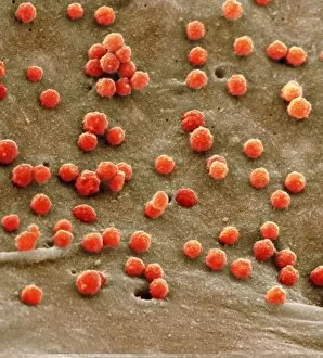
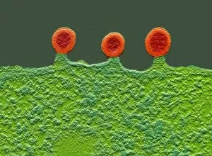
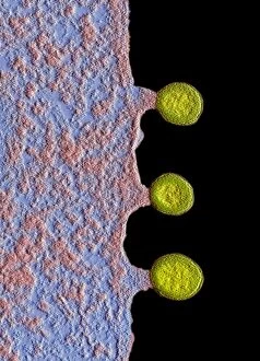
Cell infected with HIV. Coloured scanning electron micrograph (SEM) of HIV particles (round) budding from the membrane of a host cell

Background imageLeukocyte Collection: Cell infected with HIV, SEM C014 / 0580

Cell infected with HIV, SEM C014 / 0580
Cell infected with HIV. Coloured scanning electron micrograph (SEM) of HIV particles (round) budding from the membrane of a host cell

Background imageLeukocyte Collection: Cell infected with HIV, SEM C014 / 0579

Cell infected with HIV, SEM C014 / 0579
Cell infected with HIV. Coloured scanning electron micrograph (SEM) of HIV particles (round) budding from the membrane of a host cell

Background imageLeukocyte Collection: Cell infected with HIV, SEM C017 / 8338

Cell infected with HIV, SEM C017 / 8338
Cell infected with HIV. Coloured scanning electron micrograph (SEM) of HIV particles (orange) budding from the membrane of a host cell

Background imageLeukocyte Collection: Cell infected with HIV, SEM C017 / 8339

Cell infected with HIV, SEM C017 / 8339
Cell infected with HIV. Coloured scanning electron micrograph (SEM) of HIV particles (orange) budding from the membrane of a host cell

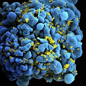
Background imageLeukocyte Collection: Cell infected with HIV, SEM C017 / 8337

Cell infected with HIV, SEM C017 / 8337
Cell infected with HIV. Coloured scanning electron micrograph (SEM) of HIV particles (yellow) budding from the membrane of a host cell

Background imageLeukocyte Collection: Cell infected with HIV, SEM C017 / 8336

Cell infected with HIV, SEM C017 / 8336
Cell infected with HIV. Coloured scanning electron micrograph (SEM) of HIV particles (pink) budding from the membrane of a host cell

Background imageLeukocyte Collection: Budding HIV particles, SEM C017 / 8302

Budding HIV particles, SEM C017 / 8302
Budding HIV particles. Coloured scanning electron micrograph (SEM) of human immunodeficiency virus (HIV) particles (yellow) budding from the surface of a T lymphocyte from the H9 cell line

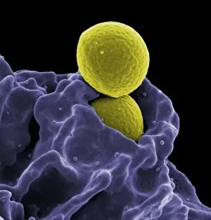
Background imageLeukocyte Collection: Neutrophil engulfing MRSA, SEM C017 / 8301

Neutrophil engulfing MRSA, SEM C017 / 8301
Neutrophil engulfing MRSA. Coloured scanning electron micrograph (SEM) of a neutrophil white blood cell (purple) engulfing methicillin-resistant Staphylococcus aureus bacteria (MRSA, yellow)

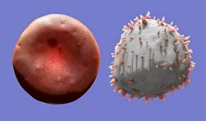
Background imageLeukocyte Collection: Red and white blood cells, artwork C016 / 4629

Red and white blood cells, artwork C016 / 4629
Red and white blood cells, computer artwork. Red blood cells are biconcave, giving them a large surface area for gas exchange, and highly elastic

Background imageLeukocyte Collection: Red and white blood cells, artwork C016 / 4630

Red and white blood cells, artwork C016 / 4630
Red and white blood cells, computer artwork. Red blood cells are biconcave, giving them a large surface area for gas exchange, and highly elastic

Background imageLeukocyte Collection: White blood cell, artwork C016 / 4628

White blood cell, artwork C016 / 4628
White blood cell, computer artwork. White blood cells are part of the bodys immune system

Background imageLeukocyte Collection: Antigen presentation, SEM C016 / 3105

Antigen presentation, SEM C016 / 3105
Antigen presentation. Coloured scanning electron micrograph (SEM) showing the interaction between a macrophage (blue) and a T helper lymphocyte (Th cell, yellow)

Background imageLeukocyte Collection: Antigen presentation, SEM C016 / 3104

Antigen presentation, SEM C016 / 3104
Antigen presentation. Coloured scanning electron micrograph (SEM) showing the interaction between a macrophage (brown) and a T helper lymphocyte (Th cell, red)

Background imageLeukocyte Collection: White blood cells, SEM C016 / 3102

White blood cells, SEM C016 / 3102
White blood cells. Coloured scanning electron micrograph (SEM) of white blood cells (leucocytes) from a sample of peripheral blood

Background imageLeukocyte Collection: Blood cells, SEM C016 / 3101

Blood cells, SEM C016 / 3101
Blood cells. Coloured scanning electron micrograph (SEM) of a variety of different white blood cells (leucocytes, blue/purple) with two red blood cells (erythrocytes)

Background imageLeukocyte Collection: Blood cells, SEM C016 / 3100

Blood cells, SEM C016 / 3100
Blood cells. Coloured scanning electron micrograph (SEM) of a variety of different white blood cells (leucocytes, yellow/green) with two red blood cells (erythrocytes)

Background imageLeukocyte Collection: Blood cells, SEM C016 / 3096

Blood cells, SEM C016 / 3096
Blood cells. Coloured scanning electron micrograph of a lymphocyte white blood cell (left) and a red blood cell (erythrocyte, right)

Background imageLeukocyte Collection: Macrophage and platelets, SEM C016 / 3094

Macrophage and platelets, SEM C016 / 3094
Macrophage and platelets. Coloured scanning electron micrograph (SEM) of platelets (green) attached to a macrophage white blood cell (turquoise)

Background imageLeukocyte Collection: Platelets, SEM C016 / 3091

Platelets, SEM C016 / 3091
Platelets, coloured scanning electron micrograph (SEM). Platelets, or thrombocytes, are fragments of white blood cells. When blood clots


For sale as Licensed Images

Choose your image, Select your licence and Download the media

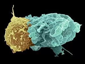
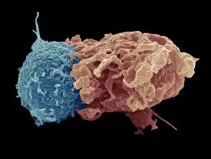
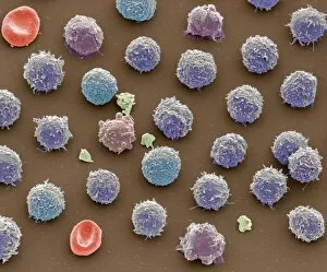
"Exploring the Mighty Defenders: Leukocytes and their Battle against Disease" In this captivating collection of images, we delve into the fascinating world of leukocytes, our body's mighty defenders against various diseases. First up, we witness the incredible power of T lymphocytes as they engage in a fierce battle with cancer cells. SEM C001 / 1679 reveals an epic showdown between these two forces. Moving on to another remarkable image, Neutrophil engulfing MRSA (SEM C018 / 8596) showcases the extraordinary ability of neutrophils to devour harmful bacteria such as Methicillin-resistant Staphylococcus aureus. Next, TEM unveils a close-up view of a human white blood cell bearing HLA antigen. This intricate image highlights the complexity and diversity within our immune system. A burst of color awaits us in Coloured SEM of a white blood cell (lymphocyte), showcasing the vibrant nature that lies within these essential cells. Meanwhile, Basophil white blood cell reminds us that even amidst chaos, there are specialized warriors ready to combat any threat. The delicate balance between red and white blood cells is beautifully captured in Red and white blood cells (SEM). This harmonious coexistence ensures our bodies remain healthy and functional. Micrograph enthusiasts will appreciate Dohle bodies in blood cell micrograph - an intriguing glimpse into abnormal cellular structures that can indicate certain health conditions like infections or inflammation. Shifting gears towards Plasma cells (TEM), we witness their crucial role in producing antibodies to fight off infections effectively. Their unique appearance underlines their significance in maintaining our overall well-being. Acute promyelocytic leukemia takes center stage next with its distinct features showcased through a powerful micrograph. This visual representation sheds light on this specific type of cancer affecting immature myeloid cells. Phagocytosis takes center stage once again as fungal spores become prey for vigilant macrophages in Phagocytosis of fungal spores (SEM).