Malignant Collection (page 2)

"Malignant: Unleashing the Battle Within" In a microscopic world, T lymphocytes stand as valiant warriors against cancer cells

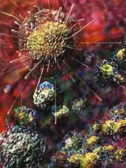
Background imageMalignant Collection: Conceptual image of cancer virus

Conceptual image of cancer virus

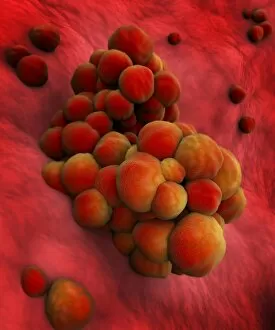
Background imageMalignant Collection: Microscopic view of a group of macrophages

Microscopic view of a group of macrophages. Macrophages contribute to tumor growth and progression. Attracted to oxygen-starved (hypoxic) and necrotic tumor cells they promote chronic inflammation

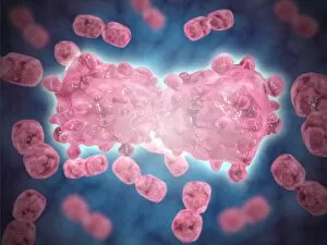
Background imageMalignant Collection: Microscopic view of a group of leukemia cells

Microscopic view of a group of leukemia cells

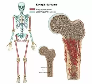
Background imageMalignant Collection: Ewings sarcoma locations on the skeleton

Ewings sarcoma locations on the skeleton and detail of tumor on head of femur

Background imageMalignant Collection: Microscopic view of cancer virus

Microscopic view of cancer virus

Background imageMalignant Collection: Isolated cancer cell Macrophage

Isolated cancer cell Macrophage. Macrophages contribute to tumor growth and progression. Attracted to oxygen-starved (hypoxic) and necrotic tumor cells they promote chronic inflammation

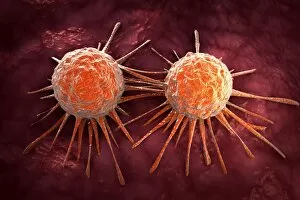
Background imageMalignant Collection: Microscopic view of cancer cells

Microscopic view of cancer cells. Cancer occurs when a cells gene mutations make the cell unable to correct DNA damage

Background imageMalignant Collection: Conceptual image illustrating the process of bone metastasis

Conceptual image illustrating the process of bone metastasis

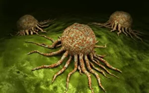
Background imageMalignant Collection: Microscopic view of tumor

Microscopic view of tumor

Background imageMalignant Collection: Microscopic view of pancreatic cancer cell

Microscopic view of pancreatic cancer cell

Background imageMalignant Collection: Microscipic view of pancreatic cancer cells

Microscipic view of pancreatic cancer cells

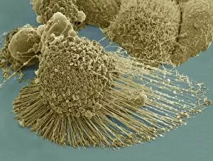
Background imageMalignant Collection: Scanning electron micrograph of an apoptotic HeLa cell

Scanning electron micrograph of an apoptotic HeLa cell. Zeiss Merlin HR-SEM

Background imageMalignant Collection: The Kingdoms Monster Uncloaked from Heaven, c. 1640-43 (woodcut) (b / w photo)

The Kingdoms Monster Uncloaked from Heaven, c. 1640-43 (woodcut) (b / w photo)
XJF106630 The Kingdoms Monster Uncloaked from Heaven, c.1640-43 (woodcut) (b/w photo) by English School, (17th century); Private Collection; English, out of copyright

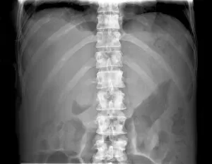
Background imageMalignant Collection: Secondary bone cancer, X-ray C017 / 7162

Secondary bone cancer, X-ray C017 / 7162
Secondary bone cancer. X-ray of the spine of a 40 year old female with metastatic (secondary) bone cancer (bright white areas). The cancer has metastasised (spread) from a primary cancer

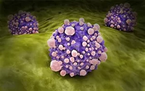
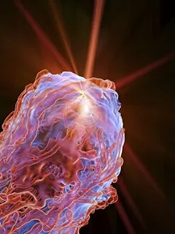
Background imageMalignant Collection: Cancer cell, artwork C016 / 9843

Cancer cell, artwork C016 / 9843
Cancer cell. Computer artwork of a cancerous (malignant) cell (top left) in tissue. The uneven surface and cytoplasmic projections are characteristic of cancer cells

Background imageMalignant Collection: Cancer treatment, artwork F007 / 4199

Cancer treatment, artwork F007 / 4199
Cancer treatment, conceptual image. Computer artwork representing the use of radiation in tumour treatment. Tumours are caused by the uncontrolled growth of previously normal cells

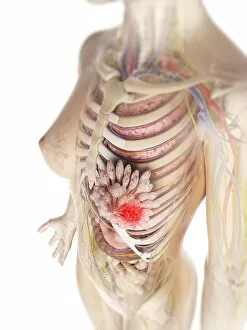
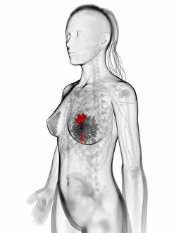
Background imageMalignant Collection: Breast cancer, artwork F006 / 7491

Breast cancer, artwork F006 / 7491
Breast cancer, computer artwork

Background imageMalignant Collection: Testicular cancer, artwork

Testicular cancer, artwork
Testicular cancer, computer artwork

Background imageMalignant Collection: Brain cancer, artwork F006 / 7482

Brain cancer, artwork F006 / 7482
Brain cancer, computer artwork

Background imageMalignant Collection: Brain cancer, artwork F006 / 7483

Brain cancer, artwork F006 / 7483
Brain cancer, computer artwork

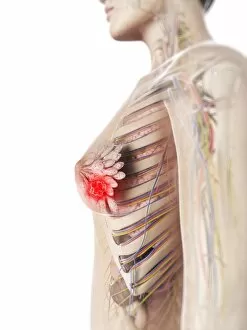
Background imageMalignant Collection: Breast cancer, artwork F006 / 7497

Breast cancer, artwork F006 / 7497
Breast cancer, computer artwork

Background imageMalignant Collection: Intestinal cancer, artwork F007 / 6664

Intestinal cancer, artwork F007 / 6664
Intestinal cancer, computer artwork

Background imageMalignant Collection: Colon cancer, artwork F007 / 6862

Colon cancer, artwork F007 / 6862
Colon cancer, computer artwork

Background imageMalignant Collection: Colon tumor, artwork F007 / 6861

Colon tumor, artwork F007 / 6861
Colon tumor, computer artwork

Background imageMalignant Collection: Colon cancer, artwork F007 / 6876

Colon cancer, artwork F007 / 6876
Colon cancer, computer artwork

Background imageMalignant Collection: Colon cancer, artwork F008 / 0425

Colon cancer, artwork F008 / 0425
Colon cancer, computer artwork

Background imageMalignant Collection: Lung cancer, artwork F008 / 0116

Lung cancer, artwork F008 / 0116
Lung cancer, computer artwork

Background imageMalignant Collection: Colon cancer, artwork F008 / 0585

Colon cancer, artwork F008 / 0585
Colon cancer, computer artwork

Background imageMalignant Collection: Intestinal cancer, artwork F008 / 1199

Intestinal cancer, artwork F008 / 1199
Intestinal cancer, computer artwork

Background imageMalignant Collection: Intestinal cancer, artwork F008 / 1197

Intestinal cancer, artwork F008 / 1197
Intestinal cancer, computer artwork

Background imageMalignant Collection: Liver cancer, artwork F008 / 0127

Liver cancer, artwork F008 / 0127
Liver cancer, computer artwork

Background imageMalignant Collection: Intestinal cancer, artwork F008 / 1198

Intestinal cancer, artwork F008 / 1198
Intestinal cancer, computer artwork

Background imageMalignant Collection: Colon cancer, artwork F008 / 0678

Colon cancer, artwork F008 / 0678
Colon cancer, computer artwork

Background imageMalignant Collection: Colon cancer, artwork F008 / 0672

Colon cancer, artwork F008 / 0672
Colon cancer, computer artwork

Background imageMalignant Collection: Colon cancer, artwork F008 / 0306

Colon cancer, artwork F008 / 0306
Colon cancer, computer artwork

Background imageMalignant Collection: Colon cancer, artwork F008 / 0050

Colon cancer, artwork F008 / 0050
Colon cancer, computer artwork

Background imageMalignant Collection: Lung cancer, artwork F008 / 0214

Lung cancer, artwork F008 / 0214
Lung cancer, computer artwork

Background imageMalignant Collection: Colon cancer, artwork F008 / 0424

Colon cancer, artwork F008 / 0424
Colon cancer, computer artwork

Background imageMalignant Collection: Colon cancer, artwork F008 / 0519

Colon cancer, artwork F008 / 0519
Colon cancer, computer artwork

Background imageMalignant Collection: Colon cancer, artwork F008 / 0142

Colon cancer, artwork F008 / 0142
Colon cancer, computer artwork

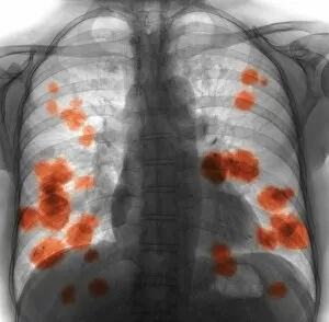
Background imageMalignant Collection: Secondary lung cancer, X-ray F008 / 3498

Secondary lung cancer, X-ray F008 / 3498
Secondary lung cancer. Coloured X-ray of the chest of a 52 year old female patient with metastatic (secondary) lung cancer (red)

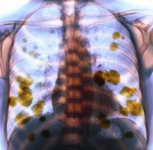
Background imageMalignant Collection: Secondary lung cancer, X-ray F008 / 3496

Secondary lung cancer, X-ray F008 / 3496
Secondary lung cancer. Coloured X-ray of the chest of a 52 year old female patient with metastatic (secondary) lung cancer (yellow)

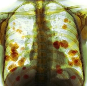
Background imageMalignant Collection: Secondary lung cancer, X-ray F008 / 3495

Secondary lung cancer, X-ray F008 / 3495
Secondary lung cancer. Coloured X-ray of the chest of a 52 year old female patient with metastatic (secondary) lung cancer (green)

Background imageMalignant Collection: Secondary lung cancer, X-ray F008 / 3497

Secondary lung cancer, X-ray F008 / 3497
Secondary lung cancer. Coloured X-ray of the chest of a 52 year old female patient with metastatic (secondary) lung cancer (orange)

Background imageMalignant Collection: Cancer cells, artwork

Cancer cells, artwork
Cancer cells, computer artwork

Background imageMalignant Collection: Breast cancer, artwork F005 / 0458

Breast cancer, artwork F005 / 0458
Breast cancer, computer artwork

Background imageMalignant Collection: Breast cancer, artwork F005 / 0473

Breast cancer, artwork F005 / 0473
Breast cancer, computer artwork

Background imageMalignant Collection: Cancer cells dividing, artwork F005 / 0540

Cancer cells dividing, artwork F005 / 0540
Cancer cells dividing, computer artwork


For sale as Licensed Images

Choose your image, Select your licence and Download the media

"Malignant: Unleashing the Battle Within" In a microscopic world, T lymphocytes stand as valiant warriors against cancer cells, their relentless pursuit captured in SEM C001 / 1679. Meanwhile, HeLa cells reveal their sinister nature under the watchful eye of a light micrograph in C017 / 8299 and C017 / 8298. A mesmerizing scene unfolds as coloured SEM showcases lymphocytes fearlessly attacking cancer cells, painting a vivid picture of hope amidst adversity. Acute promyelocytic leukemia reveals its haunting presence through an ominous micrograph while ovarian cancer exposes its treacherous grip in light micrograph C015 / 7103. Witnessing the battle at a cellular level is awe-inspiring; a coloured SEM image captures the moment when leukaemic white blood cells are confronted head-on by nanorobots determined to eradicate this malignant threat (M132 / 0488). The fight against cancer takes on otherworldly dimensions reminiscent of Amazing Stories sci-fi magazine covers featuring giant plants and fantastical creatures. Yet not all battles are fought within our bodies; sometimes they transcend into realms beyond. Like an exorcism performed by a cleric casting spells against evil spirits, we confront malignancy with unwavering determination. Just as history has witnessed struggles such as "The Condition of Ireland under the 'No Rent' Policy, " or "Johnnys Defeat at the Dock, " we too face adversity head-on. Amidst these hints lie tales of resilience and courage that remind us to never surrender to darkness. Malignant forces may try to overpower us, but united we stand ready to conquer them - be it through scientific advancements or sheer willpower. Together, let us rewrite the narrative and triumph over malignancy's grasp for brighter tomorrows await those who dare to fight back.