Malignant Collection (page 9)

"Malignant: Unleashing the Battle Within" In a microscopic world, T lymphocytes stand as valiant warriors against cancer cells

Background imageMalignant Collection: Dividing liver cancer cell, SEM

Dividing liver cancer cell, SEM
Dividing liver cancer cell. Coloured scanning electron micrograph (SEM) of a hepatocellular carcinoma (HCC) cell undergoing mitosis (nuclear division)

Background imageMalignant Collection: Stomach cancer, barium X-ray

Stomach cancer, barium X-ray
Stomach cancer. Coloured frontal X-ray of the upper gastroduodenal tract of a patient with stomach cancer (grey, upper centre)

Background imageMalignant Collection: Secondary bowel cancer, endoscopic view C016 / 8337

Secondary bowel cancer, endoscopic view C016 / 8337
Secondary bowel cancer. Endoscopic view of the interior of a patients descending duodenum (first part of the small intestine), showing cancer that has spread (metastasised) from the pancreas

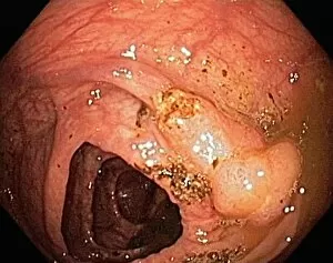
Background imageMalignant Collection: Colon cancer, endoscopic view C016 / 8307

Colon cancer, endoscopic view C016 / 8307
Colon cancer. Endoscopic view of a malignant (cancerous) adenocarcinoma (lower right) in a patients ascending colon (large intestine)

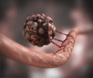
Background imageMalignant Collection: Blood vessel formation, artwork

Blood vessel formation, artwork
Blood vessel formation. Artwork showing malignant (cancerous) tumour cells promoting the formation of new blood vessels, a process known as angiogenesis

Background imageMalignant Collection: Radiotherapy, conceptual image C013 / 5587

Radiotherapy, conceptual image C013 / 5587
Radiotherapy, conceptual image. Computer artwork representing the use of ionising radiation to kill cancerous cells (centre)

Background imageMalignant Collection: Dividing cancer cell, SEM C014 / 0361

Dividing cancer cell, SEM C014 / 0361
Dividing cancer cell. Coloured scanning electron micrograph (SEM) of a colorectal cancer cell undergoing mitosis (nuclear division) and splitting into two daughter cells (left and right)

Background imageMalignant Collection: Dividing brain cancer cells, SEM C014 / 0354

Dividing brain cancer cells, SEM C014 / 0354
Dividing brain cancer cells. Coloured scanning electron micrograph (SEM) of a cancerous astrocyte brain cell that has just undergone cytokinesis (cell division)

Background imageMalignant Collection: Dividing bone cancer cell, SEM

Dividing bone cancer cell, SEM
SEM Dividing Osteosarcoma cells Osteosarcoma is an aggressive malignant neoplasm arising from primitive transformed cells of mesenchymal origin (and thus a sarcoma)

Background imageMalignant Collection: Cervical cancer, light micrograph C015 / 7000

Cervical cancer, light micrograph C015 / 7000
Cervical cancer. Light micrograph of a section through tissue from the cervix (neck of the womb) showing intraepithelial carcinoma (centre), a form of cancer

Background imageMalignant Collection: Cervical cancer, light micrograph C015 / 6999

Cervical cancer, light micrograph C015 / 6999
Cervical cancer. Light micrograph of a section through tissue from the cervix (neck of the womb) showing intraepithelial carcinoma (across centre), a form of cancer

Background imageMalignant Collection: Cervical cancer, light micrograph C015 / 6998

Cervical cancer, light micrograph C015 / 6998
Cervical cancer. Light micrograph of a section through tissue from the cervix (neck of the womb) showing intraepithelial carcinoma (dark areas), a form of cancer

Background imageMalignant Collection: Cervical cancer, light micrograph C015 / 6997

Cervical cancer, light micrograph C015 / 6997
Cervical cancer. Light micrograph of a section through tissue from the cervix (neck of the womb) showing intraepithelial carcinoma (across centre), a form of cancer

Background imageMalignant Collection: Cancer of the chorion C015 / 6905

Cancer of the chorion C015 / 6905
Cancer of the chorion. Light micrograph of a section through a sarcoma of the chorion, a malignant (cancerous) tumour of the membrane (chorion) that surrounds a developing embryo

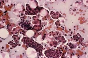
Background imageMalignant Collection: Cervical smear showing endometrial cancer C015 / 6750

Cervical smear showing endometrial cancer C015 / 6750
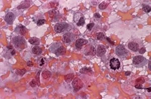
Cervical smear showing endometrial cancer. Light micrograph of cells from a cervical smear test showing endometrial sarcoma. This is a cancer of the lining of the womb, the endometrium

Background imageMalignant Collection: Cancer cell, SEM C014 / 0628

Cancer cell, SEM C014 / 0628
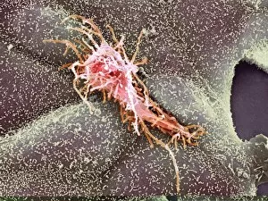
Cancer cell. Coloured scanning electron micrograph (SEM) of a carcinoma cancer cell (pink) migrating (metastasising) along a layer of normal epithelial cells (purple)

Background imageMalignant Collection: Cancer cell, SEM C014 / 0627

Cancer cell, SEM C014 / 0627
Cancer cell. Coloured scanning electron micrograph (SEM) of a carcinoma cancer cell (yellow) migrating (metastasising) along a layer of normal epithelial cells (brown)

Background imageMalignant Collection: Throat cancer, artwork C013 / 5939

Throat cancer, artwork C013 / 5939
Throat cancer. Cutaway computer artwork of a persons head, showing cancer (yellow) in the pharynx (centre) and oesophagus (lower centre)

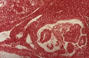
Background imageMalignant Collection: Endometrial cancer, light micrograph C015 / 6061

Endometrial cancer, light micrograph C015 / 6061
Endometrial cancer. Light micrograph of a section through a tumour in the endometrium, the lining of the uterus (womb). Endometrial cancer is the third most common cause of gynaecological cancer

Background imageMalignant Collection: Secondary endometrial cancer C015 / 6058

Secondary endometrial cancer C015 / 6058
Secondary endometrial cancer. Light micrograph of a section through a tumour in an inguinal lymph node due to a secondary endometrial cancer

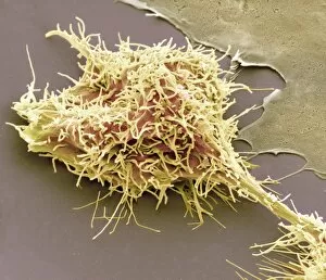
Background imageMalignant Collection: Bone cancer cell, SEM

Bone cancer cell, SEM
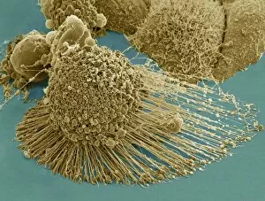
Bone cancer cell. Coloured scanning electron micrograph of a fibrosarcoma (fibroblastic sarcoma) cell, showing the numerous filopodia (hair-like) covering its surface

Background imageMalignant Collection: HeLa cells, SEM C017 / 8305

HeLa cells, SEM C017 / 8305
HeLa cells, coloured scanning electron micrograph (SEM). The cells have just replicated. HeLa cells were the first continuously cultured cell line

Background imageMalignant Collection: HeLa cell dying, SEM C017 / 8304

HeLa cell dying, SEM C017 / 8304
HeLa cell dying, coloured scanning electron micrograph (SEM). The cell in the foreground is undergoing apoptosis, or programmed cell death. Apoptosis occurs when a cell becomes old or damaged

Background imageMalignant Collection: Mesothelioma lung cancer, CT scan

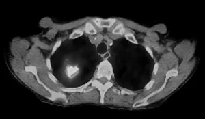
Mesothelioma lung cancer, CT scan
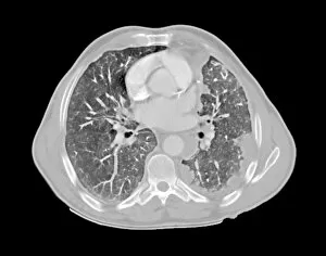
Mesothelioma lung cancer. Computed tomography (CT) scan of the lungs of a 69 year old patient with mesothelioma, a type of lung cancer

Background imageMalignant Collection: Lung cancer, CT and PET scans

Lung cancer, CT and PET scans
Lung cancer. Combined computed tomography (CT) and positron emission tomography (PET) scans of the lungs of an 84 year old woman with a malignant (cancerous) tumour (bright patch)

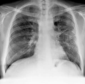
Background imageMalignant Collection: Lung cancer, X-ray C017 / 7964

Lung cancer, X-ray C017 / 7964
Lung cancer. X-ray if the chest of a patient with malignant (cancerous) tumours in middle of the left lung (seen at right). The main cause of lung cancer is cigarette smoke

Background imageMalignant Collection: Chronic myeloid leukaemia, micrograph C015 / 6227

Chronic myeloid leukaemia, micrograph C015 / 6227
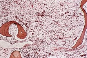
Chronic myeloid leukaemia. Light micrograph of bone marrow from a patient with chronic myeloid leukaemia (CML). This cancer affects the myeloid tissue (bone marrow)

Background imageMalignant Collection: Acute myeloid leukaemia C015 / 6225

Acute myeloid leukaemia C015 / 6225
Acute myeloid leukaemia. Light micrograph of bone marrow from a patient with acute myeloid leukaemia (AML). This cancer affects the myeloid tissue (bone marrow)

Background imageMalignant Collection: Spleen cancer, gross specimen C015 / 6421

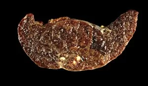
Spleen cancer, gross specimen C015 / 6421
Spleen cancer. Gross specimen of a spleen showing a large lymphosarcoma, or non-Hodgkins lymphoma, a cancer of lymphoid tissue. The lymphatic system is a major part of the immune system

Background imageMalignant Collection: Tumour embolism, light micrograph C015 / 6397

Tumour embolism, light micrograph C015 / 6397
Tumour embolism. Light micrograph of a section through an embolism (blood clot) caused by cells from a cervical cancer. The cells have left the primary tumour and are metastasising (spreading)

Background imageMalignant Collection: Angiosarcoma of the spleen C015 / 6398

Angiosarcoma of the spleen C015 / 6398
Angiosarcoma. Gross specimen of a sectioned spleen affected by angiosarcoma. Malignant (cancerous) tumours (yellow) can be seen at bottom centre

Background imageMalignant Collection: Secondary pancreatic cancer, ultrasound C017 / 7765

Secondary pancreatic cancer, ultrasound C017 / 7765
Secondary pancreatic cancer. Ultrasound scan of a 63 year old male patients pancreas showing metastatic (secondary) tumours. The cancer has metastasised (spread) from a primary cancer

Background imageMalignant Collection: Secondary thyroid cancer C015 / 6170

Secondary thyroid cancer C015 / 6170
Secondary thyroid cancer. Light micrograph of a section through a tumour in a lymph node due to a secondary thyroid cancer

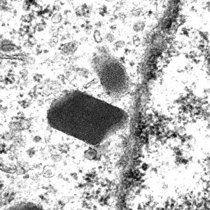
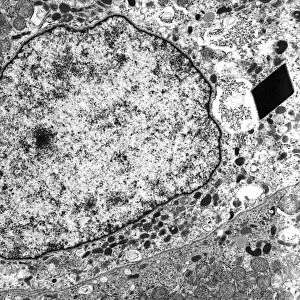
Background imageMalignant Collection: Alveolar soft part sarcoma, TEM C016 / 5826

Alveolar soft part sarcoma, TEM C016 / 5826
Alveolar soft part sarcoma. Transmission electron micrograph (TEM) of a section through an alveolar soft part sarcoma (ASPS) showing a characteristic rhomboid crystal (black, centre)

Background imageMalignant Collection: Alveolar soft part sarcoma, TEM C016 / 5816

Alveolar soft part sarcoma, TEM C016 / 5816
Alveolar soft part sarcoma. Transmission electron micrograph (TEM) of a section through an alveolar soft part sarcoma (ASPS) showing a characteristic rhomboid crystal (black)

Background imageMalignant Collection: Brain cancer, MRI scan C016 / 4438

Brain cancer, MRI scan C016 / 4438
Brain cancer. Coloured coronal magnetic resonance imaging (MRI) scan through the head of a 48 year old male patient with a glioblastoma (red)

Background imageMalignant Collection: Brain cancer, MRI scan C016 / 4436

Brain cancer, MRI scan C016 / 4436
Brain cancer. Coloured coronal magnetic resonance imaging (MRI) scan through the head of a 48 year old male patient with a glioblastoma (centre right)

Background imageMalignant Collection: Brain cancer, MRI scan C016 / 4437

Brain cancer, MRI scan C016 / 4437
Brain cancer. Coloured coronal magnetic resonance imaging (MRI) scan through the head of a 48 year old male patient with a glioblastoma (orange)

Background imageMalignant Collection: Lung cancer, X-ray C016 / 4435

Lung cancer, X-ray C016 / 4435
Lung cancer. Coloured chest X-ray of a 64 year old male patient with lung cancer. The tumour is seen as the red mass in the right lung (bright area at left)

Background imageMalignant Collection: Lung cancer, X-ray C016 / 4432

Lung cancer, X-ray C016 / 4432
Lung cancer. Coloured chest X-ray of a 64 year old male patient with lung cancer. The tumour is seen as the orange mass in the right lung (dark area at left)

Background imageMalignant Collection: Lung cancer, X-ray C016 / 4433

Lung cancer, X-ray C016 / 4433
Lung cancer. Coloured chest X-ray of a 64 year old male patient with lung cancer. The tumour is seen as the orange mass in the right lung (bright area at left)

Background imageMalignant Collection: Preventing cancer spreading, artwork C013 / 8816

Preventing cancer spreading, artwork C013 / 8816
Preventing cancer spreading, conceptual artwork. Cancer cells inside a geodesic cage, representing the prevention of metastasis - spreading to other tissues of the body

Background imageMalignant Collection: Angiogenesis inhibitors, conceptual image C013 / 7789

Angiogenesis inhibitors, conceptual image C013 / 7789
Angiogenesis inhibitors, conceptual image. Computer artwork showing a brick wall preventing new blood vessels from reaching a cancerous tumour

Background imageMalignant Collection: Lung cancer cell division, artwork C013 / 4630

Lung cancer cell division, artwork C013 / 4630
Lung cancer cell division. Computer artwork of a lung cancer cell during cell division (cytokinesis). The two daughter cells remain temporarily joined by a cytoplasmic bridge (centre)

Background imageMalignant Collection: Cancer cell, artwork C013 / 4622

Cancer cell, artwork C013 / 4622
Cancer cell. Computer artwork of a single typical cancer cell in the human body. The uneven surface and cytoplasmic projections are characteristic of cancer cells

Background imageMalignant Collection: Blood cancer, conceptual image C013 / 4618

Blood cancer, conceptual image C013 / 4618
Blood cancer, conceptual image. Computer artwork of the interior of a blood vessel, showing cancer cells (dark) amongst normal red blood cells (red)

Background imageMalignant Collection: Blood cancer, conceptual image C013 / 4614

Blood cancer, conceptual image C013 / 4614
Blood cancer, conceptual image. Computer artwork of the interior of a blood vessel, showing cancer cells (white) amongst normal red blood cells (red)

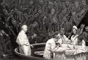
Background imageMalignant Collection: Agnews surgical clinic, 1889

Agnews surgical clinic, 1889
Agnews surgical clinic. At left, holding a scalpel, is US surgeon David Hayes Agnew (1818-1892), supervising a partial breast removal operation (mastectomy)


For sale as Licensed Images

Choose your image, Select your licence and Download the media

"Malignant: Unleashing the Battle Within" In a microscopic world, T lymphocytes stand as valiant warriors against cancer cells, their relentless pursuit captured in SEM C001 / 1679. Meanwhile, HeLa cells reveal their sinister nature under the watchful eye of a light micrograph in C017 / 8299 and C017 / 8298. A mesmerizing scene unfolds as coloured SEM showcases lymphocytes fearlessly attacking cancer cells, painting a vivid picture of hope amidst adversity. Acute promyelocytic leukemia reveals its haunting presence through an ominous micrograph while ovarian cancer exposes its treacherous grip in light micrograph C015 / 7103. Witnessing the battle at a cellular level is awe-inspiring; a coloured SEM image captures the moment when leukaemic white blood cells are confronted head-on by nanorobots determined to eradicate this malignant threat (M132 / 0488). The fight against cancer takes on otherworldly dimensions reminiscent of Amazing Stories sci-fi magazine covers featuring giant plants and fantastical creatures. Yet not all battles are fought within our bodies; sometimes they transcend into realms beyond. Like an exorcism performed by a cleric casting spells against evil spirits, we confront malignancy with unwavering determination. Just as history has witnessed struggles such as "The Condition of Ireland under the 'No Rent' Policy, " or "Johnnys Defeat at the Dock, " we too face adversity head-on. Amidst these hints lie tales of resilience and courage that remind us to never surrender to darkness. Malignant forces may try to overpower us, but united we stand ready to conquer them - be it through scientific advancements or sheer willpower. Together, let us rewrite the narrative and triumph over malignancy's grasp for brighter tomorrows await those who dare to fight back.