Malignant Collection (page 5)

"Malignant: Unleashing the Battle Within" In a microscopic world, T lymphocytes stand as valiant warriors against cancer cells

Background imageMalignant Collection: Tumour, computer artwork F007 / 0218

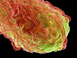
Tumour, computer artwork F007 / 0218
Tumour, computer artwork. Tumours are caused by the uncontrolled growth of previously normal cells. The resulting growth (green) can invade and damage surrounding tissue
Background imageMalignant Collection: Tumour, computer artwork F007 / 0216

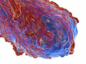
Tumour, computer artwork F007 / 0216
Tumour, computer artwork. Tumours are caused by the uncontrolled growth of previously normal cells. The resulting growth (blue) can invade and damage surrounding tissue

Background imageMalignant Collection: Tumour, computer artwork F007 / 0214

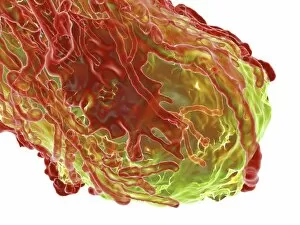
Tumour, computer artwork F007 / 0214
Tumour, computer artwork. Tumours are caused by the uncontrolled growth of previously normal cells. The resulting growth (green) can invade and damage surrounding tissue

Background imageMalignant Collection: Tumour, computer artwork F007 / 0212

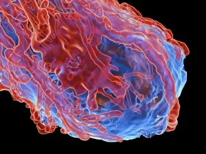
Tumour, computer artwork F007 / 0212
Tumour, computer artwork. Tumours are caused by the uncontrolled growth of previously normal cells. The resulting growth (blue) can invade and damage surrounding tissue

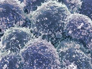
Background imageMalignant Collection: Lung cancer cells, SEM F006 / 8641

Lung cancer cells, SEM F006 / 8641
Lung cancer cells, coloured scanning electron micrograph (SEM). Magnification: x12, 000 when printed at 10 centimetres wide

Background imageMalignant Collection: Bone cancer cell, SEM F006 / 8639

Bone cancer cell, SEM F006 / 8639
Bone cancer cell. Coloured scanning electron micrograph of an osteosarcoma cancer cell. Osteosarcoma is an aggressive malignant neoplasm arising from primitive transformed mesenchymal cells

Background imageMalignant Collection: Lung cancer cells, SEM F006 / 8640

Lung cancer cells, SEM F006 / 8640
Lung cancer cells, coloured scanning electron micrograph (SEM). Magnification: x12, 000 when printed at 10 centimetres wide

Background imageMalignant Collection: Bone cancer cell, SEM F006 / 8638

Bone cancer cell, SEM F006 / 8638
Bone cancer cell. Coloured scanning electron micrograph of an osteosarcoma cancer cell. Osteosarcoma is an aggressive malignant neoplasm arising from primitive transformed mesenchymal cells

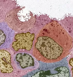
Background imageMalignant Collection: Breast cancer, TEM F006 / 8633

Breast cancer, TEM F006 / 8633
Breast cancer. Coloured transmission electron micrograph (TEM) of a section through a ductal carcinoma (cancer) of the breast showing malignant epithelial cells

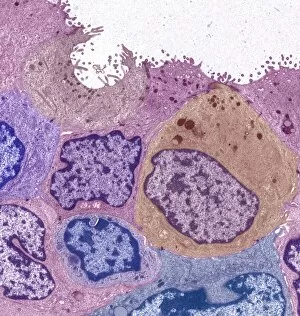
Background imageMalignant Collection: Breast cancer, TEM F006 / 8632

Breast cancer, TEM F006 / 8632
Breast cancer. Coloured transmission electron micrograph (TEM) of a section through a ductal carcinoma (cancer) of the breast showing malignant epithelial cells

Background imageMalignant Collection: Bone cancer, light micrograph F006 / 9810

Bone cancer, light micrograph F006 / 9810
Bone cancer. Light micrograph of a section through an undifferentiated pleomorphic sarcoma of bone. This is a malignant (cancerous) tumour. Magnification: x200 when printed at 10 centimetres wide

Background imageMalignant Collection: Metastatic breast cancer F006 / 9802

Metastatic breast cancer F006 / 9802
Metastatic breast cancer. Light micrograph of a section through a lymph node showing a malignant (cancerous) tumour (purple) that originated in the breast

Background imageMalignant Collection: Uterine cancer, artwork F006 / 8325

Uterine cancer, artwork F006 / 8325
Uterine cancer, computer artwork

Background imageMalignant Collection: Stomach cancer, artwork F006 / 8321

Stomach cancer, artwork F006 / 8321
Stomach cancer, computer artwork

Background imageMalignant Collection: Stomach cancer, artwork F006 / 8318

Stomach cancer, artwork F006 / 8318
Stomach cancer, computer artwork

Background imageMalignant Collection: Stomach cancer, artwork F006 / 8317

Stomach cancer, artwork F006 / 8317
Stomach cancer, computer artwork

Background imageMalignant Collection: Pancreatic cancer, artwork F006 / 7997

Pancreatic cancer, artwork F006 / 7997
Pancreatic cancer, computer artwork

Background imageMalignant Collection: Female stomach, artwork

Female stomach, artwork
Female stomach, computer artwork

Background imageMalignant Collection: Kidney cancer, artwork F006 / 7943

Kidney cancer, artwork F006 / 7943
Kidney cancer, computer artwork

Background imageMalignant Collection: Kidney cancer, artwork F006 / 7941

Kidney cancer, artwork F006 / 7941
Kidney cancer, computer artwork

Background imageMalignant Collection: Female lung cancer, artwork

Female lung cancer, artwork
Female lung cancer, computer artwork

Background imageMalignant Collection: Lung cancer, artwork

Lung cancer, artwork
Lung cancer, computer artwork

Background imageMalignant Collection: NK cells attacking cancer cells, artwork

NK cells attacking cancer cells, artwork
NK cells attacking cancer cells. Computer artwork of human Natural Killer (NK) cells (small, round) attacking cancer cells (large, round)

Background imageMalignant Collection: Immunotherapy, artwork

Immunotherapy, artwork
Immunotherapy. Computer artwork of immunotherapy being used to destroy a tumour (right). Immunotherapy uses the bodys immune system to fight a disease

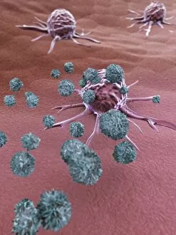
Background imageMalignant Collection: Cancer cells spreading, artwork

Cancer cells spreading, artwork
Cancer cells spreading. Computer artwork showing cancerous cells (blue) migrating from a tumour (upper centre) into the blood stream (centre)

Background imageMalignant Collection: Spinal cancer, MRI scan

Spinal cancer, MRI scan
Spinal cancer. Coloured magnetic resonance imaging (MRI) scan of a sagittal section through the cervical (neck) region of the spine of a 22-year-old patient

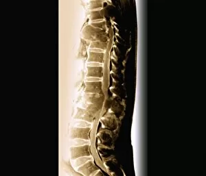
Background imageMalignant Collection: Secondary spinal cancer, MRI scan

Secondary spinal cancer, MRI scan
Secondary spinal cancer. Magnetic resonance imaging (MRI) scan of a section through the spine of a 48-year-old patient, showing malignant (cancerous) tumours

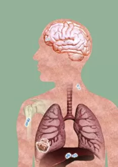
Background imageMalignant Collection: Metastatic cancer, artwork

Metastatic cancer, artwork
Metastatic cancer. Computer artwork of a section through a persons upper body and head, showing a primary cancer in the right liver (lower left) that has spread (metastasised)

Background imageMalignant Collection: Cancer blood supply, conceptual image C016 / 7505

Cancer blood supply, conceptual image C016 / 7505
Cancer blood supply, conceptual image. Computer artwork representing the blood supply to a cancerous (malignant) tumour

Background imageMalignant Collection: Total mesorectal excision, artwork C016 / 6996

Total mesorectal excision, artwork C016 / 6996
Total mesorectal excision (TME). Artwork of the pelvis and the area of the colon and rectum that is removed in total mesorectal excision

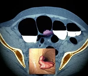
Background imageMalignant Collection: Colon cancer, barium contrast CT scan

Colon cancer, barium contrast CT scan
Colon cancer. Coloured barium contrast computed tomography (CT) scan, and 3D CT colonoscopy image (inset), of an axial section through the sigmoid colon (large intestine) of a 54-year-old patient

Background imageMalignant Collection: Colon cancer, 3D colonoscopy image

Colon cancer, 3D colonoscopy image
Colon cancer. 3D colonoscopy image showing the location of a cancerous growth (centre right) in the colon of a 59-year-old patient. Here, the growth has caused a narrowing (stenosis) of the colon

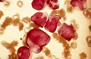
Background imageMalignant Collection: Acute myeloid leukaemia, micrograph

Acute myeloid leukaemia, micrograph
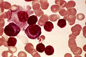
Acute myeloid leukaemia. Light micrograph of blood cells from bone marrow in a case of acute myeloid leukaemia. These cells include a monocytoid myeloblast stem cell

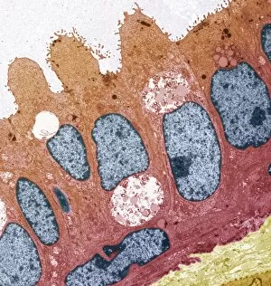
Background imageMalignant Collection: Ductal breast cancer, TEM

Ductal breast cancer, TEM
Ductal breast cancer. Transmission electron micrograph (TEM) of a section through breast tissue, showing malignant (cancerous) epithelial cells arising from the milk ducts of the breast

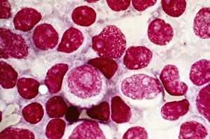
Background imageMalignant Collection: Blood cell cancer, light micrograph

Blood cell cancer, light micrograph
Blood cell cancer. Light micrograph of blood cells from a lymphatic ganglion in a case of a blood cancer of a mixed cell type

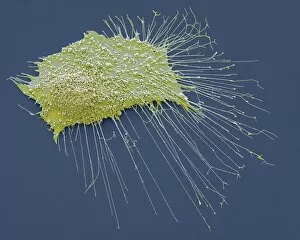
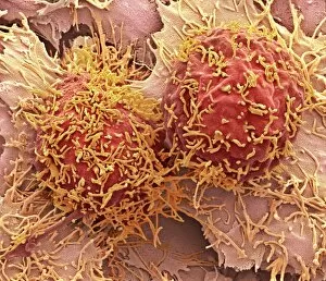
Background imageMalignant Collection: Liver cancer cells, SEM

Liver cancer cells, SEM
Liver cancer cells. Coloured scanning electron micrograph (SEM) of two hepatocellular carcinoma (HCC) cells, showing the numerous filopodia (hair-like) covering their surface

Background imageMalignant Collection: Lymphoblastic sarcoma, light micrograph

Lymphoblastic sarcoma, light micrograph
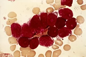
Lymphoblastic sarcoma. Light micrograph of blood cells from bone marrow in a case of lymphoblastic sarcoma. These small lymphoblasts (dark red) characteristically have conspicuous nucleoli

Background imageMalignant Collection: Multiple myeloma, light micrograph

Multiple myeloma, light micrograph
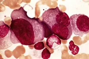
Multiple myeloma. Light micrograph of dystrophy (abnormality) of plasmocyte blood cells from bone marrow in a case of multiple myeloma, also known as Kahlers disease or plasma cell myeloma

Background imageMalignant Collection: Lymphoplasmacytic lymphoma, micrograph

Lymphoplasmacytic lymphoma, micrograph
Lymphoplasmacytic lymphoma. Light micrograph of a mixture of small lymphocytes and plasma cells from bone marrow in a case of lymphoplasmacytic lymphoma, also known as Waldenstroms macroglobulinemia

Background imageMalignant Collection: Hairy cell leukaemia, light micrograph

Hairy cell leukaemia, light micrograph
Hairy cell leukaemia. Light micrograph of blood cells in a case of hairy cell leukaemia. This sample, treated with May-Grunwald-Giemsa (MGG) stain

Background imageMalignant Collection: Brain tumour, MRI scans

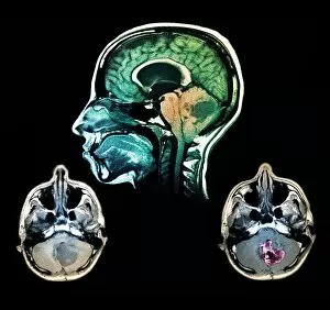
Brain tumour, MRI scans
Brain tumour. Coloured magnetic resonance imaging (MRI) scans of sections through the head of a 23-year-old patient with a lesion (dark patches)


For sale as Licensed Images

Choose your image, Select your licence and Download the media

"Malignant: Unleashing the Battle Within" In a microscopic world, T lymphocytes stand as valiant warriors against cancer cells, their relentless pursuit captured in SEM C001 / 1679. Meanwhile, HeLa cells reveal their sinister nature under the watchful eye of a light micrograph in C017 / 8299 and C017 / 8298. A mesmerizing scene unfolds as coloured SEM showcases lymphocytes fearlessly attacking cancer cells, painting a vivid picture of hope amidst adversity. Acute promyelocytic leukemia reveals its haunting presence through an ominous micrograph while ovarian cancer exposes its treacherous grip in light micrograph C015 / 7103. Witnessing the battle at a cellular level is awe-inspiring; a coloured SEM image captures the moment when leukaemic white blood cells are confronted head-on by nanorobots determined to eradicate this malignant threat (M132 / 0488). The fight against cancer takes on otherworldly dimensions reminiscent of Amazing Stories sci-fi magazine covers featuring giant plants and fantastical creatures. Yet not all battles are fought within our bodies; sometimes they transcend into realms beyond. Like an exorcism performed by a cleric casting spells against evil spirits, we confront malignancy with unwavering determination. Just as history has witnessed struggles such as "The Condition of Ireland under the 'No Rent' Policy, " or "Johnnys Defeat at the Dock, " we too face adversity head-on. Amidst these hints lie tales of resilience and courage that remind us to never surrender to darkness. Malignant forces may try to overpower us, but united we stand ready to conquer them - be it through scientific advancements or sheer willpower. Together, let us rewrite the narrative and triumph over malignancy's grasp for brighter tomorrows await those who dare to fight back.