Malignant Collection (page 7)

"Malignant: Unleashing the Battle Within" In a microscopic world, T lymphocytes stand as valiant warriors against cancer cells

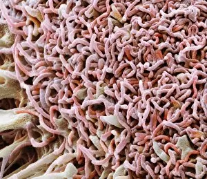
Background imageMalignant Collection: Colorectal cancer cell, SEM

Colorectal cancer cell, SEM
Colorectal cancer cell. Coloured scanning electron micrograph (SEM) of a cancer cell from the human colon (large intestine). Cancer of the colon is also known as colorectal cancer

Background imageMalignant Collection: Acute erythroid leukaemia, micrograph

Acute erythroid leukaemia, micrograph
Acute erythroid leukaemia. Light micrograph of blood cells from bone marrow in a case of acute erythroid leukaemia. The cells include dystrophic (degenerated) erythroblasts (nuclei stained dark red)

Background imageMalignant Collection: Acute lymphoblastic leukaemia, micrograph

Acute lymphoblastic leukaemia, micrograph
Acute lymphoblastic leukaemia. Light micrograph of blood cells from bone marrow in a case of acute lymphoblastic leukaemia. Leukaemia is a cancer where certain blood cells form in excess

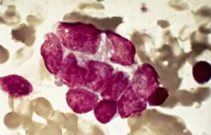
Background imageMalignant Collection: Metastatic breast cancer, micrograph

Metastatic breast cancer, micrograph
Metastatic breast cancer. Light micrograph of blood cells in a case of metastatic (secondary) medullary breast cancer. Medullary breast cancer is a rare form of breast cancer that is diagnosed by

Background imageMalignant Collection: Metastatic lung cancer, micrograph

Metastatic lung cancer, micrograph
Metastatic lung cancer. Light micrograph of blood cells in a case of metastatic (secondary) medullary lung cancer. Medullary cancer (carcinoma)

Background imageMalignant Collection: Secondary lung cancer, X-ray

Secondary lung cancer, X-ray
Secondary lung cancers. Coloured X-ray of malignant (cancerous) tumours (red patches) in the lungs of a 60-year-old female patient

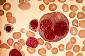
Background imageMalignant Collection: Acute leukaemia, light micrograph

Acute leukaemia, light micrograph
Acute leukaemia. Light micrograph of blood cells from bone marrow in a case of acute leukaemia. These cells include a polynuclear degranulated basophil and myeloblasts

Background imageMalignant Collection: Colon cancer, CT scans

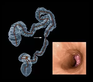
Colon cancer, CT scans
Colon cancer. 2D computed tomography (CT) rendering (left) and 3D colonoscopy image (right) showing the location of a cancerous growth in the colon of a 59-year-old patient

Background imageMalignant Collection: Leukaemia cell, SEM

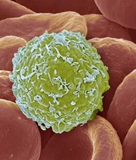
Leukaemia cell, SEM
Leukaemia cell. Coloured scanning electron micrograph (SEM) of a leukaemic (cancerous) lymphocyte white blood cell (green), amongst normal red blood cells (erythrocytes, red)

Background imageMalignant Collection: Chronic myeloid leukaemia, micrograph

Chronic myeloid leukaemia, micrograph
Chronic myeloid leukaemia. Light micrograph of blood cells from bone marrow in a case of chronic myeloid leukaemia. Leukaemia is a cancer where certain blood cells form in excess

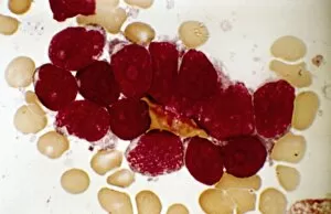
Background imageMalignant Collection: Reticulosarcoma, light micrograph

Reticulosarcoma, light micrograph
Reticulosarcoma. Light micrograph of reticulocyte blood cells (red, one at upper left) from a lymphatic ganglion in a case of reticulosarcoma

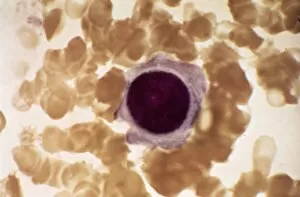
Background imageMalignant Collection: Hodgkins disease, light micrograph

Hodgkins disease, light micrograph
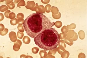
Hodgkins disease. Light micrograph of two non-dystrophic histiocyte blood cells (centre) in a case of Hodgkins disease, also called Hodgkins lymphoma

Background imageMalignant Collection: Metastatic cancer, artwork C016 / 6258

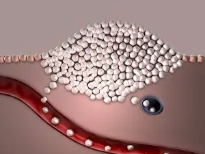
Metastatic cancer, artwork C016 / 6258
Metastatic cancer. Computer artwork showing malignant cancer cells (white) migrating into the blood stream (red) and lymphatic system (blue)

Background imageMalignant Collection: Oncogenes, artwork C016 / 6262

Oncogenes, artwork C016 / 6262
Oncogenes. Computer artwork comparing the DNA (deoxyribonucleic acid, coiled strand) of a normal cell (top) with that of a cancer cell (bottom)

Background imageMalignant Collection: Normal and leukaemic blood, artwork C016 / 6261

Normal and leukaemic blood, artwork C016 / 6261
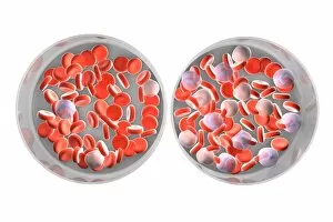
Normal and leukaemic blood. Computer artwork comparing the appearance of normal blood (left) and that affected by leukaemia (right)

Background imageMalignant Collection: Stages of cancer, artwork C016 / 6257

Stages of cancer, artwork C016 / 6257
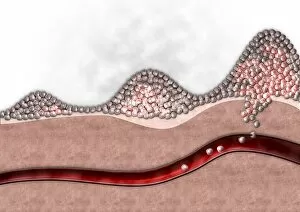
Stages of cancer. Computer artwork showing three stages of cancer (whiteish cells). From left to right: benign (non-cancerous)

Background imageMalignant Collection: Reticulosarcoma, light micrograph C015 / 7133

Reticulosarcoma, light micrograph C015 / 7133
Reticulosarcoma. Light micrograph of reticulocyte blood cells (red, one at upper left) from a lymphatic ganglion in a case of reticulosarcoma

Background imageMalignant Collection: Bowel cancer, light micrograph C016 / 0511

Bowel cancer, light micrograph C016 / 0511
Bowel cancer. Light micrograph of a section through a carcinoid tumour in the small bowel. The tumour has invaded the submucosa of the bowel

Background imageMalignant Collection: Thymus gland cancer, light micrograph C015 / 7128

Thymus gland cancer, light micrograph C015 / 7128
Thymus cancer. Light micrograph of a section through a thymus gland with thymoma cancer. The thymus is a gland in the upper chest that plays an important role in the immune system

Background imageMalignant Collection: Rectal cancer, MRI scans C018 / 0508

Rectal cancer, MRI scans C018 / 0508
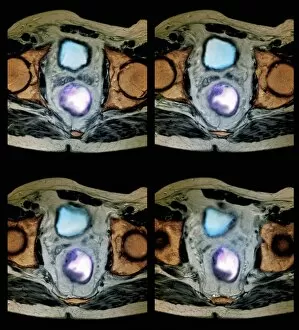
Rectal cancer. Coloured magnetic resonance imaging (MRI) scans of the pelvis of a 65 year old patient with a malignant (cancerous) tumour (purple) in the rectum, the last part of the large intestine

Background imageMalignant Collection: Rectal cancer, MRI scans C018 / 0510

Rectal cancer, MRI scans C018 / 0510
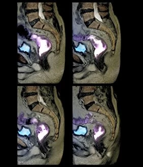
Rectal cancer. Coloured magnetic resonance imaging (MRI) scans of the pelvis of a 65 year old patient with a malignant (cancerous) tumour in the rectum, the last part of the large intestine

Background imageMalignant Collection: Secondary bone cancer, X-ray C018 / 0594

Secondary bone cancer, X-ray C018 / 0594
Secondary bone cancer. X-ray of the shoulders of a 39 year old patient with metastatic (secondary) bone cancer. This cancer has spread from a primary thyroid cancer

Background imageMalignant Collection: Secondary bone cancer, X-ray C018 / 0531

Secondary bone cancer, X-ray C018 / 0531
Secondary bone cancer. X-ray of the knees of a 68 year old patient with metastatic (secondary) bone cancer of the left patella (knee cap). This cancer has spread from a primary lung cancer

Background imageMalignant Collection: Liver cancer, CT scan C018 / 0461

Liver cancer, CT scan C018 / 0461
Liver cancer. Coloured computed tomography (CT) scan of the abdomen of a 50 year old alcoholic patient with liver cancer. The liver (orange) is enlarged due to the presence of the tumour

Background imageMalignant Collection: Rectal cancer, MRI scans C018 / 0511

Rectal cancer, MRI scans C018 / 0511
Rectal cancer. Coloured magnetic resonance imaging (MRI) scans of the pelvis of a 65 year old patient with a malignant (cancerous) tumour (purple) in the rectum, the last part of the large intestine

Background imageMalignant Collection: Secondary bone cancer, X-ray C018 / 0595

Secondary bone cancer, X-ray C018 / 0595
Secondary bone cancer. X-ray of the left shoulder of a 39 year old patient with metastatic (secondary) bone cancer. This cancer has spread from a primary thyroid cancer

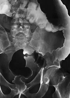
Background imageMalignant Collection: Colon cancer, barium X-ray C018 / 0574

Colon cancer, barium X-ray C018 / 0574
Colon cancer. Coloured X-ray of the large intestine of a 62 year old patient with cancer of the sigmoid colon. The patient was given a barium enema prior to the X-ray to show up any abnormalities

Background imageMalignant Collection: Liver cancer, CT scan C018 / 0462

Liver cancer, CT scan C018 / 0462
Liver cancer. Computed tomography (CT) scan of the abdomen of a 50 year old alcoholic patient with liver cancer. The liver (left to upper centre) is enlarged due to the presence of the tumour

Background imageMalignant Collection: Animal models for cancer research C018 / 0748

Animal models for cancer research C018 / 0748
Animal models for cancer research, conceptual illustration. Fruit fly (Drosophila melanogaster) with malignant (cancerous) tumours in one of its eyes

Background imageMalignant Collection: Liver cancer, CT scan C018 / 0460

Liver cancer, CT scan C018 / 0460
Liver cancer. Coloured computed tomography (CT) scan of the abdomen of a 50 year old alcoholic patient with liver cancer. The liver (orange) is enlarged due to the presence of the tumour

Background imageMalignant Collection: Rectal cancer, MRI scan C018 / 0512

Rectal cancer, MRI scan C018 / 0512
Rectal cancer. Coloured magnetic resonance imaging (MRI) scan of the pelvis of a 65 year old patient with a malignant (cancerous) tumour in the rectum, the last part of the large intestine

Background imageMalignant Collection: Secondary bone cancer, X-ray C018 / 0596

Secondary bone cancer, X-ray C018 / 0596
Secondary bone cancer. Coloured X-ray of the left shoulder of a 39 year old patient with metastatic (secondary) bone cancer. This cancer has spread from a primary thyroid cancer

Background imageMalignant Collection: Secondary bone cancer, X-ray C017 / 7161

Secondary bone cancer, X-ray C017 / 7161
Secondary bone cancer. X-ray of the abdomen of a 40 year old female with metastatic (secondary) bone cancer (bright white areas). The cancer has metastasised (spread) from a primary cancer

Background imageMalignant Collection: Glioblastoma brain tumour, DTI MRI scan C017 / 7052

Glioblastoma brain tumour, DTI MRI scan C017 / 7052
Glioblastoma brain tumour. 3D diffusion tensor imaging (DTI) magnetic resonance imaging (MRI) scan of nerve pathways (coloured) around a glioblastoma (upper right) in the brain

Background imageMalignant Collection: Mouth cancer cell, SEM C014 / 1460

Mouth cancer cell, SEM C014 / 1460
Mouth cancer cell. Coloured scanning electron micrograph (SEM) of a squamous cell carcinoma (cancer) cell from a human mouth

Background imageMalignant Collection: HeLa cells, SEM C014 / 0366

HeLa cells, SEM C014 / 0366
HeLa cells. Coloured scanning electron micrograph (SEM) of HeLa cells (round) grown in a 3D matrix (background). HeLa cells are a continuously cultured cell line of immortal human cancer cells

Background imageMalignant Collection: Mouth cancer cell, SEM C014 / 1459

Mouth cancer cell, SEM C014 / 1459
Mouth cancer cell. Coloured scanning electron micrograph (SEM) of a squamous cell carcinoma (cancer) cell from a human mouth

Background imageMalignant Collection: Dividing cancer cell, SEM C014 / 0362

Dividing cancer cell, SEM C014 / 0362
Dividing cancer cell. Coloured scanning electron micrograph (SEM) of a colorectal cancer cell undergoing mitosis (nuclear division) and splitting into two daughter cells (left and right)

Background imageMalignant Collection: HeLa cells, SEM C014 / 0369

HeLa cells, SEM C014 / 0369
HeLa cells. Coloured scanning electron micrograph (SEM) of HeLa cells (round) grown in a 3D matrix (background). HeLa cells are a continuously cultured cell line of immortal human cancer cells

Background imageMalignant Collection: Mouth cancer cells, SEM

Mouth cancer cells, SEM
Mouth cancer cells. Coloured scanning electron micrograph (SEM) of squamous cell carcinoma (cancer) cells from a human mouth

Background imageMalignant Collection: Mouth cancer cell dividing, SEM

Mouth cancer cell dividing, SEM
Mouth cancer dividing. Coloured scanning electron micrograph (SEM) of a squamous cell carcinoma (cancer) cell from a human mouth undergoing mitosis (nuclear division)

Background imageMalignant Collection: Mouth cancer cell, SEM

Mouth cancer cell, SEM
Mouth cancer cell. Coloured scanning electron micrograph (SEM) showing the surface of a squamous cell carcinoma (cancer) cell from a human mouth

Background imageMalignant Collection: Liver cancer cell, SEM

Liver cancer cell, SEM
Liver cancer cell. Coloured scanning electron micrograph (SEM) of a hepatocellular carcinoma (HCC) cell, showing the numerous filopodia (hair-like) covering its surface


For sale as Licensed Images

Choose your image, Select your licence and Download the media

"Malignant: Unleashing the Battle Within" In a microscopic world, T lymphocytes stand as valiant warriors against cancer cells, their relentless pursuit captured in SEM C001 / 1679. Meanwhile, HeLa cells reveal their sinister nature under the watchful eye of a light micrograph in C017 / 8299 and C017 / 8298. A mesmerizing scene unfolds as coloured SEM showcases lymphocytes fearlessly attacking cancer cells, painting a vivid picture of hope amidst adversity. Acute promyelocytic leukemia reveals its haunting presence through an ominous micrograph while ovarian cancer exposes its treacherous grip in light micrograph C015 / 7103. Witnessing the battle at a cellular level is awe-inspiring; a coloured SEM image captures the moment when leukaemic white blood cells are confronted head-on by nanorobots determined to eradicate this malignant threat (M132 / 0488). The fight against cancer takes on otherworldly dimensions reminiscent of Amazing Stories sci-fi magazine covers featuring giant plants and fantastical creatures. Yet not all battles are fought within our bodies; sometimes they transcend into realms beyond. Like an exorcism performed by a cleric casting spells against evil spirits, we confront malignancy with unwavering determination. Just as history has witnessed struggles such as "The Condition of Ireland under the 'No Rent' Policy, " or "Johnnys Defeat at the Dock, " we too face adversity head-on. Amidst these hints lie tales of resilience and courage that remind us to never surrender to darkness. Malignant forces may try to overpower us, but united we stand ready to conquer them - be it through scientific advancements or sheer willpower. Together, let us rewrite the narrative and triumph over malignancy's grasp for brighter tomorrows await those who dare to fight back.