Mitochondria Collection (#3)

Mitochondria, the powerhouse of the cell, play a vital role in various organisms

Background imageMitochondria Collection: TFAM transcription factor bound to DNA C015 / 7060

TFAM transcription factor bound to DNA C015 / 7060
TFAM transcription factor bound to DNA, molecular model. Human mitochondrial transcription factor A (TFAM, pink) bound to a strand of DNA (deoxyribonucleic acid, orange and green)

Background imageMitochondria Collection: Animal cell organelles, artwork C015 / 6793

Animal cell organelles, artwork C015 / 6793
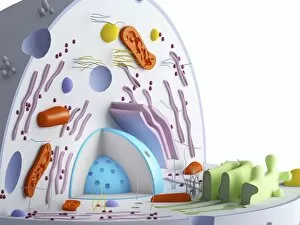
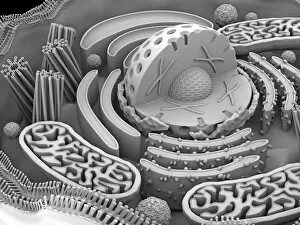
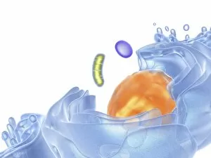
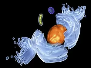
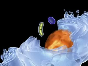
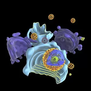
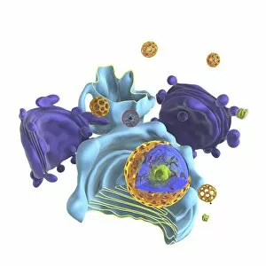
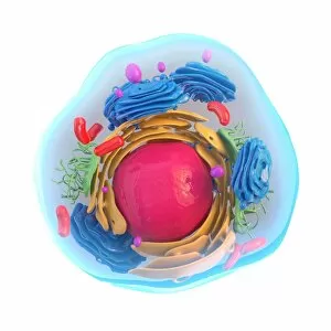
Animal cell organelles. Computer artwork showing the organelles in a eukaryotic cell. This is an animal cell. Structures include the nucleus (centre) which has a membrane with nuclear pores

Background imageMitochondria Collection: Animal cell organelles, artwork C015 / 6792

Animal cell organelles, artwork C015 / 6792
Animal cell organelles. Computer artwork showing the organelles in a eukaryotic cell. This is an animal cell. Structures include the nucleus (centre) which has a membrane with nuclear pores

Background imageMitochondria Collection: Mitochondrial structure, artwork C015 / 6784

Mitochondrial structure, artwork C015 / 6784
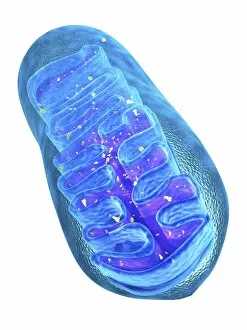
Mitochondrial structure. Computer artwork of a mitochondrion, showing a loop of mitochondrial DNA (deoxyribonucleic acid, mtDNA), the organelles genetic material

Background imageMitochondria Collection: Animal cell organelles, artwork C015 / 6775

Animal cell organelles, artwork C015 / 6775
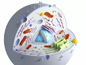
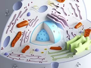
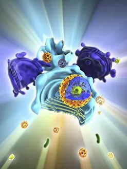
Animal cell organelles. Computer artwork showing the organelles in a eukaryotic cell. This is an animal cell. Structures include the nucleus (centre) which has a membrane with nuclear pores

Background imageMitochondria Collection: Animal cell organelles, artwork C015 / 6778

Animal cell organelles, artwork C015 / 6778
Animal cell organelles. Computer artwork showing the organelles in a eukaryotic cell. This is an animal cell. Structures include the nucleus (centre) which has a membrane with nuclear pores

Background imageMitochondria Collection: Animal cell organelles, artwork C015 / 6777

Animal cell organelles, artwork C015 / 6777
Animal cell organelles. Computer artwork showing the organelles in a eukaryotic cell. This is an animal cell. Structures include the nucleus (centre) which has a membrane with nuclear pores

Background imageMitochondria Collection: Mitochondrial structure, artwork C015 / 6765

Mitochondrial structure, artwork C015 / 6765
Mitochondrial structure. Computer artwork of a mitochondrion, showing a loop of mitochondrial DNA (deoxyribonucleic acid, mtDNA), the organelles genetic material

Background imageMitochondria Collection: Animal cell organelles, artwork C016 / 0619

Animal cell organelles, artwork C016 / 0619
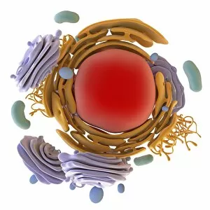
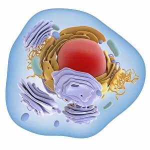
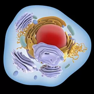
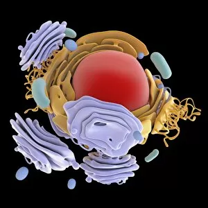
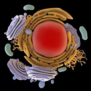
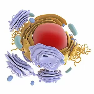
Computer artwork showing the organelles in a eukaryotic cell. This is an animal cell. Structures include the nucleus (centre, red) which has a membrane with nuclear pores

Background imageMitochondria Collection: Animal cell organelles, artwork C016 / 0621

Animal cell organelles, artwork C016 / 0621
Computer artwork showing the organelles in a eukaryotic cell. This is an animal cell. Structures include the nucleus (centre, red) which has a membrane with nuclear pores

Background imageMitochondria Collection: Animal cell organelles, artwork C016 / 0620

Animal cell organelles, artwork C016 / 0620
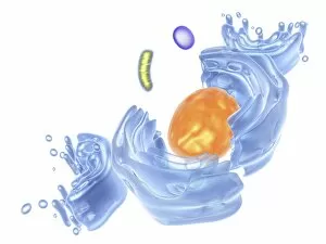
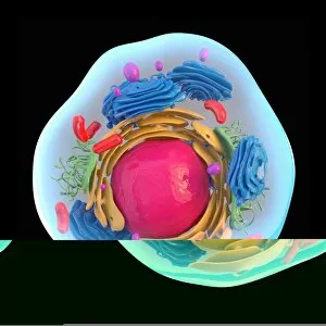
Computer artwork showing the organelles in a eukaryotic cell. This is an animal cell. Structures include the nucleus (centre, orange) which has a membrane with nuclear pores

Background imageMitochondria Collection: Animal cell organelles, artwork C016 / 0617

Animal cell organelles, artwork C016 / 0617
Computer artwork showing the organelles in a eukaryotic cell. This is an animal cell. Structures include the nucleus (centre, red) which has a membrane with nuclear pores

Background imageMitochondria Collection: Animal cell organelles, artwork C016 / 0618

Animal cell organelles, artwork C016 / 0618
Computer artwork showing the organelles in a eukaryotic cell. This is an animal cell. Structures include the nucleus (centre, orange) which has a membrane with nuclear pores

Background imageMitochondria Collection: Animal cell organelles, artwork C016 / 0615

Animal cell organelles, artwork C016 / 0615
Computer artwork showing the organelles in a eukaryotic cell. This is an animal cell. Structures include the nucleus (centre, orange) which has a membrane with nuclear pores

Background imageMitochondria Collection: Animal cell organelles, artwork C016 / 0616

Animal cell organelles, artwork C016 / 0616
Computer artwork showing the organelles in a eukaryotic cell. This is an animal cell. Structures include the nucleus (centre, orange) which has a membrane with nuclear pores

Background imageMitochondria Collection: Animal cell organelles, artwork C016 / 0611

Animal cell organelles, artwork C016 / 0611
Computer artwork showing the organelles in a eukaryotic cell. This is an animal cell. Structures include the nucleus (centre, red) which has a membrane with nuclear pores

Background imageMitochondria Collection: Animal cell organelles, artwork C016 / 0612

Animal cell organelles, artwork C016 / 0612
Computer artwork showing the organelles in a eukaryotic cell. This is an animal cell. Structures include the nucleus (centre, orange) which has a membrane with nuclear pores

Background imageMitochondria Collection: Animal cell organelles, artwork C016 / 0605

Animal cell organelles, artwork C016 / 0605
Computer artwork showing the organelles in a eukaryotic cell. This is an animal cell. Structures include the nucleus (centre, red) which has a membrane with nuclear pores

Background imageMitochondria Collection: Animal cell organelles, artwork C016 / 0610

Animal cell organelles, artwork C016 / 0610
Computer artwork showing the organelles in a eukaryotic cell. This is an animal cell. Structures include the nucleus (centre, orange) which has a membrane with nuclear pores

Background imageMitochondria Collection: Animal cell organelles, artwork C016 / 0604

Animal cell organelles, artwork C016 / 0604
Computer artwork showing the organelles in a eukaryotic cell. This is an animal cell. Structures include the nucleus (centre, red) which has a membrane with nuclear pores

Background imageMitochondria Collection: Animal cell organelles, artwork C016 / 0606

Animal cell organelles, artwork C016 / 0606
Computer artwork showing the organelles in a eukaryotic cell. This is an animal cell. Structures include the nucleus (centre, orange) which has a membrane with nuclear pores

Background imageMitochondria Collection: Fibroblast cell, fluorescent micrograph

Fibroblast cell, fluorescent micrograph
Fibroblast cell. Confocal laser scanning micrograph of a fibroblast cell. The cell nucleus, which contains the cells genetic information, is green

Background imageMitochondria Collection: Mitochondrial inclusions, TEM C016 / 5807

Mitochondrial inclusions, TEM C016 / 5807
Mitochondrial inclusions. Transmission electron micrograph (TEM) of a section through a mitochondrion showing crystalline inclusions (thick parallel rods) in the intermembrane spaces

Background imageMitochondria Collection: Mitochondrial inclusions, TEM C016 / 5806

Mitochondrial inclusions, TEM C016 / 5806
Mitochondrial inclusions. Transmission electron micrograph (TEM) of a section through a mitochondrion showing crystalline inclusions (thick parallel rods) in the intermembrane spaces

Background imageMitochondria Collection: NADH dehydrogenase molecule, artwork C014 / 0009

NADH dehydrogenase molecule, artwork C014 / 0009
NADH dehydrogenase molecule. Computer artwork showing the structure of a molecule of NADH dehydrogenase (NADH:ubiquinone reductase or Complex I)

Background imageMitochondria Collection: Animal cell, artwork C013 / 9985

Animal cell, artwork C013 / 9985
Animal cell. Computer artwork showing the cell organelles found inside a typical animal cell. The nucleus (large round) can be seen at centre

Background imageMitochondria Collection: Animal cell, artwork C013 / 9984

Animal cell, artwork C013 / 9984
Animal cell. Computer artwork showing the cell organelles found inside a typical animal cell. The nucleus (large round) can be seen at centre

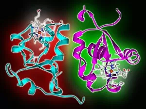
Background imageMitochondria Collection: Cytochrome b5 molecules C013 / 8874

Cytochrome b5 molecules C013 / 8874
Cytochrome b5 molecules. Computer artwork showing the secondary structure of two molecules of cytochrome B5. This protein plays a crucial role in metabolism in animals (including humans), fungi

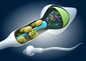
Background imageMitochondria Collection: Sperm cell anatomy, artwork C013 / 4648

Sperm cell anatomy, artwork C013 / 4648
Sperm cell anatomy. Cutaway computer artwork showing the internal structure of a sperm cell (spermatozoon), the male sex cell

Background imageMitochondria Collection: Mitochondria, artwork C013 / 4993

Mitochondria, artwork C013 / 4993
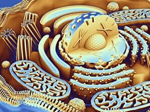
Mitochondria, computer artwork. Mitochondria are a type of organelle found in the cytoplasm of eukaryotic cells. They oxidise sugars and fats to produce energy in a process called respiration

Background imageMitochondria Collection: Mitochondria, artwork C013 / 4991

Mitochondria, artwork C013 / 4991
Mitochondria, computer artwork. Mitochondria are a type of organelle found in the cytoplasm of eukaryotic cells. They oxidise sugars and fats to produce energy in a process called respiration

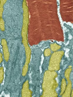
Background imageMitochondria Collection: Retinal rod cell, TEM C013 / 4805

Retinal rod cell, TEM C013 / 4805
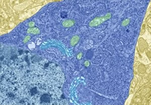
Retinal rod cell. Transmission electron micrograph (TEM) of a section through a rod cell from the retina of an eye, showing the inner segment (bottom) filled with mitochondria (green)

Background imageMitochondria Collection: Retinal rod cell, TEM C013 / 4804

Retinal rod cell, TEM C013 / 4804
Retinal rod cell. Transmission electron micrograph (TEM) of a section through a rod cell from the retina of an eye, showing the inner segment (bottom) filled with mitochondria (green)

Background imageMitochondria Collection: Brain cells, TEM C013 / 4801

Brain cells, TEM C013 / 4801
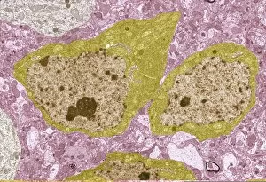
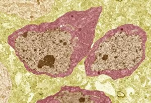
Brain cells. Transmission electron micrograph (TEM) of a section through oligodendrocytes (dark) in human brain tissue, showing free ribosomes (dark green dots), golgi apparatus (curved brown lines)

Background imageMitochondria Collection: Brain cells, TEM C013 / 4800

Brain cells, TEM C013 / 4800
Brain cells. Transmission electron micrograph (TEM) of a section through oligodendrocytes in human brain tissue, showing free ribosomes (dark pink dots), golgi apparatus (curved brown lines)

Background imageMitochondria Collection: Nerve cell, TEM C013 / 4797

Nerve cell, TEM C013 / 4797
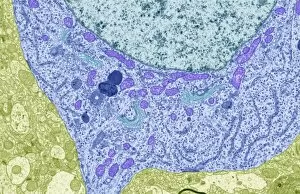
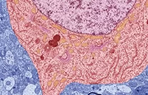
Nerve cell. Transmission electron micrograph (TEM) of a section through a neuron (nerve cell), showing characteristic Nissl body (dark blue lines), numerous golgi apparatus (curved green lines)

Background imageMitochondria Collection: Brain cell, TEM C013 / 4799

Brain cell, TEM C013 / 4799
Brain cell. Transmission electron micrograph (TEM) of a section through an oligodendrocyte in human brain tissue, showing free ribosomes (dark brown dots), golgi apparatus (curved orange lines)

Background imageMitochondria Collection: Nerve cell, TEM C013 / 4796

Nerve cell, TEM C013 / 4796
Nerve cell. Transmission electron micrograph (TEM) of a section through a neuron (nerve cell), showing characteristic Nissl body (dark red lines), numerous golgi apparatus (curved pink lines)

Background imageMitochondria Collection: Brain cell, TEM C013 / 4798

Brain cell, TEM C013 / 4798
Brain cell. Transmission electron micrograph (TEM) of a section through an oligodendrocyte in human brain tissue, showing free ribosomes (dark blue dots), golgi apparatus (curved light blue lines)

Background imageMitochondria Collection: Sperm in the testis, TEM

Sperm in the testis, TEM
Sperm cells. Coloured transmission electron micrograph (TEM) of a section through the testis, showing sperm cells. The heads of the sperm, which contain the genetic material, are red

Background imageMitochondria Collection: Sperm tails, TEM

Sperm tails, TEM
Sperm tails. Coloured transmission electron micrograph (TEM) of cross sections through sperm tails. A sperms tails ability to move is partly due to the arrangement of microtubules

Background imageMitochondria Collection: Sperm cell, computer artwork

Sperm cell, computer artwork
Sperm cell. Computer artwork of a human sperm cell, showing its inner structure. The sperm head (orange) carries genetic material

Background imageMitochondria Collection: Pancreatic cells, TEM

Pancreatic cells, TEM
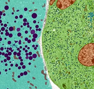
Pancreatic cells. Coloured transmission electron micrograph (TEM) of an acinar (exocrine) pancreatic cell (blue) adjacent to an hormone- secreting (endocrine) Islet of Langerhans cell (green)

Background imageMitochondria Collection: Pancreatic cell, TEM

Pancreatic cell, TEM
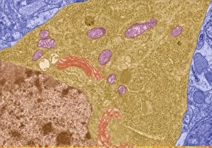
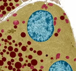
Pancreatic cell. Coloured transmission electron micrograph (TEM) of part of an acinar (exocrine) pancreatic cell. Mitochondria (red) are seen in the cells endoplasmic reticulum (yellow)

Background imageMitochondria Collection: Alveolar cell, TEM

Alveolar cell, TEM
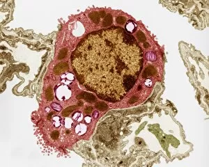
Alveolar cell. Coloured transmission electron micrograph (TEM) of a section through a type II alveolar cell. This cell is found in the alveoli (air sacs) of the lungs

Background imageMitochondria Collection: Illustration of structure of human hepatocyte

Illustration of structure of human hepatocyte
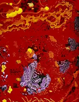
Artwork illustrating the compact internal structure of a human hepatocyte, a liver cell. The cytoplasm of a hepatocyte is crowded with organelles; the density relects its numerous metabolic

Background imageMitochondria Collection: Insulin-secreting pancreatic cells

Insulin-secreting pancreatic cells
Insulin production. Artwork of a section through beta cells secreting insulin (blue spheres) into a capillary (small blood vessel, grey tube). Beta cells are found in the pancreas

Background imageMitochondria Collection: Artwork of a nerve synapse

Artwork of a nerve synapse
Synapse. Illustration of a synapse, the junction between two nerve cells. The green structures in the terminal swelling (bouton) of the pre-synaptic cell are synaptic vesicles


For sale as Licensed Images

Choose your image, Select your licence and Download the media

Mitochondria, the powerhouse of the cell, play a vital role in various organisms. From budding yeast cells to ovarian cells, their presence is visually striking under scanning electron microscopy (SEM). The colored SEM image in ovarian cells showcases their intricate structure and diversity across different cell types. In nerve cells, transmission electron microscopy (TEM) reveals the fascinating architecture of mitochondria. These elongated organelles are strategically positioned along axons and dendrites, ensuring efficient energy supply for neuronal functions. The TEM images capture the complexity and importance within nerve cells. Moving beyond nerve cells, plasma cells also exhibit distinct mitochondrial features when observed through TEM. These specialized immune system components rely on robust energy production to carry out antibody synthesis efficiently. Their unique morphology highlights how mitochondria adapt to meet specific cellular demands. The eye muscle's TEM image further emphasizes the significance in highly active tissues. With an abundance of these organelles, eye muscles can sustain continuous contractions required for precise vision movements. Macrophage cells also showcase intriguing mitochondrial structures when examined using TEM techniques. As key players in our immune defense system, macrophages require substantial energy reserves to engulf pathogens effectively. Mitochondrial dynamics contribute significantly to this process by providing ample ATP generation. Under SEM imaging techniques specifically focused on mitochondria themselves, their distinctive shape becomes apparent—ranging from elongated tubules to spherical forms that vary across species and tissue types. Understanding animal cell structure necessitates studying not only individual organelles but also their interactions with other cellular components like DNA-binding proteins such as TFAM transcription factor bound to DNA shown in C015 / 7059 micrograph. Exploring various microscopic views allows us to appreciate the remarkable diversity and essential roles played by mitochondria across different cell types and organisms alike—a testament to their fundamental significance within biological systems.