
Mitochondrion Collection
The mitochondrion, often referred to as the powerhouse of the cell, is a fascinating and vital organelle found in various cell types


For sale as Licensed Images
Choose your image, Select your licence and Download the media
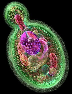
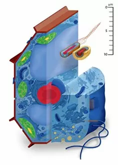
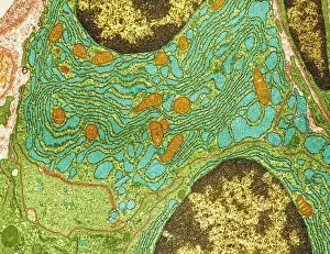
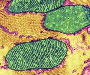
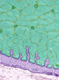
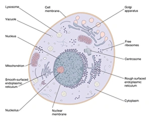
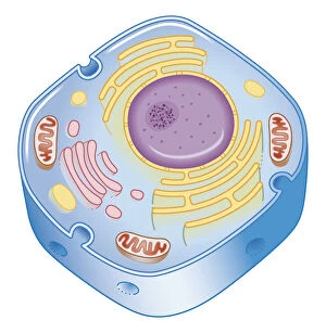
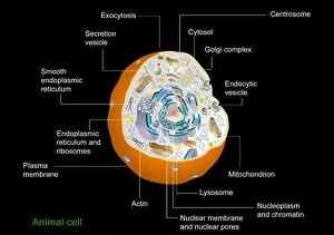
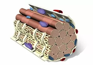
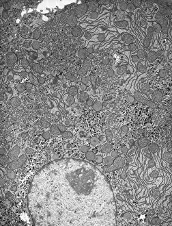
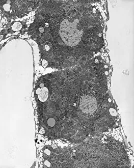
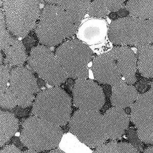
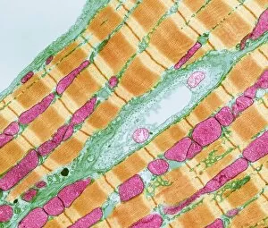
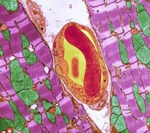
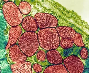
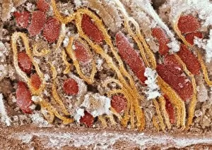
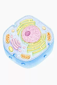
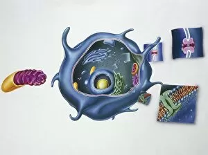
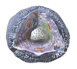
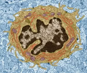
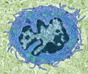
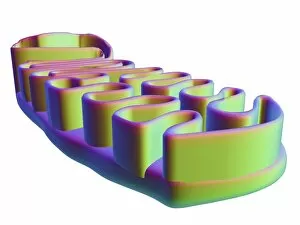
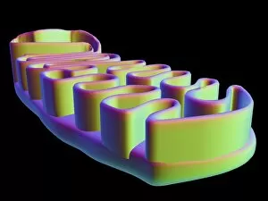
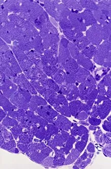
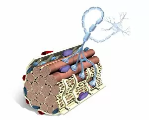
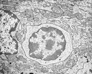
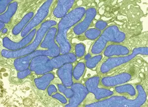
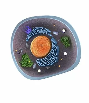
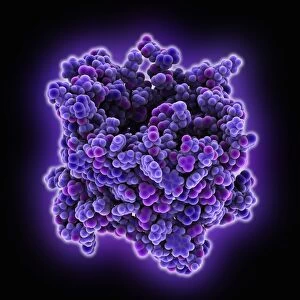
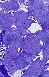
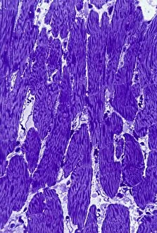
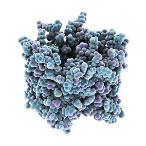
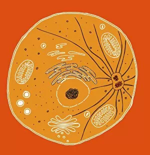
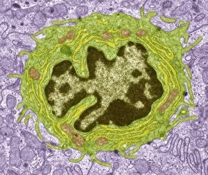
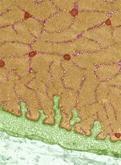
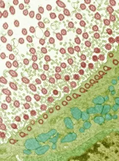
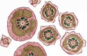
The mitochondrion, often referred to as the powerhouse of the cell, is a fascinating and vital organelle found in various cell types. In budding yeast cells, it plays a crucial role in energy production through cellular respiration. This tiny structure can be visualized using transmission electron microscopy (TEM), revealing its intricate details. Not limited to yeast cells, mitochondria are present in different cell types throughout our bodies. TEM images showcase their distinct shapes and sizes within plasma cells, eye muscles, macrophage cells, and hepatocyte liver cells. These microscopic snapshots allow scientists to study their functions and understand how they contribute to overall cellular health. Scanning electron microscopy (SEM) provides another perspective on mitochondria's appearance within animal cell structures. These high-resolution images capture the surface of these organelles with stunning detail. Illustrations further enhance our understanding of mitochondria's complex composition. They depict an animal cell nucleus containing cytoplasm filled with numerous mitochondria units intertwined with DNA strands and chromosomes. Such illustrations also highlight TFAM transcription factors bound to DNA molecules—an essential process for regulating mitochondrial gene expression. Artwork showcasing muscle fiber structure emphasizes the significance of mitochondria in muscle function. Their abundance within muscle fibers ensures efficient energy supply during physical activity.