Molecular Model Collection (page 4)

Molecular models offer a glimpse into the intricate world of science and medicine, revealing the hidden secrets of life at a microscopic level

Background imageMolecular Model Collection: DNA molecule, artwork F006 / 3711

DNA molecule, artwork F006 / 3711
DNA molecule, computer artwork

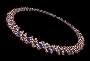
Background imageMolecular Model Collection: Circular DNA molecule, artwork F006 / 7083

Circular DNA molecule, artwork F006 / 7083
Circular DNA (deoxyribonucleic acid) molecule, computer artwork. Circular DNA has no ends, but consists of a ring structure

Background imageMolecular Model Collection: Circular DNA molecule, space artwork F006 / 7077

Circular DNA molecule, space artwork F006 / 7077
Circular DNA (deoxyribonucleic acid) molecule, computer artwork and space nebula artwork, depicting origin of life

Background imageMolecular Model Collection: Circular DNA molecule, artwork F006 / 7084

Circular DNA molecule, artwork F006 / 7084
Circular DNA (deoxyribonucleic acid) molecule, computer artwork. Circular DNA has no ends, but consists of a ring structure

Background imageMolecular Model Collection: Circular DNA molecule, space artwork F006 / 7087

Circular DNA molecule, space artwork F006 / 7087
Circular DNA (deoxyribonucleic acid) molecule, computer artwork and space nebula artwork, depicting origin of life

Background imageMolecular Model Collection: DNA molecule, artwork F006 / 7127

DNA molecule, artwork F006 / 7127
DNA (deoxyribonucleic acid) molecule, computer artwork

Background imageMolecular Model Collection: DNA molecule F007 / 6423

DNA molecule F007 / 6423
DNA (deoxyribonucleic acid) molecule

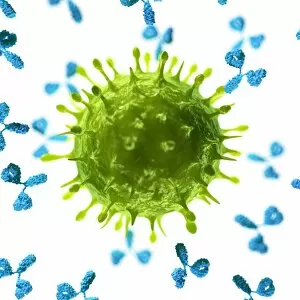
Background imageMolecular Model Collection: Antibodies attacking a virus, artwork F007 / 6623

Antibodies attacking a virus, artwork F007 / 6623
Antibodies attacking a virus, computer artwork

Background imageMolecular Model Collection: Antibodies attacking a virus, artwork F007 / 6624

Antibodies attacking a virus, artwork F007 / 6624
Antibodies attacking a virus, computer artwork

Background imageMolecular Model Collection: Antibodies attacking a virus, artwork F007 / 6622

Antibodies attacking a virus, artwork F007 / 6622
Antibodies attacking a virus, computer artwork

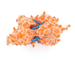
Background imageMolecular Model Collection: Restriction enzyme and DNA, artwork F007 / 6436

Restriction enzyme and DNA, artwork F007 / 6436
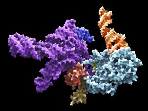
Restriction enzyme. Compute artwork of a restriction enzyme (orange) complexed with DNA (deoxyribonucleic acid, blue). Restriction enzymes, also known as restriction endonucleases

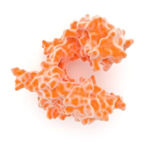
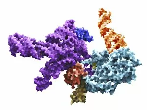
Background imageMolecular Model Collection: DNA polymerase molecule F007 / 6422

DNA polymerase molecule F007 / 6422
DNA polymerase molecule. DNA polymerases are enzymes that synthesise new strands of DNA from a complementary template strand

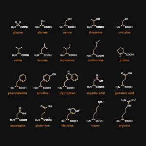
Background imageMolecular Model Collection: Amino acid structures F007 / 6424

Amino acid structures F007 / 6424
Amino acid structures. Chemical structures of 20 of the 22 standard amino acids

Background imageMolecular Model Collection: DNA nucleosome, artwork F007 / 6435

DNA nucleosome, artwork F007 / 6435
DNA nucleosome. Computer artwork of a nucleosome, the fundamental repeating unit used to package DNA (deoxyribonucleic acid) inside cell nuclei

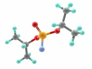
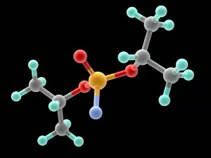
Background imageMolecular Model Collection: Sarin nerve gas molecule F007 / 9936

Sarin nerve gas molecule F007 / 9936
Sarin nerve gas, molecular model. Atoms are represented as spheres and are colour-coded: carbon (grey), hydrogen (green), oxygen (red), phosphorous (orange) and fluorine (blue)

Background imageMolecular Model Collection: Heat shock factor protein F007 / 9885

Heat shock factor protein F007 / 9885
Molecular model of a Heat Shock Protein (HSP).HSPs are a group of proteins whose levels increase when cells are exposed to raised temperatures or other stress

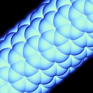
Background imageMolecular Model Collection: Carbon nanotube F007 / 9900

Carbon nanotube F007 / 9900
Buckytube. Molecular model of part of the cage structure of a bucky- or nanotube. The spheres represent carbon atoms. In this structure hundreds of atoms form hexagon shapes along a tube

Background imageMolecular Model Collection: Carbon nanotube F007 / 9915

Carbon nanotube F007 / 9915
Buckytube. Molecular model of part of the cage structure of a bucky- or nanotube. The spheres represent carbon atoms. In this structure hundreds of atoms form hexagon shapes along a tube

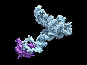
Background imageMolecular Model Collection: Glycine riboswitch molecule F007 / 9921

Glycine riboswitch molecule F007 / 9921
Molecular model of the bacterial glycine riboswitch. This is an RNA element that can bind the amino acid glycine. Glycine riboswitches usually consist of two metabolite-binding aptamer domains tandem

Background imageMolecular Model Collection: Heat shock factor 70 protein F007 / 9895

Heat shock factor 70 protein F007 / 9895
Molecular model of the Heat Shock Protein 70 (HSP).HSPs are a group of proteins whose levels increase when cells are exposed to raised temperatures or other stress

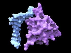
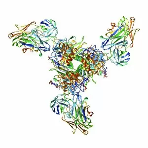
Background imageMolecular Model Collection: Haemagglutinin viral surface protein F007 / 9932

Haemagglutinin viral surface protein F007 / 9932
Haemagglutinin viral surface protein. Molecular model of haemagglutinin, a surface protein from the influenza virus, complexed with a neutralising antibody

Background imageMolecular Model Collection: Carbon nanotube F007 / 9910

Carbon nanotube F007 / 9910
Buckytube. Molecular model of part of the cage structure of a bucky- or nanotube. The spheres represent carbon atoms. In this structure hundreds of atoms form hexagon shapes along a tube

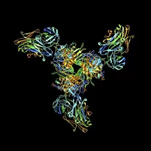
Background imageMolecular Model Collection: Haemagglutinin viral surface protein F007 / 9931

Haemagglutinin viral surface protein F007 / 9931
Haemagglutinin viral surface protein. Molecular model of haemagglutinin, a surface protein from the influenza virus, complexed with a neutralising antibody

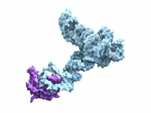
Background imageMolecular Model Collection: Glycine riboswitch molecule F007 / 9906

Glycine riboswitch molecule F007 / 9906
Molecular model of the bacterial glycine riboswitch. This is an RNA element that can bind the amino acid glycine. Glycine riboswitches usually consist of two metabolite-binding aptamer domains tandem

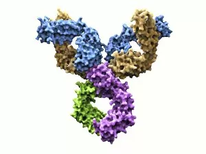
Background imageMolecular Model Collection: Immunoglobulin G antibody molecule F007 / 9901

Immunoglobulin G antibody molecule F007 / 9901
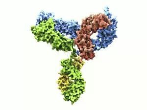
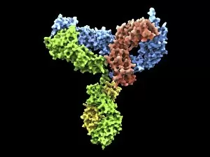
Immunoglobulin G antibody molecule. Computer model of the secondary structure of immunoglobulin G (IgG). This is the most abundant immunoglobulin and is found in all body fluids

Background imageMolecular Model Collection: DNA nucleosome, molecular model F007 / 9883

DNA nucleosome, molecular model F007 / 9883
DNA nucleosome. Molecular model of a nucleosome, the fundamental repeating unit used to package DNA (deoxyribonucleic acid) inside cell nuclei

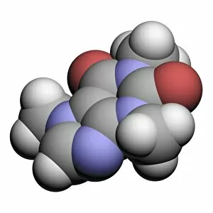
Background imageMolecular Model Collection: Caffeine drug molecule F007 / 9899

Caffeine drug molecule F007 / 9899
Caffeine. Computer model of a molecule of the alkaloid, stimulant and legal drug caffeine. Caffeine is most often consumed in drinks like tea and coffee

Background imageMolecular Model Collection: Sarin nerve gas molecule F007 / 9933

Sarin nerve gas molecule F007 / 9933
Sarin nerve gas, molecular model. Atoms are represented as spheres and are colour-coded: carbon (grey), hydrogen (green), oxygen (red), phosphorous (orange) and fluorine (blue)

Background imageMolecular Model Collection: DNA nucleosome, molecular model F007 / 9888

DNA nucleosome, molecular model F007 / 9888
DNA nucleosome. Molecular model of a nucleosome, the fundamental repeating unit used to package DNA (deoxyribonucleic acid) inside cell nuclei

Background imageMolecular Model Collection: Flap endonuclease protein F007 / 9914

Flap endonuclease protein F007 / 9914
Molecular model of the flap endonuclease protein. This is a class of nucleolytic enzymes that act as both 5 -3 exonucleases

Background imageMolecular Model Collection: Heat shock factor 70 protein F007 / 9903

Heat shock factor 70 protein F007 / 9903
Molecular model of the Heat Shock Protein 70 (HSP).HSPs are a group of proteins whose levels increase when cells are exposed to raised temperatures or other stress

Background imageMolecular Model Collection: Human serum albumin molecule F007 / 9904

Human serum albumin molecule F007 / 9904
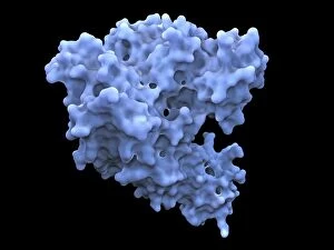
Human serum albumin, molecular model. Albumin is the most abundant protein in human blood plasma. One of albumins functions is to transport fatty acids to the liver

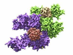
Background imageMolecular Model Collection: Human 80S ribosome F007 / 9902

Human 80S ribosome F007 / 9902
Ribosomal subunit. Computer model showing the structure of the RNA (ribonucleic acid) molecules in an 80S (large) ribosomal sub-unit. Ribosomes are composed of protein and RNA

Background imageMolecular Model Collection: Flap endonuclease protein F007 / 9916

Flap endonuclease protein F007 / 9916
Molecular model of the flap endonuclease protein. This is a class of nucleolytic enzymes that act as both exonucleases and structure-specific endonucleases on specialised DNA structures that occur

Background imageMolecular Model Collection: Immunoglobulin G antibody molecule F007 / 9920

Immunoglobulin G antibody molecule F007 / 9920
Immunoglobulin G antibody molecule. Computer model of the secondary structure of immunoglobulin G (IgG). This is the most abundant immunoglobulin and is found in all body fluids

Background imageMolecular Model Collection: Heat shock factor protein F007 / 9892

Heat shock factor protein F007 / 9892
Molecular model of a Heat Shock Protein (HSP).HSPs are a group of proteins whose levels increase when cells are exposed to raised temperatures or other stress

Background imageMolecular Model Collection: Human serum albumin molecule F007 / 9905

Human serum albumin molecule F007 / 9905
Human serum albumin, molecular model. Albumin is the most abundant protein in human blood plasma. One of albumins functions is to transport fatty acids to the liver

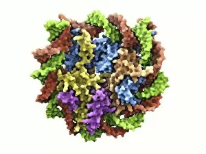
Background imageMolecular Model Collection: Yeast enzyme, molecular model F007 / 9913

Yeast enzyme, molecular model F007 / 9913
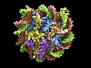
Yeast enzyme. Molecular model of an enzyme from bakers yeast (Saccharomyces cerevisiae). This is the 20S proteasome. A proteasome is a complex type of proteinase (protein-digesting enzyme)

Background imageMolecular Model Collection: Sarin nerve gas molecule F007 / 9938

Sarin nerve gas molecule F007 / 9938
Leucine, molecular model. Essential alpha-amino acid contained in eggs, soy protein, seaweed, turkey, chicken, lamb, cheese, and fish

Background imageMolecular Model Collection: Immunoglobulin G antibody molecule F007 / 9889

Immunoglobulin G antibody molecule F007 / 9889
Immunoglobulin G antibody molecule. Computer model of the secondary structure of immunoglobulin G (IgG). This is the most abundant immunoglobulin and is found in all body fluids

Background imageMolecular Model Collection: Human 80S ribosome F007 / 9898

Human 80S ribosome F007 / 9898
Ribosomal subunit. Computer model showing the structure of the RNA (ribonucleic acid) molecules in an 80S (large) ribosomal sub-unit. Ribosomes are composed of protein and RNA

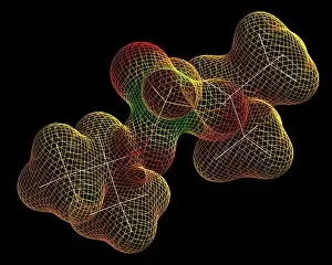
Background imageMolecular Model Collection: Sarin nerve gas molecule F007 / 9935

Sarin nerve gas molecule F007 / 9935
Sarin nerve gas, molecular model, The wire-frame map represents the electrostatic potential across the molecules surface. The atoms carbon, hydrogen, oxygen

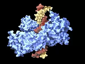
Background imageMolecular Model Collection: Type I topoisomerase protein bound to DNA F007 / 9893

Type I topoisomerase protein bound to DNA F007 / 9893
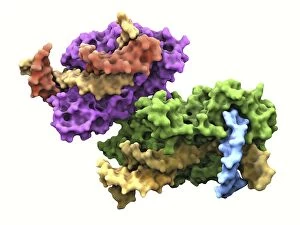
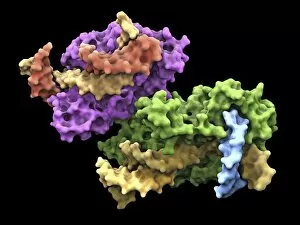
Type I topoisomerase bound to DNA. Molecular model showing a type I topoisomerase molecule (blue) bound to a strand of DNA (deoxyribonucleic acid, yellow and red)

Background imageMolecular Model Collection: Ubiquitin activating enzyme protein E1 F007 / 9919

Ubiquitin activating enzyme protein E1 F007 / 9919
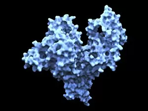
Molecular model of a ubiquitin-activating enzyme, also known as E1 enzymes. These catalyse the first step in the ubiquitination reaction, which targets a protein for degradation via a proteasome

Background imageMolecular Model Collection: Yeast enzyme, molecular model F007 / 9887

Yeast enzyme, molecular model F007 / 9887
Yeast enzyme. Molecular model of an enzyme from bakers yeast (Saccharomyces cerevisiae). This is the 20S proteasome. A proteasome is a complex type of proteinase (protein-digesting enzyme)
Background imageMolecular Model Collection: DNA molecule, artwork F008 / 3371

DNA molecule, artwork F008 / 3371
DNA molecule and chromosome, artwork

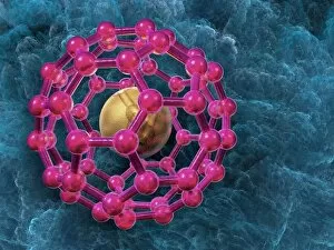
Background imageMolecular Model Collection: Buckyball, artwork F008 / 3355

Buckyball, artwork F008 / 3355
Buckyball, computer artwork

Background imageMolecular Model Collection: Buckyball, artwork F008 / 3347

Buckyball, artwork F008 / 3347
Buckyball, computer artwork


For sale as Licensed Images

Choose your image, Select your licence and Download the media

Molecular models offer a glimpse into the intricate world of science and medicine, revealing the hidden secrets of life at a microscopic level. In one captivating image, an anaesthetic molecule is seen inhibiting an ion channel C015/6718, unlocking new possibilities for pain management. Another striking model showcases the complex structure of a double-stranded RNA molecule, shedding light on its crucial role in gene regulation and viral defense mechanisms. Delving deeper into genetics, we explore DNA transcription through a mesmerizing molecular model that unravels the intricate process of genetic information transfer. The spotlight then shifts to Immunoglobulin G antibody molecules - powerful defenders against pathogens - as their elegant structures are unveiled with precision. From F007/9894 variant to artwork-inspired representations, these models showcase the diversity within our immune system's arsenal. Venturing beyond traditional boundaries, we encounter 2C-B psychedelic drug's molecular model – offering insights into its unique chemical composition and potential therapeutic applications. Art meets science once again as we marvel at an artistic interpretation showcasing secondary structures of proteins; highlighting their vital roles in cellular functions. Inorganic wonders take center stage with the perovskite crystal structure model – unveiling its remarkable properties that revolutionize solar energy technology. Returning to genetics, we witness a computer-generated DNA molecule model providing us with invaluable insights into our blueprint for life. The complexity continues with the intricately designed nucleosome molecule – unraveling how DNA is packaged within our cells' nucleus while maintaining accessibility for essential processes. Finally, awe-inspiring artwork captures antibodies' beauty and significance as they stand tall against invading antigens. These captivating molecular models serve as windows into worlds unseen by the naked eye; bridging gaps between scientific exploration and artistic expression. They inspire curiosity and ignite imagination while propelling breakthroughs in fields ranging from medicine to materials science.