Molecular Model Collection (page 3)

Molecular models offer a glimpse into the intricate world of science and medicine, revealing the hidden secrets of life at a microscopic level

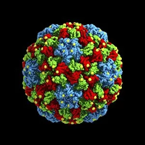
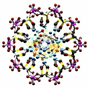
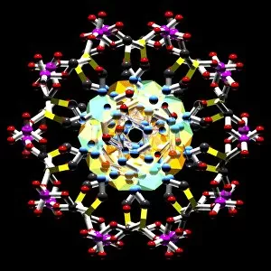
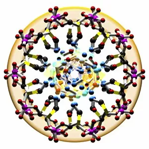
Background imageMolecular Model Collection: Cucumber mosaic virus, computer model

Cucumber mosaic virus, computer model
Cucumber mosaic virus (CMV), computer model. This image was created using molecular modelling software and data from X-ray crystallography

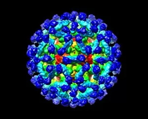
Background imageMolecular Model Collection: Murine norovirus with antibody fragments

Murine norovirus with antibody fragments
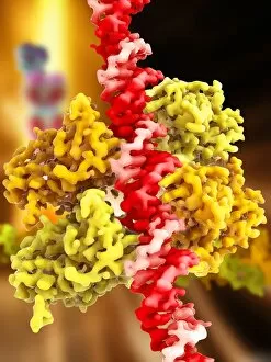
Murine norovirus (MNV) with antibody fragments, computer model. This image was created using molecular modelling software and data from cryo- electron microscopy

Background imageMolecular Model Collection: Biohazard symbol and virus

Biohazard symbol and virus. Computer artwork of the symbol for a biohazard (red) superimposed on a virus (blue). A biohazard is an organism or biological substance that is harmful to human health

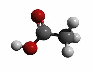
Background imageMolecular Model Collection: Acetic acid molecule

Acetic acid molecule
Acetic acid, molecular model. Acetic acid, also called ethanoic acid, is the component of vinegar that gives it its sour taste and pungent smell

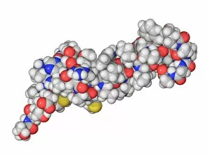
Background imageMolecular Model Collection: Parathyroid hormone molecule

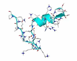
Parathyroid hormone molecule. Computer model showing the structure of parathyroid hormone (PTH), or parathormone. Atoms are colour-coded (carbon: dark grey, hydrogen: light grey, oxygen: red)
Background imageMolecular Model Collection: Ghrelin hormone molecule

Ghrelin hormone molecule. Computer model showing the crystal structure of the human hormone ghrelin. The crystal structure consists of both the secondary structure

Background imageMolecular Model Collection: Alanine, molecular model

Alanine, molecular model
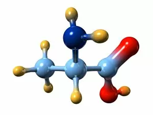
Alanine. Molecular model of the amino acid alanine. Its chemical formula is C3.H7.N.O3. Atoms are represented as balls and are colour-coded: carbon (blue), hydrogen (gold)

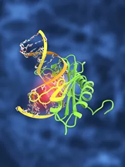
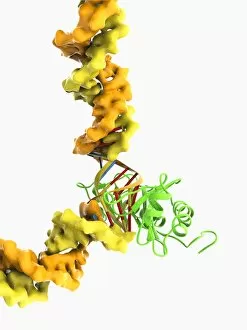
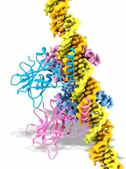
Background imageMolecular Model Collection: Tumour suppressor protein and DNA C017 / 3647

Tumour suppressor protein and DNA C017 / 3647
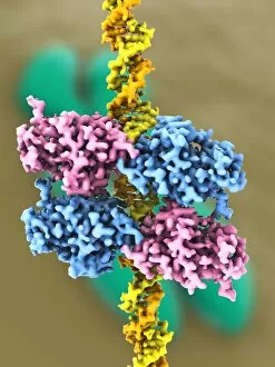
Tumour suppressor protein and DNA. Computer artwork showing a molecule of the tumour suppressor protein p53 (blue and pink) bound to a molecule of DNA (deoxyribonucleic acid, yellow and orange)

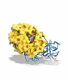
Background imageMolecular Model Collection: Ricin A-chain, artwork C017 / 3653

Ricin A-chain, artwork C017 / 3653
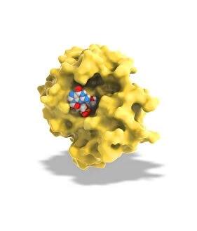
Ricin A-chain. Computer artwork showing the enzymatically active A-chain from a molecule of the toxic protein ricin. Ricin comprises two entwined amino acid chains; A (seen here) and B (not shown)

Background imageMolecular Model Collection: TATA box-binding protein complex C017 / 7082

TATA box-binding protein complex C017 / 7082
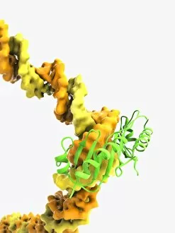
TATA box-binding protein complex. Molecular model showing a TATA box-binding protein (TBP, green) complexed with a strand of DNA (deoxyribonucleic acid, yellow) and transcription factor IIB

Background imageMolecular Model Collection: Water molecule C017 / 3605

Water molecule C017 / 3605
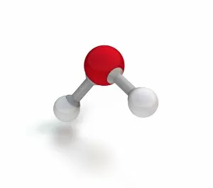
Water molecule. Computer artwork showing the structure of a molecule of water (H2O). Atoms are colour coded: oxygen (red) and hydrogen (white), with the bonds between them as bars (grey)

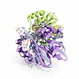
Background imageMolecular Model Collection: Antibiotic resistance enzyme molecule C017 / 2272

Antibiotic resistance enzyme molecule C017 / 2272
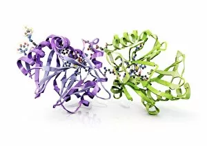
Antibiotic resistance enzyme. Molecular model of the New Delhi metallo-beta-lactamase 1 enzyme. This bacterial enzyme confers antibiotic resistance on cells that carry it

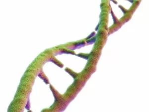
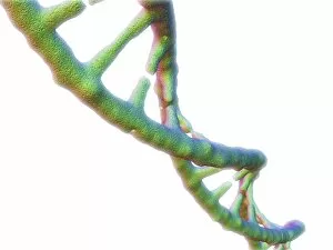
Background imageMolecular Model Collection: DNA molecule, artwork C017 / 7217

DNA molecule, artwork C017 / 7217
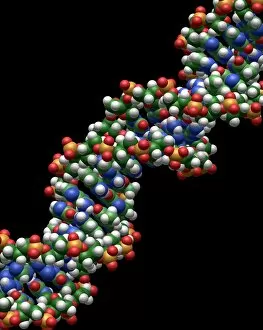
DNA molecule. Computer artwork showing a double stranded DNA (deoxyribonucleic acid) molecule. DNA is composed of two strands twisted into a double helix

Background imageMolecular Model Collection: DNA molecule, artwork C017 / 0616

DNA molecule, artwork C017 / 0616
DNA molecule. Computer artwork looking along the interior of a double stranded DNA (deoxyribonucleic acid) molecule. DNA is composed of two strands twisted into a double helix

Background imageMolecular Model Collection: TATA box-binding protein complex C017 / 7088

TATA box-binding protein complex C017 / 7088
TATA box-binding protein complex. Molecular model showing a TATA box-binding protein (TBP, green) complexed with a strand of DNA (deoxyribonucleic acid, yellow) and transcription factor IIB

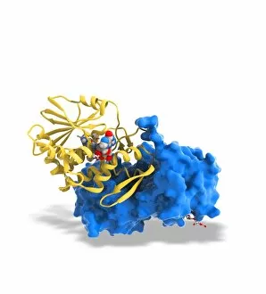
Background imageMolecular Model Collection: Ricin molecule, artwork C017 / 3652

Ricin molecule, artwork C017 / 3652
Ricin molecule. Computer artwork showing the structure of a molecule of the toxic protein ricin. Ricin comprises two entwined amino acid chains; A (yellow) and B (blue)

Background imageMolecular Model Collection: DNA molecule, artwork C017 / 0615

DNA molecule, artwork C017 / 0615
DNA molecule. Computer artwork looking along the interior of a double stranded DNA (deoxyribonucleic acid) molecule. DNA is composed of two strands twisted into a double helix

Background imageMolecular Model Collection: DNA molecule, artwork C017 / 0617

DNA molecule, artwork C017 / 0617
DNA molecule. Computer artwork looking along the interior of a double stranded DNA (deoxyribonucleic acid) molecule. DNA is composed of two strands twisted into a double helix

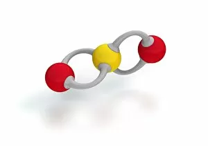
Background imageMolecular Model Collection: Sulphur dioxide molecule, artwork C017 / 3617

Sulphur dioxide molecule, artwork C017 / 3617
Sulphur dioxide molecule. Computer artwork showing the structure of a molecule of sulphur dioxide (SO2). Atoms are colour coded: sulphur (yellow) and oxygen (red)

Background imageMolecular Model Collection: Antibiotic resistance enzyme molecule C017 / 2271

Antibiotic resistance enzyme molecule C017 / 2271
Antibiotic resistance enzyme. Molecular model of the New Delhi metallo-beta-lactamase 1 enzyme. This bacterial enzyme confers antibiotic resistance on cells that carry it

Background imageMolecular Model Collection: TATA box-binding protein complex C017 / 7084

TATA box-binding protein complex C017 / 7084
TATA box-binding protein complex. Molecular model showing a TATA box-binding protein (TBP, green) complexed with a strand of DNA (deoxyribonucleic acid, yellow) and transcription factor IIB

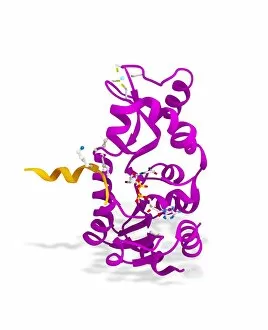
Background imageMolecular Model Collection: Sirtuin enzyme and p53, artwork C017 / 3659

Sirtuin enzyme and p53, artwork C017 / 3659
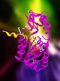
Sirtuin enzyme and p53. Computer artwork of a sirtuin (Sir2) enzyme (pink) bound to a p53 peptide (orange). Sir2 enzymes form a unique class of NAD(+)

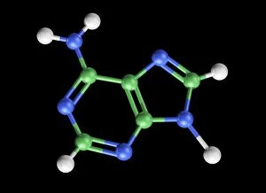
Background imageMolecular Model Collection: Adenine molecule, artwork C017 / 7200

Adenine molecule, artwork C017 / 7200
Adenine molecule. Computer artwork showing the structure of a molecule of the nucleobase adenine. Atoms are colour-coded spheres: carbon (green), nitrogen (blue), and oxygen (white)

Background imageMolecular Model Collection: Tumour suppressor protein and DNA C017 / 3644

Tumour suppressor protein and DNA C017 / 3644
Tumour suppressor protein and DNA. Computer artwork showing a molecule of the tumour suppressor protein p53 (blue and pink) bound to a molecule of DNA (deoxyribonucleic acid, yellow and orange)

Background imageMolecular Model Collection: Methane molecule, artwork C017 / 3613

Methane molecule, artwork C017 / 3613
Methane molecule. Computer artwork showing the structure of a molecule of methane (CH4). Atoms are colour coded: carbon (black) and hydrogen (white), with the bonds between them as rods (grey)

Background imageMolecular Model Collection: Ricin molecule, artwork C017 / 3651

Ricin molecule, artwork C017 / 3651
Ricin molecule. Computer artwork showing the structure of a molecule of the toxic protein ricin. Ricin comprises two entwined amino acid chains; A (yellow) and B (blue)

Background imageMolecular Model Collection: Ricin molecule, artwork C017 / 3650

Ricin molecule, artwork C017 / 3650
Ricin molecule. Computer artwork showing the structure of a molecule of the toxic protein ricin. Ricin comprises two entwined amino acid chains; A (yellow) and B (blue)

Background imageMolecular Model Collection: Sirtuin enzyme and p53, artwork C017 / 3658

Sirtuin enzyme and p53, artwork C017 / 3658
Sirtuin enzyme and p53. Computer artwork of a sirtuin (Sir2) enzyme (pink) bound to a p53 peptide (orange). Sir2 enzymes form a unique class of NAD(+)

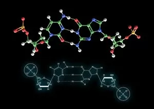
Background imageMolecular Model Collection: Cytosine-guanine interaction, artwork C017 / 7215

Cytosine-guanine interaction, artwork C017 / 7215
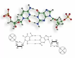
Cytosine-guanine interaction. Computer artwork showing the structure of bound cytosine (left) and guanine molecules (right)

Background imageMolecular Model Collection: SIRT3 molecule, artwork C017 / 3657

SIRT3 molecule, artwork C017 / 3657
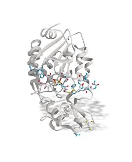
SIRT3 molecule. Computer artwork showing the structure of a molecule of NAD-dependent deacetylase sirtuin-3, mitochondrial (SIRT3)

Background imageMolecular Model Collection: Tumour suppressor protein and DNA C017 / 3646

Tumour suppressor protein and DNA C017 / 3646
Tumour suppressor protein and DNA. Computer artwork showing a molecule of the tumour suppressor protein p53 (blue and pink) bound to a molecule of DNA (deoxyribonucleic acid, yellow and orange)

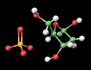
Background imageMolecular Model Collection: DNA components, artwork C017 / 7350

DNA components, artwork C017 / 7350
DNA components. Computer artwork showing the structure of the two molecules that make up the backbone of DNA (deoxyribonucleic acid), phosphate (left) and deoxyribose (right)

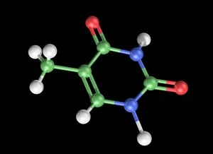
Background imageMolecular Model Collection: Thymine molecule, artwork C017 / 7366

Thymine molecule, artwork C017 / 7366
Thymine molecule. Computer artwork showing the structure of a molecule of the nucleobase thymine. Atoms are colour-coded spheres: carbon (green), nitrogen (blue), oxygen (red), and hydrogen (white)

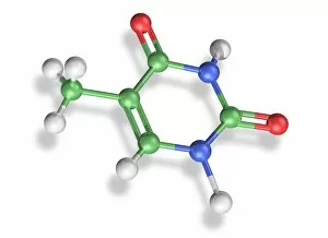
Background imageMolecular Model Collection: Thymine molecule, artwork C017 / 7365

Thymine molecule, artwork C017 / 7365
Thymine molecule. Computer artwork showing the structure of a molecule of the nucleobase thymine. Atoms are colour-coded spheres: carbon (green), nitrogen (blue), oxygen (red), and hydrogen (white)

Background imageMolecular Model Collection: Cytosine-guanine interaction, artwork C017 / 7216

Cytosine-guanine interaction, artwork C017 / 7216
Cytosine-guanine interaction. Computer artwork showing the structure of bound cytosine (left) and guanine molecules (right)

Background imageMolecular Model Collection: Thymine-adenine interaction, artwork C017 / 7367

Thymine-adenine interaction, artwork C017 / 7367
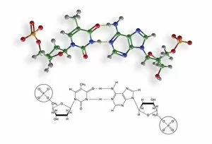
Thymine-adenine interaction. Computer artwork showing the structure of bound thymine and adenine molecules. Atoms are shown as colour-coded spheres: carbon (green), hydrogen (white)

Background imageMolecular Model Collection: DNA molecule, artwork F007 / 4200

DNA molecule, artwork F007 / 4200
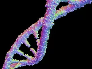
DNA molecule, computer artwork

Background imageMolecular Model Collection: DNA molecule, artwork F007 / 4196

DNA molecule, artwork F007 / 4196
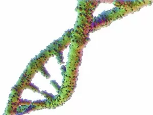
DNA molecule, computer artwork

Background imageMolecular Model Collection: DNA molecule, artwork F007 / 4203

DNA molecule, artwork F007 / 4203
DNA molecule, computer artwork

Background imageMolecular Model Collection: DNA molecule, artwork F007 / 4207

DNA molecule, artwork F007 / 4207
DNA molecule, computer artwork

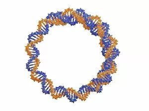
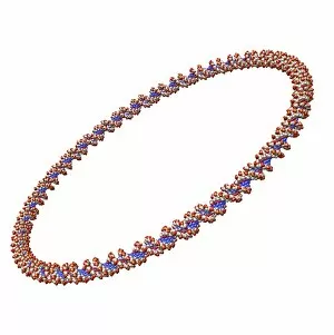
Background imageMolecular Model Collection: Circular DNA molecule, artwork F006 / 7088

Circular DNA molecule, artwork F006 / 7088
Circular DNA (deoxyribonucleic acid) molecule, computer artwork. Circular DNA has no ends, but consists of a ring structure

Background imageMolecular Model Collection: Tablet computer showing a DNA molecule F006 / 6310

Tablet computer showing a DNA molecule F006 / 6310
Tablet computer showing artwork of a DNA molecule

Background imageMolecular Model Collection: Circular DNA molecule, space artwork F006 / 7089

Circular DNA molecule, space artwork F006 / 7089
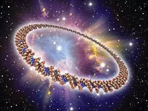
Circular DNA (deoxyribonucleic acid) molecule, computer artwork and space nebula artwork, depicting origin of life

Background imageMolecular Model Collection: Circular DNA molecule, artwork F006 / 7072

Circular DNA molecule, artwork F006 / 7072
Circular DNA (deoxyribonucleic acid) molecule, computer artwork. Circular DNA has no ends, but consists of a ring structure

Background imageMolecular Model Collection: DNA molecule, artwork F006 / 3715

DNA molecule, artwork F006 / 3715
DNA molecule, computer artwork

Background imageMolecular Model Collection: DNA molecule, artwork F006 / 7147

DNA molecule, artwork F006 / 7147
DNA (deoxyribonucleic acid) molecule, computer artwork

Background imageMolecular Model Collection: Circular DNA molecule, artwork F006 / 7095

Circular DNA molecule, artwork F006 / 7095
Circular DNA (deoxyribonucleic acid) molecule, computer artwork. Circular DNA has no ends, but consists of a ring structure

Background imageMolecular Model Collection: Circular DNA molecule, artwork F006 / 7086

Circular DNA molecule, artwork F006 / 7086
Circular DNA (deoxyribonucleic acid) molecule, computer artwork. Circular DNA has no ends, but consists of a ring structure


For sale as Licensed Images

Choose your image, Select your licence and Download the media

Molecular models offer a glimpse into the intricate world of science and medicine, revealing the hidden secrets of life at a microscopic level. In one captivating image, an anaesthetic molecule is seen inhibiting an ion channel C015/6718, unlocking new possibilities for pain management. Another striking model showcases the complex structure of a double-stranded RNA molecule, shedding light on its crucial role in gene regulation and viral defense mechanisms. Delving deeper into genetics, we explore DNA transcription through a mesmerizing molecular model that unravels the intricate process of genetic information transfer. The spotlight then shifts to Immunoglobulin G antibody molecules - powerful defenders against pathogens - as their elegant structures are unveiled with precision. From F007/9894 variant to artwork-inspired representations, these models showcase the diversity within our immune system's arsenal. Venturing beyond traditional boundaries, we encounter 2C-B psychedelic drug's molecular model – offering insights into its unique chemical composition and potential therapeutic applications. Art meets science once again as we marvel at an artistic interpretation showcasing secondary structures of proteins; highlighting their vital roles in cellular functions. Inorganic wonders take center stage with the perovskite crystal structure model – unveiling its remarkable properties that revolutionize solar energy technology. Returning to genetics, we witness a computer-generated DNA molecule model providing us with invaluable insights into our blueprint for life. The complexity continues with the intricately designed nucleosome molecule – unraveling how DNA is packaged within our cells' nucleus while maintaining accessibility for essential processes. Finally, awe-inspiring artwork captures antibodies' beauty and significance as they stand tall against invading antigens. These captivating molecular models serve as windows into worlds unseen by the naked eye; bridging gaps between scientific exploration and artistic expression. They inspire curiosity and ignite imagination while propelling breakthroughs in fields ranging from medicine to materials science.