
Molecular Model Collection (page 2)
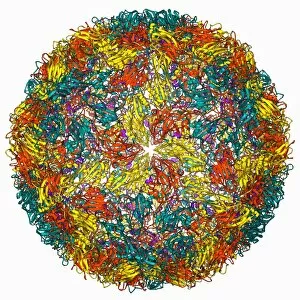
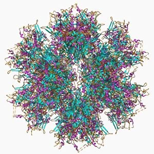
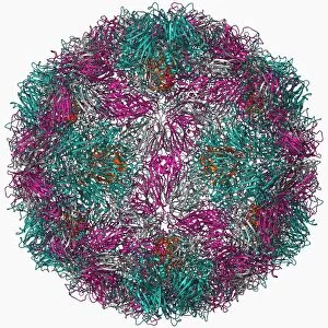
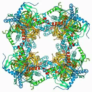
Molecular models offer a glimpse into the intricate world of science and medicine, revealing the hidden secrets of life at a microscopic level


For sale as Licensed Images
Choose your image, Select your licence and Download the media
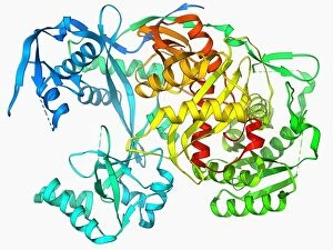
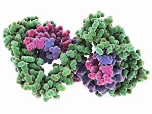
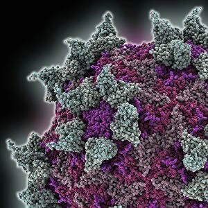
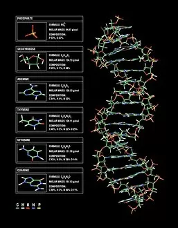
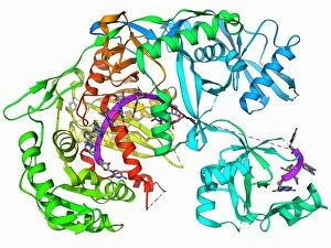
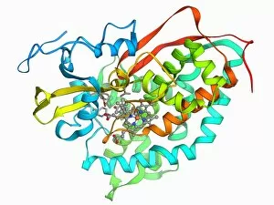
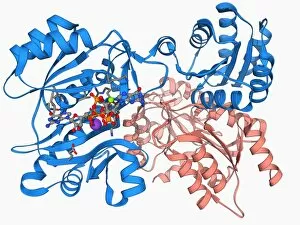
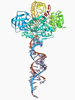
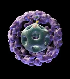
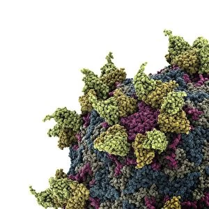
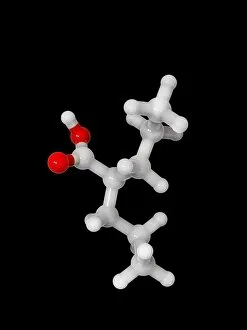
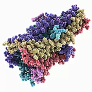
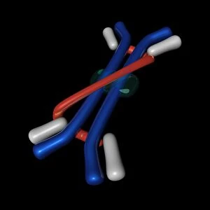
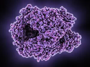
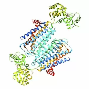
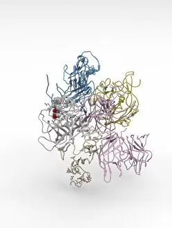
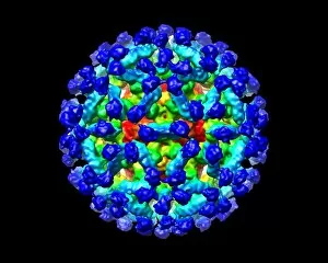
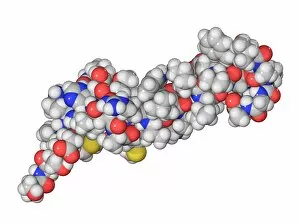
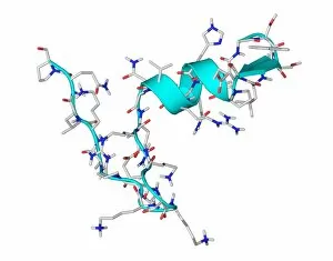
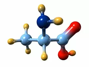
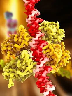
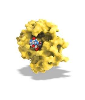
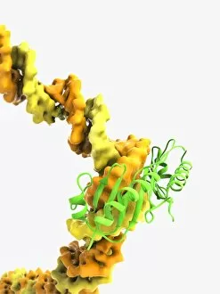
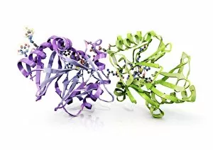
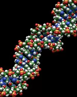
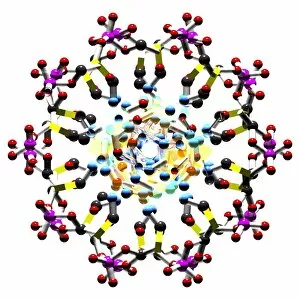
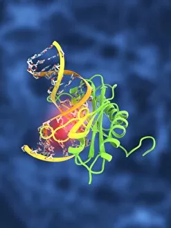
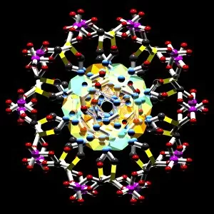
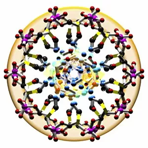
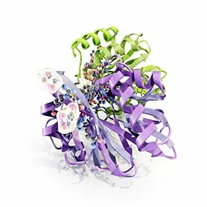
Molecular models offer a glimpse into the intricate world of science and medicine, revealing the hidden secrets of life at a microscopic level. In one captivating image, an anaesthetic molecule is seen inhibiting an ion channel C015/6718, unlocking new possibilities for pain management. Another striking model showcases the complex structure of a double-stranded RNA molecule, shedding light on its crucial role in gene regulation and viral defense mechanisms. Delving deeper into genetics, we explore DNA transcription through a mesmerizing molecular model that unravels the intricate process of genetic information transfer. The spotlight then shifts to Immunoglobulin G antibody molecules - powerful defenders against pathogens - as their elegant structures are unveiled with precision. From F007/9894 variant to artwork-inspired representations, these models showcase the diversity within our immune system's arsenal. Venturing beyond traditional boundaries, we encounter 2C-B psychedelic drug's molecular model – offering insights into its unique chemical composition and potential therapeutic applications. Art meets science once again as we marvel at an artistic interpretation showcasing secondary structures of proteins; highlighting their vital roles in cellular functions. Inorganic wonders take center stage with the perovskite crystal structure model – unveiling its remarkable properties that revolutionize solar energy technology. Returning to genetics, we witness a computer-generated DNA molecule model providing us with invaluable insights into our blueprint for life. The complexity continues with the intricately designed nucleosome molecule – unraveling how DNA is packaged within our cells' nucleus while maintaining accessibility for essential processes. Finally, awe-inspiring artwork captures antibodies' beauty and significance as they stand tall against invading antigens. These captivating molecular models serve as windows into worlds unseen by the naked eye; bridging gaps between scientific exploration and artistic expression. They inspire curiosity and ignite imagination while propelling breakthroughs in fields ranging from medicine to materials science.