Molecular Model Collection (page 9)

Molecular models offer a glimpse into the intricate world of science and medicine, revealing the hidden secrets of life at a microscopic level

Background imageMolecular Model Collection: Glutamine molecule

Glutamine molecule
Proline, molecular model. Non-essential alpha-amino acid, one of the 20 DNA-encoded amino acids. Atoms are represented as spheres and are colour-coded: carbon (grey), hydrogen (blue-green)

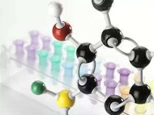
Background imageMolecular Model Collection: Chemical research, conceptual image

Chemical research, conceptual image

Background imageMolecular Model Collection: Isoleucine molecule

Isoleucine molecule
Isoleucine, molecular model. Essential alpha-amino acid contained in eggs, soy protein, seaweed, turkey, chicken, lamb, cheese, and fish

Background imageMolecular Model Collection: Threonine molecule

Threonine molecule
Threonine, molecular model. Essential alpha-amino acid and one of the 20 proteinogenic amino acids. Atoms are represented as spheres and are colour-coded: carbon (grey), hydrogen (blue-green)

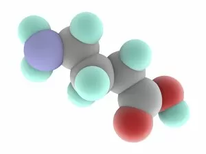
Background imageMolecular Model Collection: Gamma-aminobutyric acid GABA molecule

Gamma-aminobutyric acid GABA molecule
Gamma-aminobutyric acid (GABA), molecular model. Main inhibitory neurotransmitter in the central nervous system of mammalians

Background imageMolecular Model Collection: Valine molecule

Valine molecule
Valine, molecular model. Essential alpha-amino acid and one of the 20 proteinogenic amino acids. Atoms are represented as spheres and are colour-coded: carbon (grey), hydrogen (blue-green)

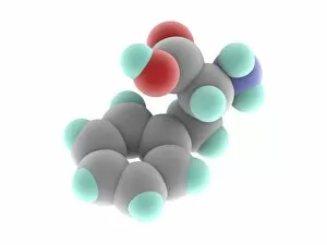
Background imageMolecular Model Collection: Phenylalanine molecule

Phenylalanine molecule
Phenylalanine, molecular model. Essential alpha-amino acid, one of the 20 common amino acids used to form proteins. Atoms are represented as spheres and are colour-coded: carbon (grey)

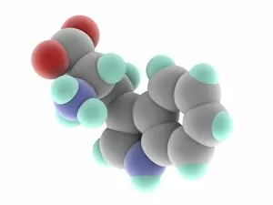
Background imageMolecular Model Collection: Tryptophan molecule

Tryptophan molecule
Tryptophan, molecular model. Essential amino acid and one of the 20 standard amino acids. Atoms are represented as spheres and are colour-coded: carbon (grey), hydrogen (blue-green)

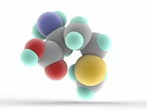
Background imageMolecular Model Collection: Methionine molecule

Methionine molecule
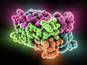
Methionine, molecular model. Essential alpha-amino acid. Atoms are represented as spheres and are colour-coded: carbon (grey), hydrogen (blue-green), nitrogen (blue), oxygen (red) and sulfur (yellow)

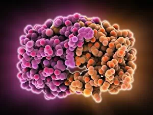
Background imageMolecular Model Collection: HIV-1 protease molecule

HIV-1 protease molecule
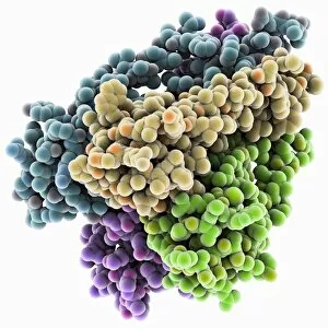
HIV-1 protease, molecular model. This enzyme, from HIV (human immunodeficiency virus), cleaves viral polyproteins into functional proteins that are essential for viral assembly and infectivity

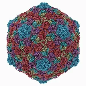
Background imageMolecular Model Collection: HK97 bacteriophage capsid

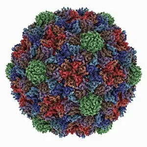
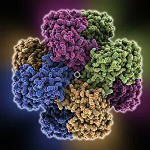
HK97 bacteriophage capsid, molecular model. Bacteriophages are viruses that infect bacteria, in this case enterobacteria such as E. coli (Escherichia coli), with the phage head shown here

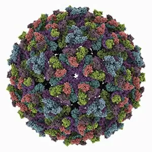
Background imageMolecular Model Collection: Chikungunya virus capsid

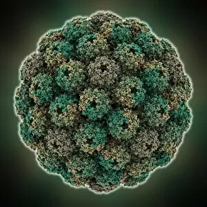
Chikungunya virus capsid, molecular model. This virus, transmitted by mosquitoes in tropical Africa and Asia, causes fever and joint pain in humans, similar to dengue fever

Background imageMolecular Model Collection: TATA box-binding protein complex C014 / 0867

TATA box-binding protein complex C014 / 0867
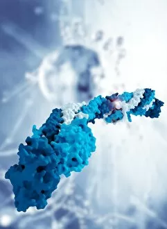
TATA box-binding protein complex. Molecular model showing a TATA box-binding protein (TBP, khaki) complexed with a strand of DNA (deoxyribonucleic acid)

Background imageMolecular Model Collection: HK97 bacteriophage procapsid

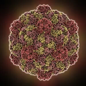
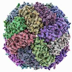
HK97 bacteriophage procapsid. Molecular model showing the structure of the prohead-I procapsid of the HK97 bacteriophage. Bacteriophages are viruses that infect bacteria
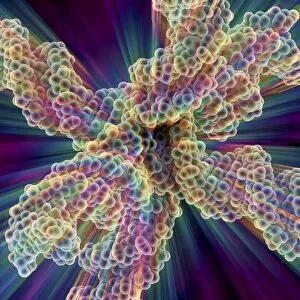
Background imageMolecular Model Collection: Turnip yellow mosaic virus capsid

Turnip yellow mosaic virus capsid, molecular model. This virus infects a wide variety of plants, including crops such as turnips and cabbages, causing yellow patches on the leaves

Background imageMolecular Model Collection: E. coli Holliday junction complex C014 / 0878

E. coli Holliday junction complex C014 / 0878
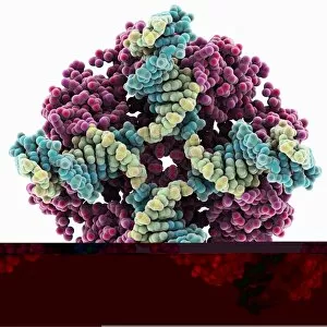
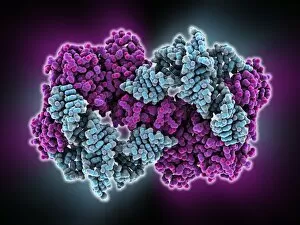
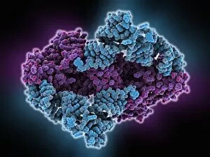
E. coli Holliday junction complex. Molecular model of a RuvA protein (dark pink) in complex with a Holliday junction between homologous strands of DNA (deoxyribonucleic acid)

Background imageMolecular Model Collection: Sindbis virus capsid, molecular model

Sindbis virus capsid, molecular model. This virus, transmitted by mosquitoes, causes sindbis fever in humans. In viruses, the capsid is the protein shell that encloses the genetic material

Background imageMolecular Model Collection: Fibrinogen molecule C014 / 0473

Fibrinogen molecule C014 / 0473
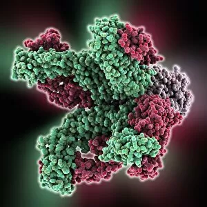
Fibrinogen. Molecular model showing the structure of the blood clotting glycoprotein fibrinogen (factor I). The molecule consists of two sets of alpha (grey)

Background imageMolecular Model Collection: Murine polyomavirus capsid

Murine polyomavirus capsid, molecular model. This virus, one of a range named for their potential to cause multiple tumours, infects mice

Background imageMolecular Model Collection: Human antitumour antibody molecule

Human antitumour antibody molecule
Human antitumour antibody. Molecular model showing the antitumour antibody BR96 complexed with part of the Lewis antigen. The Lewis antigen is expressed on the surface of human carcinoma cells

Background imageMolecular Model Collection: Iron containing protein, molecular model

Iron containing protein, molecular model
Iron containing protein. Molecular model showing the structure of a bacterial homolog of the animal iron containing protein ferritin

Background imageMolecular Model Collection: Brome mosaic virus capsid

Brome mosaic virus capsid, molecular model. This plant virus infects grasses, especially brome grasses, and also barley. It causes mosaic patches of discolouration

Background imageMolecular Model Collection: Synthetic DNA molecule

Synthetic DNA molecule
Synthetic DNA. Molecule model of a synthetic form of DNA (deoxyribonucleic acid). DNA is composed of two strands twisted into a double helix

Background imageMolecular Model Collection: Cowpea chlorotic mottle virus capsid

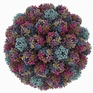
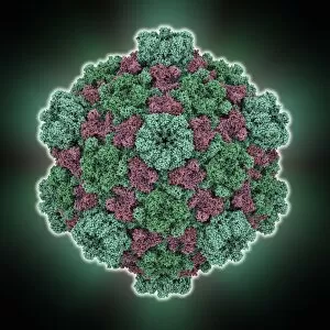
Cowpea chlorotic mottle virus capsid, molecular model. This virus (CCMV) infects the cowpea plant (Vigna unguiculata), causing yellow spots of discolouration

Background imageMolecular Model Collection: Potassium ion channel protein structure

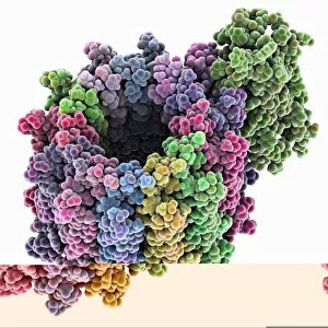
Potassium ion channel protein structure. Molecular model of a KcsA potassium ion (K+) channel from Streptomyces lividans bacteria

Background imageMolecular Model Collection: Streptavidin-biotin molecular complex

Streptavidin-biotin molecular complex. Molecular model of a single-strand binding complex of streptavidin (ribbons) and biotin (space-filled model, centre). Biotin is also known as vitamin B7

Background imageMolecular Model Collection: DNA supercoil, artwork

DNA supercoil, artwork
DNA supercoils. Computer artwork showing a supercoiled strand of DNA (deoxyribonucleic acid). Supercoiling is important in a number of biological processes

Background imageMolecular Model Collection: Potassium ion channel beta subunit

Potassium ion channel beta subunit. Molecular model showing the structure a beta subunit of a voltage-dependent potassium (K+) channel

Background imageMolecular Model Collection: Tyrosyl-tRNA synthetase molecule

Tyrosyl-tRNA synthetase molecule
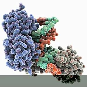
Tyrosyl-tRNA synthetase protein molecule. Molecular model showing bacterial tyrosyl-tRNA synthetase complexed with tyrosyl tRNA (transfer ribonucleic acid)

Background imageMolecular Model Collection: KCNQ ion channel protein structure

KCNQ ion channel protein structure. Molecular model showing the protein structure of an ion channel domain. Ion channels are membrane-spanning proteins that form a pathway for the movement of

Background imageMolecular Model Collection: Proliferating cell nuclear antigen

Proliferating cell nuclear antigen molecule. Molecular model of human proliferating cell nuclear antigen (PCNA, blue, green and red), complexed with its loader protein (purple, orange)

Background imageMolecular Model Collection: Potassium ion channel cavity structure

Potassium ion channel cavity structure. Molecular model showing the structure of a cavity formed by potassium ion channel proteins

Background imageMolecular Model Collection: Endonuclease IV molecule

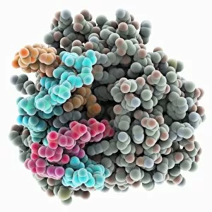
Endonuclease IV molecule. Molecular model of the endonuclease IV restriction enzyme EcoRV (grey) bound to a cleaved section of DNA (deoxyribonucleic acid, blue, orange and pink)

Background imageMolecular Model Collection: Tumour suppressor protein and DNA C017 / 3645

Tumour suppressor protein and DNA C017 / 3645
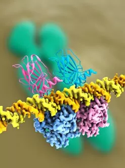
Tumour suppressor protein and DNA. Computer artwork showing a molecule of the tumour suppressor protein p53 (blue and pink) bound to a molecule of DNA (deoxyribonucleic acid, yellow and orange)

Background imageMolecular Model Collection: ATP synthase molecule C014 / 0880

ATP synthase molecule C014 / 0880
ATP synthase molecule. Molecular model showing the structure of ATP synthase (ATPase) subunit C. ATPase is an important enzyme that provides energy for cells through the synthesis of adenosine

Background imageMolecular Model Collection: Avian polyomavirus capsid

Avian polyomavirus capsid, molecular model. This virus, one of a range named for their potential to cause multiple tumours, infects birds. Discovered in budgerigars in 1981, it is often fatal

Background imageMolecular Model Collection: DNA 6-way junction, artwork C014 / 2587

DNA 6-way junction, artwork C014 / 2587
DNA 6-way junction. Computer artwork of a synthetic assemblage of nucleic acids which are useful in the design of nanostructures

Background imageMolecular Model Collection: Anthrax protective antigen molecule C014 / 0886

Anthrax protective antigen molecule C014 / 0886
Anthrax protective antigen molecule. Computer model showing the structure of a molecule of protective antigen (PA) produced by anthrax (Bacillus anthracis) bacteria

Background imageMolecular Model Collection: HIV enzyme protein, molecular model C014 / 0876

HIV enzyme protein, molecular model C014 / 0876
HIV enzyme protein. Computer model showing the structure of the catalytic domain of a molecule of HIV-1 retroviral integrase (IN) from the human immunodeficiency virus (HIV)

Background imageMolecular Model Collection: Tryptophanyl-tRNA synthetase molecule

Tryptophanyl-tRNA synthetase molecule
Tryptophanyl-tRNA synthetase protein molecule. Molecular model showing human tryptophanyl-tRNA synthetase complexed with tryptophan tRNA (transfer ribonucleic acid)

Background imageMolecular Model Collection: Cytoplasmic polyhedrosis virus capsid

Cytoplasmic polyhedrosis virus capsid, molecular model. Part of the Cypovirus genus and invariably fatal, this insect virus is transmitted by contamination of leaves eaten (examples include silkworms)

Background imageMolecular Model Collection: Pho4 transcription factor bound to DNA C014 / 0861

Pho4 transcription factor bound to DNA C014 / 0861
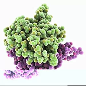
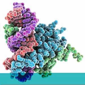
Pho4 transcription factor bound to DNA. Molecular model showing phosphate system positive regulatory protein (Pho4) (blue and green) bound to a strand of DNA (deoxyribonucleic acid, red and purple)

Background imageMolecular Model Collection: EcoRV restriction enzyme molecule C014 / 2117

EcoRV restriction enzyme molecule C014 / 2117
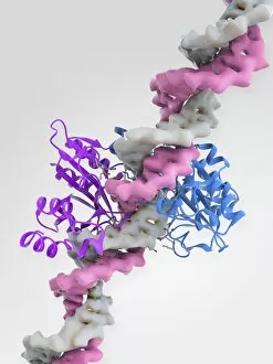
EcoRV restriction enzyme. Molecular model of the type II restriction enzyme EcoRV (purple and blue) bound to a DNA molecule (deoxyribonucleic acid, pink and white)

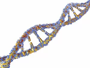
Background imageMolecular Model Collection: DNA molecule, artwork F007 / 1996

DNA molecule, artwork F007 / 1996
DNA molecule, computer artwork


For sale as Licensed Images

Choose your image, Select your licence and Download the media

Molecular models offer a glimpse into the intricate world of science and medicine, revealing the hidden secrets of life at a microscopic level. In one captivating image, an anaesthetic molecule is seen inhibiting an ion channel C015/6718, unlocking new possibilities for pain management. Another striking model showcases the complex structure of a double-stranded RNA molecule, shedding light on its crucial role in gene regulation and viral defense mechanisms. Delving deeper into genetics, we explore DNA transcription through a mesmerizing molecular model that unravels the intricate process of genetic information transfer. The spotlight then shifts to Immunoglobulin G antibody molecules - powerful defenders against pathogens - as their elegant structures are unveiled with precision. From F007/9894 variant to artwork-inspired representations, these models showcase the diversity within our immune system's arsenal. Venturing beyond traditional boundaries, we encounter 2C-B psychedelic drug's molecular model – offering insights into its unique chemical composition and potential therapeutic applications. Art meets science once again as we marvel at an artistic interpretation showcasing secondary structures of proteins; highlighting their vital roles in cellular functions. Inorganic wonders take center stage with the perovskite crystal structure model – unveiling its remarkable properties that revolutionize solar energy technology. Returning to genetics, we witness a computer-generated DNA molecule model providing us with invaluable insights into our blueprint for life. The complexity continues with the intricately designed nucleosome molecule – unraveling how DNA is packaged within our cells' nucleus while maintaining accessibility for essential processes. Finally, awe-inspiring artwork captures antibodies' beauty and significance as they stand tall against invading antigens. These captivating molecular models serve as windows into worlds unseen by the naked eye; bridging gaps between scientific exploration and artistic expression. They inspire curiosity and ignite imagination while propelling breakthroughs in fields ranging from medicine to materials science.