
Osteology Collection (#2)
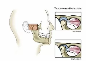
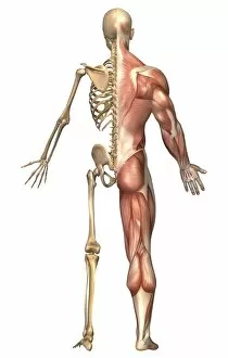
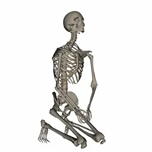
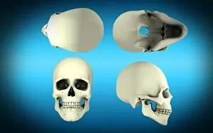
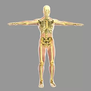
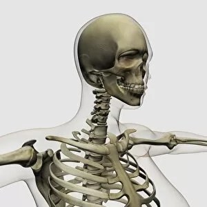
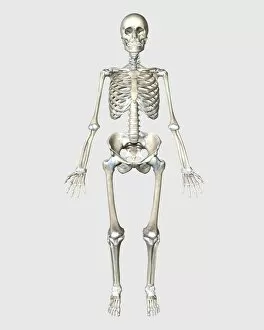
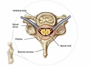
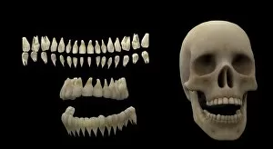
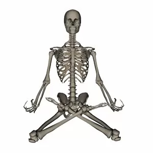
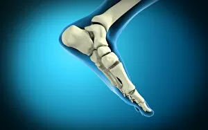
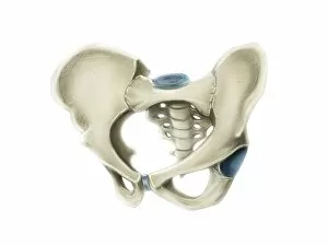
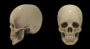
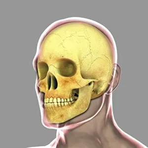
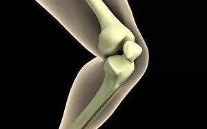
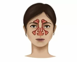
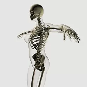
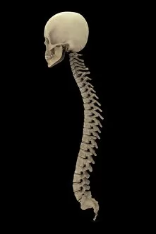
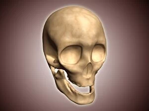
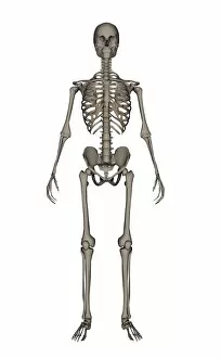
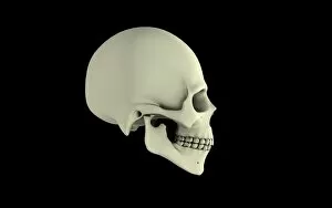
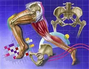
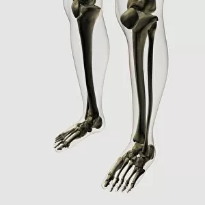
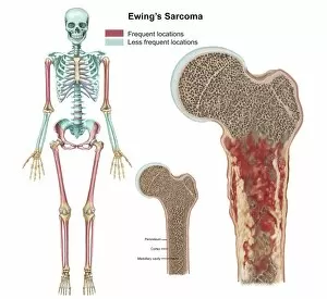
Osteology, the fascinating study of bones and skeletal structures, unveils a world of intricate details and medical advancements


For sale as Licensed Images
Choose your image, Select your licence and Download the media
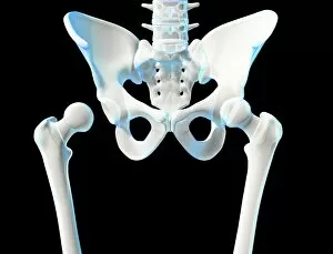
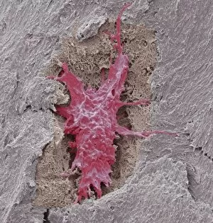
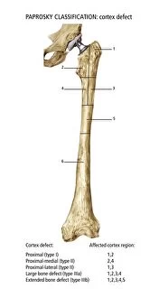
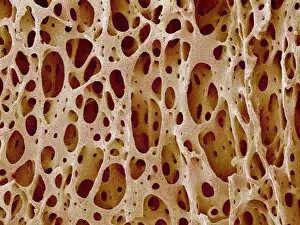
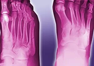
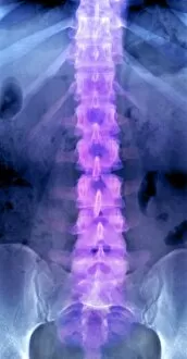
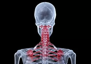
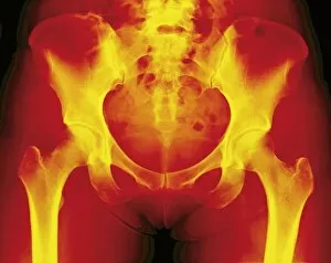
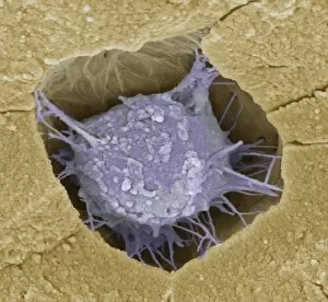
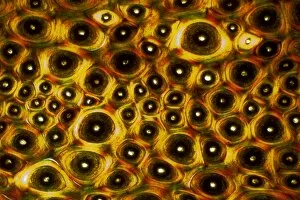
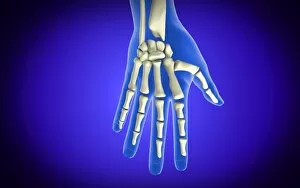
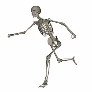
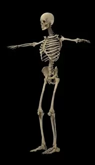
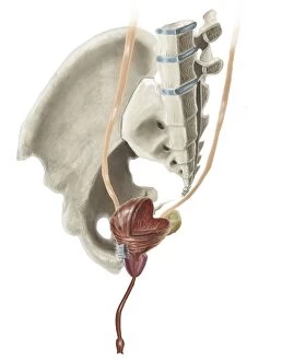
Osteology, the fascinating study of bones and skeletal structures, unveils a world of intricate details and medical advancements. From total hip replacements to X-ray imaging, this field has revolutionized the way we understand and treat bone-related conditions. In the realm of orthopedics, total hip replacement surgeries have become a game-changer for those suffering from debilitating joint pain. With cutting-edge techniques and prosthetic materials, these procedures restore mobility and improve quality of life. Delving into history, Calots spinal surgery in the 19th century stands as a testament to human resilience. Despite limited resources and knowledge at that time, surgeons courageously ventured into complex spinal operations with remarkable outcomes. Artwork captures both the beauty and vulnerability of our skeletal system. A damaged knee ligament depicted in brushstrokes reminds us of how delicate our bodies can be. Meanwhile, a running skeleton portrayed through art showcases strength and agility even in its bare form. X-ray artwork takes us beneath the surface to reveal hidden stories within our bones. Skeletons captured by this technique offer an ethereal glimpse into their structure while evoking curiosity about their past lives. An X-ray artwork showcasing a skeleton from below adds another dimension to our understanding of anatomy. It invites contemplation on how every angle tells a different story about our body's framework. Sometimes even mundane ailments find artistic expression through X-rays. A headache immortalized as an artwork serves as a reminder that even seemingly minor discomforts can leave traces within us. Fractured ankle X-rays remind us that bones are not invincible; they can break under pressure or unfortunate circumstances. Yet with modern medicine's intervention, healing is possible once again. Microscopic exploration reveals wonders too small for the naked eye to behold. Compact bone viewed under light micrograph exposes its intricate structure like never before – reminding us that there is more than meets the eye when it comes to osteology.