Osteology Collection (#8)

Osteology, the fascinating study of bones and skeletal structures, unveils a world of intricate details and medical advancements

Background imageOsteology Collection: Georges Pouchet, French naturalist

Georges Pouchet, French naturalist
Georges Pouchet (1833-1894), French naturalist. Pouchet, the son of the naturalist Felix Pouchet, worked as an anatomist at the Museum of Natural History in Paris, France

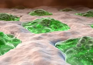
Background imageOsteology Collection: Osteoclast bone cells, artwork

Osteoclast bone cells, artwork
Osteoclast bone cells. Computer artwork of normal osteoclasts (green) in the lacunae (spaces) of bone tissue. Osteoclasts remodel bone by degrading

Background imageOsteology Collection: Arthritic hands

Arthritic hands
MODEL RELEASED. Arthritic hands. Hands of a person that have become deformed due to arthritis. Arthritis is a degenerative condition that affects the joints causing inflammation and pain

Background imageOsteology Collection: Hip fracture, digital X-ray

Hip fracture, digital X-ray
Hip fracture. Digital frontal X-ray of the pelvis and hips of a patient in their mid eighties. This patient has a fracture of the neck of their left femur (upper leg bone)

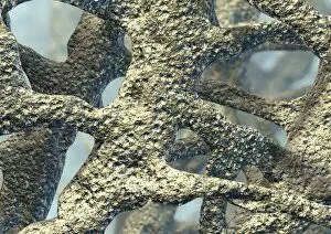
Background imageOsteology Collection: Osteoporotic bone, artwork

Osteoporotic bone, artwork
Osteoporosis. Computer artwork of osteoporotic bone. Osteoporosis (brittle bone disease) causes a reduction in overall bone mass and an increase in the bones porosity

Background imageOsteology Collection: Amputated fingertip, X-ray

Amputated fingertip, X-ray
Amputated fingertip. Coloured X-ray of the 4th and 5th fingers of a hand where the end of the 5th (little) finger has been amputated (red)

Background imageOsteology Collection: Bone growth, artwork

Bone growth, artwork
Bone growth. Computer artwork showing the development and growth of a long bone. Behind the epiphysis (head) is an area of spongy bone consisting of trabeculae filled with bone marrow (green)

Background imageOsteology Collection: Main in pain, artwork

Main in pain, artwork
Main in pain, computer artwork

Background imageOsteology Collection: Bone death, artwork

Bone death, artwork
Bone death. Computer artwork of a human hip and knee joint affected by osteonecrosis - death of the bone tissue. This condition is caused by an impaired blood supply to the bone cells

Background imageOsteology Collection: Osteoporosis, artwork

Osteoporosis, artwork
Osteoporosis. Artwork of vertical sections comparing the upper parts of a healthy femur (left) and one with reduced bone mass (right)

Background imageOsteology Collection: Osteoporosis in the skull, X-ray

Osteoporosis in the skull, X-ray
Osteoporosis in the skull. Coloured X-ray of the skull of a 71-year-old person with osteoporosis. Osteoporosis (brittle bone disease)

Background imageOsteology Collection: Melorheostosis of the shoulder, X-ray

Melorheostosis of the shoulder, X-ray
Melorheostosis of the shoulder. X-ray of 59-year-old patients shoulder, showing damage (dense white, upper right) caused by melorheostosis

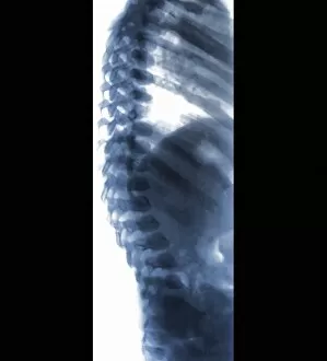
Background imageOsteology Collection: Spine in Hurler Syndrome, X-ray

Spine in Hurler Syndrome, X-ray
Spine in Hurler Syndrome. Coloured X-ray of the spine of a 26-month-old child with Hurler syndrome (also called mucopolysaccharidosis type I, MPS I)

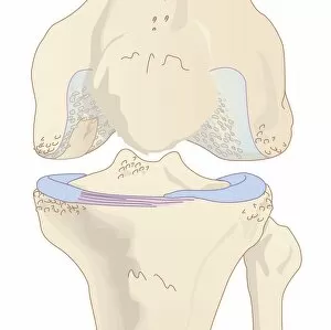
Background imageOsteology Collection: Osteoarthritis of the knee, artwork

Osteoarthritis of the knee, artwork
Osteoarthritis of the knee. Computer artwork showing a knee joint affected by osteoarthritis. Osteoarthritis is a degenerative disease that involves degradation of the bone

Background imageOsteology Collection: Skull bones, artwork

Skull bones, artwork
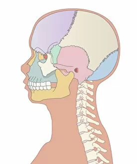
Skull bones. Computer artwork showing the major bones of the human skull. These are: frontal (purple, upper left), parietal (green/beige, upper right), occipital (blue, lower right), temporal (pink)

Background imageOsteology Collection: Shin splints, artwork

Shin splints, artwork
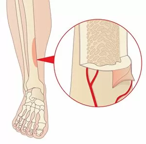
Shin splints. Computer artwork of a lower leg showing an area of the tibia (shin bone) affected by medial tibial stress syndrome, or shin splints

Background imageOsteology Collection: Fractured pelvis, X-ray

Fractured pelvis, X-ray
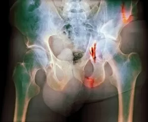
Fractured pelvis. Coloured X-ray of fractures (red) in the pelvis. The head of the femur (thigh bone) articulates with the acetabulum to form the hip joint

Background imageOsteology Collection: Human spine model

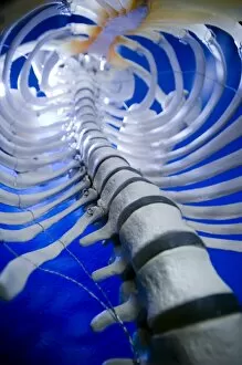
Human spine model. Model of a human vertebral column (spine) showing a number of its vertebrae (white discs). The spine is the central supporting structure of the human body

Background imageOsteology Collection: Fractured arm, X-ray

Fractured arm, X-ray
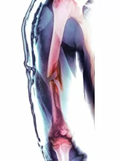
Fractured arm. Coloured X-ray of the front view of a fractured upper arm bone (humerus). The fracture is at centre (red), half way down the bone

Background imageOsteology Collection: Football kick, skeleton artwork

Football kick, skeleton artwork
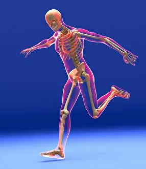
Football kick. Computer artwork showing a skeleton in the process of kicking a football (not seen). The outline of the body can also be seen

Background imageOsteology Collection: Fractured femur, X-ray

Fractured femur, X-ray
Fractured femur. X-ray of a patient with a total hip replacement who has fractured their femur (thigh bone). The fracture (centre right) has occurred towards the end of the implant (solid white)

Background imageOsteology Collection: Dislocated hip replacement, X-ray

Dislocated hip replacement, X-ray
Dislocated hip replacement. Frontal X-ray of the pelvis of a patient with bi-lateral (both sides) total hip replacements. The right joint replacement (right) has become dislocated

Background imageOsteology Collection: Skeletons head, artwork

Skeletons head, artwork
Skeletons head. Computer artwork showing the bones of the head, neck and shoulders of a human skeleton that is lying down

Background imageOsteology Collection: Skeleton X-ray, artwork

Skeleton X-ray, artwork
Skeleton X-ray, computer artwork. This depiction of a full body X-ray shows the 206 bones of the human skeleton in a normal standing position

Background imageOsteology Collection: Skeletons feet, artwork

Skeletons feet, artwork
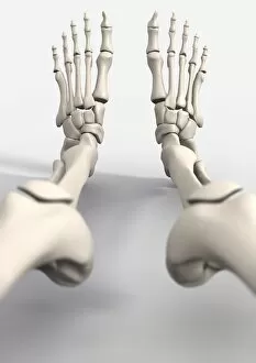
Skeletons feet. Computer artwork showing the bones of the feet of a human skeleton that is lying down

Background imageOsteology Collection: Skeletons, artwork

Skeletons, artwork
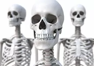
Skeletons. Computer artwork of the skull and neck of a human skeleton, flanked by two other skeletons

Background imageOsteology Collection: Skeleton and ligaments, artwork

Skeleton and ligaments, artwork
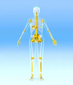
Female skeleton, computer artwork. The human skeleton has 206 bones. The skull (at top) protects the brain. The ribs of the chest (at upper centre) enclose the heart and lungs

Background imageOsteology Collection: Skeletons torso, artwork

Skeletons torso, artwork
Skeletons torso. Computer artwork showing the bones of the torso of a human skeleton that is lying down

Background imageOsteology Collection: Compact bone, light micrograph

Compact bone, light micrograph
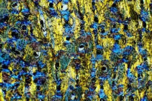
Compact bone. Polarised light micrograph of a transverse section through compact bone tissue, showing Haversian canals (circular regions)

Background imageOsteology Collection: Osteoclasts in bone lacunae, SEM

Osteoclasts in bone lacunae, SEM
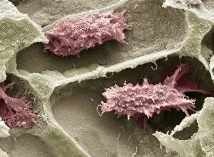
Osteoclasts in bone lacunae, coloured scanning electron micrograph (SEM). These osteoclasts are seen in Howslips lacunae, spaces in the bone where they are responsible for remodeling the bone


For sale as Licensed Images

Choose your image, Select your licence and Download the media

Osteology, the fascinating study of bones and skeletal structures, unveils a world of intricate details and medical advancements. From total hip replacements to X-ray imaging, this field has revolutionized the way we understand and treat bone-related conditions. In the realm of orthopedics, total hip replacement surgeries have become a game-changer for those suffering from debilitating joint pain. With cutting-edge techniques and prosthetic materials, these procedures restore mobility and improve quality of life. Delving into history, Calots spinal surgery in the 19th century stands as a testament to human resilience. Despite limited resources and knowledge at that time, surgeons courageously ventured into complex spinal operations with remarkable outcomes. Artwork captures both the beauty and vulnerability of our skeletal system. A damaged knee ligament depicted in brushstrokes reminds us of how delicate our bodies can be. Meanwhile, a running skeleton portrayed through art showcases strength and agility even in its bare form. X-ray artwork takes us beneath the surface to reveal hidden stories within our bones. Skeletons captured by this technique offer an ethereal glimpse into their structure while evoking curiosity about their past lives. An X-ray artwork showcasing a skeleton from below adds another dimension to our understanding of anatomy. It invites contemplation on how every angle tells a different story about our body's framework. Sometimes even mundane ailments find artistic expression through X-rays. A headache immortalized as an artwork serves as a reminder that even seemingly minor discomforts can leave traces within us. Fractured ankle X-rays remind us that bones are not invincible; they can break under pressure or unfortunate circumstances. Yet with modern medicine's intervention, healing is possible once again. Microscopic exploration reveals wonders too small for the naked eye to behold. Compact bone viewed under light micrograph exposes its intricate structure like never before – reminding us that there is more than meets the eye when it comes to osteology.