Osteology Collection (#4)

Osteology, the fascinating study of bones and skeletal structures, unveils a world of intricate details and medical advancements

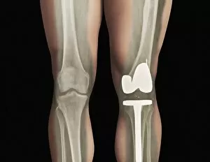
Background imageOsteology Collection: Total knee replacement, X-ray

Total knee replacement, X-ray
Total knee replacement. X-ray of the legs of a 49-year-old patient who has had a total knee replacement (white) of the left knee (right)

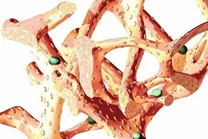
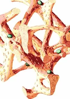
Background imageOsteology Collection: Bone structure, artwork C016 / 7504

Bone structure, artwork C016 / 7504
Bone structure. Computer artwork showing bone trabeculae. Trabeculae are the bars of bone that form a honeycombed hollow network, and make up the structure of spongy bone, the inner layer of a bone

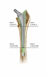
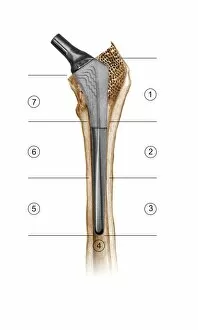
Background imageOsteology Collection: Prosthetic hip joint and Gruen zones C016 / 6778

Prosthetic hip joint and Gruen zones C016 / 6778
Prosthetic hip joint and Gruen zones. Cutaway diagram of a femur (thigh bone) showing a femoral component of a hip prosthesis and labels indicating Gruen zones

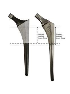
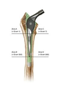
Background imageOsteology Collection: Prosthetic hip joints and Gruen zones C016 / 6781

Prosthetic hip joints and Gruen zones C016 / 6781
Prosthetic hip joints and Gruen zones. Diagram of the femoral components of two types of hip prosthesis with labels indicating standard and modified Gruen zones

Background imageOsteology Collection: Prosthetic hip joint, artwork C016 / 6782

Prosthetic hip joint, artwork C016 / 6782
Prosthetic hip joint. Cutaway artwork of a femur (thigh bone) showing a femoral component of a hip prosthesis. This component is implanted in the femur after the head of the femur has been surgically

Background imageOsteology Collection: Bone drill and grafter, artwork C016 / 6845

Bone drill and grafter, artwork C016 / 6845
Bone drill and grafter. Artwork of a cross-section through a bone drill and grafter that is being used in a hip socket bone grafting operation

Background imageOsteology Collection: Hip socket bone grafting, diagram C016 / 6786

Hip socket bone grafting, diagram C016 / 6786
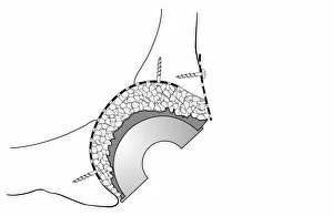
Hip socket bone grafting. Diagram showing completed hip socket bone grafting as part of a total hip replacement operation

Background imageOsteology Collection: Hip socket bone grafting, diagram C016 / 6784

Hip socket bone grafting, diagram C016 / 6784
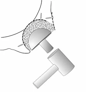
Hip socket bone grafting. Diagram showing the second stage in hip socket bone grafting as part of a total hip replacement operation

Background imageOsteology Collection: Broken finger bone, SEM C016 / 9027

Broken finger bone, SEM C016 / 9027
Broken finger bone. Coloured scanning electron micrograph (SEM) showing the internal structure of a broken finger bone (phalanx)

Background imageOsteology Collection: Hip socket bone grafting, diagram C016 / 6785

Hip socket bone grafting, diagram C016 / 6785
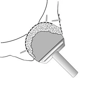
Hip socket bone grafting. Diagram showing the third stage in hip socket bone grafting as part of a total hip replacement operation

Background imageOsteology Collection: Hip socket bone grafting, diagram C016 / 6783

Hip socket bone grafting, diagram C016 / 6783
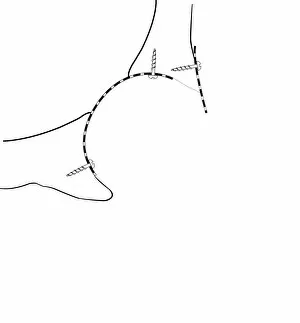
Hip socket bone grafting. Diagram showing the first stage in hip socket bone grafting as part of a total hip replacement operation

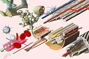
Background imageOsteology Collection: Human bone structure, artwork C016 / 7494

Human bone structure, artwork C016 / 7494
Human bone structure. Computer artwork showing all the different parts that make up a human bone, including the bone marrow (bottom left) where blood cells (round, white and red) are formed

Background imageOsteology Collection: Prosthetic hip joint, diagram C016 / 6775

Prosthetic hip joint, diagram C016 / 6775
Prosthetic hip joint. Cutaway diagram of a femur (thigh bone) showing a femoral component of a hip prosthesis. This component is implanted in the femur after the head of the femur has been surgically

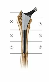
Background imageOsteology Collection: Prosthetic hip joint and Gruen zones C016 / 6777

Prosthetic hip joint and Gruen zones C016 / 6777
Prosthetic hip joint and Gruen zones. Cutaway diagram of a femur (thigh bone) showing a femoral component of a hip prosthesis and labels indicating Gruen zones

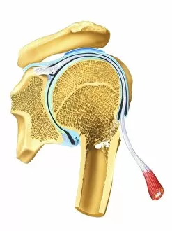
Background imageOsteology Collection: Broken shoulder, artwork C016 / 6568

Broken shoulder, artwork C016 / 6568
Broken shoulder. Artwork of a vertical (coronal) section through the bones of the shoulder, showing a fracture (centre right) of the upper part of the humerus (upper arm bone)

Background imageOsteology Collection: Prosthetic hip joint and Gruen zones C016 / 6780

Prosthetic hip joint and Gruen zones C016 / 6780
Prosthetic hip joint and Gruen zones. Diagram of the femoral component of a hip prosthesis with labels indicating the hip connection (upper left) and the seven Gruen zones

Background imageOsteology Collection: Prosthetic hip joint and Gruen zones C016 / 6776

Prosthetic hip joint and Gruen zones C016 / 6776
Prosthetic hip joint and Gruen zones. Cutaway diagram of a femur (thigh bone) showing a femoral component of a hip prosthesis and labels indicating Gruen zones

Background imageOsteology Collection: Prosthetic hip joint and Gruen zones C016 / 6779

Prosthetic hip joint and Gruen zones C016 / 6779
Prosthetic hip joint and Gruen zones. Cutaway diagram of a femur (thigh bone) showing a femoral component of a hip prosthesis and labels indicating Gruen zones

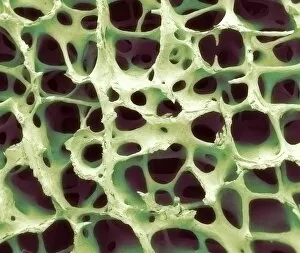
Background imageOsteology Collection: Bone tissue, SEM

Bone tissue, SEM
Bone tissue. Coloured scanning electron micrograph (SEM) of cancellous (spongy) bone. Bone tissue can be either cortical (compact) or cancellous

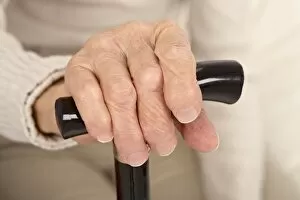
Background imageOsteology Collection: Arthritic hand and walking stick C015 / 8808

Arthritic hand and walking stick C015 / 8808
MODEL RELEASED. Arthritic hand and walking stick. Hand of an 82-year-old woman holding a walking stick. The joints are deformed by polyarthritis

Background imageOsteology Collection: Holding arthritic hands C015 / 8818

Holding arthritic hands C015 / 8818
MODEL RELEASED. Holding arthritic hands. Arthritic hands of an 82-year-old woman being held by the hands of the young woman caring for her

Background imageOsteology Collection: Elderly woman and carer C015 / 8814

Elderly woman and carer C015 / 8814
MODEL RELEASED. Elderly woman and carer. Woman caring for an 82-year-old woman who is using a walking stick. The joints of the elderly womans hands are deformed by polyarthritis

Background imageOsteology Collection: Elderly woman and carer C015 / 8815

Elderly woman and carer C015 / 8815
MODEL RELEASED. Elderly woman and carer. Woman caring for an 82-year-old woman who is using a walking stick. The joints of the elderly womans hands are deformed by polyarthritis

Background imageOsteology Collection: Arthritic hand C015 / 8813

Arthritic hand C015 / 8813
MODEL RELEASED. Arthritic hand. Hand of an 82-year-old woman resting on her leg. The joints are deformed by polyarthritis, a term for arthritis that has affected multiple joints

Background imageOsteology Collection: Elderly woman and carer C015 / 8819

Elderly woman and carer C015 / 8819
MODEL RELEASED. Elderly woman and carer. Woman caring for an 82-year-old woman who is using a walking stick. The joints of the elderly womans hands are deformed by polyarthritis

Background imageOsteology Collection: Arthritic hand and walking stick C015 / 8809

Arthritic hand and walking stick C015 / 8809
MODEL RELEASED. Arthritic hand and walking stick. Hand of an 82-year-old woman holding a walking stick. The joints are deformed by polyarthritis

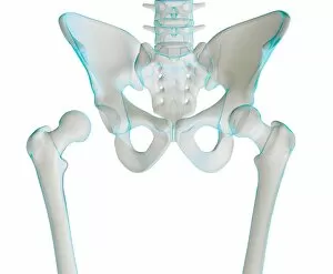
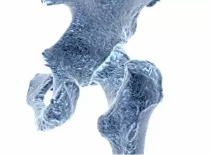
Background imageOsteology Collection: Hip joint bones and anatomy, artwork C014 / 2031

Hip joint bones and anatomy, artwork C014 / 2031
Hip joint bones and anatomy. Artwork of the bones that form the two hip joints (right and left). The hip is a ball-and-socket joint that provides the mobility needed for walking and running
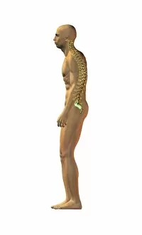
Background imageOsteology Collection: Normal spinal posture, artwork

Normal spinal posture, artwork
Normal spinal posture. Artwork of a man standing in an upright posture with normal spinal alignment. The bones (vertebrae) of the spine are shown

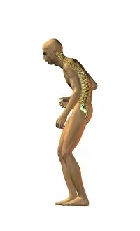
Background imageOsteology Collection: Spinal disorder onset, artwork

Spinal disorder onset, artwork
Spinal disorder onset. Artwork of a man standing in a hunched posture demonstrating the onset of deteriorating spinal alignment

Background imageOsteology Collection: Spinal disorder, artwork

Spinal disorder, artwork
Spinal disorder. Artwork of a man standing in a markedly hunched posture demonstrating the onset of deteriorating spinal alignment

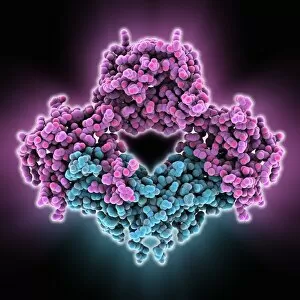
Background imageOsteology Collection: Bone morphogenetic protein complex

Bone morphogenetic protein complex, molecular model. Bone Morphogenetic Protein-7 (BMP-7, blue) in complex with the secreted antagonist Noggin (pink)

Background imageOsteology Collection: Bones of the human body, artwork C016 / 7493

Bones of the human body, artwork C016 / 7493
Bones of the human body, computer artwork

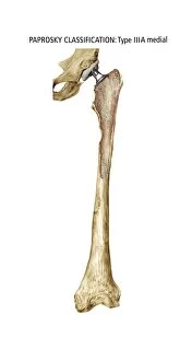
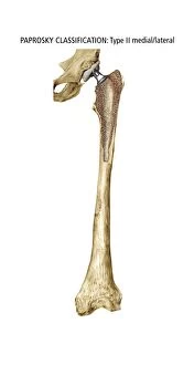
Background imageOsteology Collection: Paprosky femur defect, type IIIA lateral

Paprosky femur defect, type IIIA lateral
Paprosky femur defect. Cutaway artwork of bone degradation in a type IV medial-lateral femur cortex defect (Paprosky classification system)

Background imageOsteology Collection: Paprosky femur defect, type IIIA med-lat

Paprosky femur defect, type IIIA med-lat
Paprosky femur defect. Cutaway artwork of bone degradation in a type IV lateral femur cortex defect (Paprosky classification system)

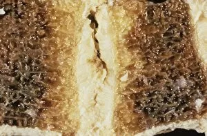
Background imageOsteology Collection: Backbone fat deposition C015 / 6053

Backbone fat deposition C015 / 6053
Backbone fat deposition. Sectioned gross clinical specimen of a vertebra (spinal bone) that displays adiposis (fat deposition, yellow) in the medullary (inner) region

Background imageOsteology Collection: Homo heidelbergensis arthritic jaw C015 / 6562

Homo heidelbergensis arthritic jaw C015 / 6562
Homo heidelbergensis arthritic jaw. Close-up of the condyloid process of the lower jaw (mandible) from fossilised remains of Homo heidelbergensis

Background imageOsteology Collection: Bone cancer cell, SEM

Bone cancer cell, SEM
Bone cancer cell. Coloured scanning electron micrograph of a fibrosarcoma (fibroblastic sarcoma) cell, showing the numerous filopodia (hair-like) covering its surface

Background imageOsteology Collection: Bone structure, artwork C016 / 7545

Bone structure, artwork C016 / 7545
Bone structure. Computer artwork showing bone trabeculae. Trabeculae are the bars of bone that form a honeycombed hollow network, and make up the structure of spongy bone, the inner layer of a bone

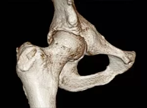
Background imageOsteology Collection: Hip, 3D bone reconstruction from MRI Data C016 / 4694

Hip, 3D bone reconstruction from MRI Data C016 / 4694
Male hip, showing slight arthrosis. 3D bone reconstruction from MRI Data

Background imageOsteology Collection: Hip, 3D bone reconstruction from MRI Data C016 / 4614

Hip, 3D bone reconstruction from MRI Data C016 / 4614
Male hip, showing slight arthrosis. 3D bone reconstruction from MRI Data

Background imageOsteology Collection: Hip, 3D bone reconstruction from MRI Data C016 / 4611

Hip, 3D bone reconstruction from MRI Data C016 / 4611
Male hip, showing slight arthrosis. 3D bone reconstruction from MRI Data

Background imageOsteology Collection: Hip, 3D bone reconstruction from MRI Data C016 / 4607

Hip, 3D bone reconstruction from MRI Data C016 / 4607
Male hip, showing slight arthrosis. 3D bone reconstruction from MRI Data

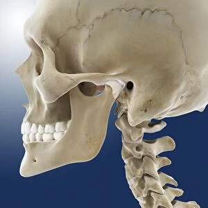
Background imageOsteology Collection: Articular disk of the jaw, artwork C016 / 2889

Articular disk of the jaw, artwork C016 / 2889
Articular disk of the jaw. Artwork of a side view of a human skull with the jaws closed, showing the articular disk (white) and other cartilage of the temporomandibular joint

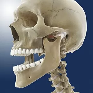
Background imageOsteology Collection: Articular disk of the jaw, artwork C016 / 2890

Articular disk of the jaw, artwork C016 / 2890
Articular disk of the jaw. Artwork of a side view of a human skull with the jaws open, showing the articular disk (white) and other cartilage of the temporomandibular joint

Background imageOsteology Collection: Humeral bone marrow cavity, artwork C016 / 2888

Humeral bone marrow cavity, artwork C016 / 2888
Humeral bone marrow cavity. Artwork of a sectioned upper arm bone (humerus), one of the long bones of the skeleton. These carry most of the load in the arms and legs

Background imageOsteology Collection: Humeral bone marrow cavity, artwork C016 / 2887

Humeral bone marrow cavity, artwork C016 / 2887
Humeral bone marrow cavity. Artwork of a sectioned upper arm bone (humerus), one of the long bones of the skeleton. These carry most of the load in the arms and legs

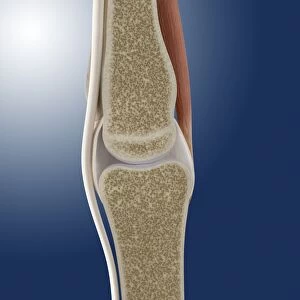
Background imageOsteology Collection: Phalanx joint anatomy, artwork C016 / 2884

Phalanx joint anatomy, artwork C016 / 2884
Phalanx joint anatomy. Artwork of a sectioned joint between two phalanx bones (phalanges). These are found in the fingers and toes

Background imageOsteology Collection: Long bone interior, artwork C016 / 2886

Long bone interior, artwork C016 / 2886
Long bone interior. Artwork of a cutaway sectioned portion of the upper arm bone (humerus). This is one of the long bones of the skeleton, the ones that carry most of the load in the arms and legs


For sale as Licensed Images

Choose your image, Select your licence and Download the media

Osteology, the fascinating study of bones and skeletal structures, unveils a world of intricate details and medical advancements. From total hip replacements to X-ray imaging, this field has revolutionized the way we understand and treat bone-related conditions. In the realm of orthopedics, total hip replacement surgeries have become a game-changer for those suffering from debilitating joint pain. With cutting-edge techniques and prosthetic materials, these procedures restore mobility and improve quality of life. Delving into history, Calots spinal surgery in the 19th century stands as a testament to human resilience. Despite limited resources and knowledge at that time, surgeons courageously ventured into complex spinal operations with remarkable outcomes. Artwork captures both the beauty and vulnerability of our skeletal system. A damaged knee ligament depicted in brushstrokes reminds us of how delicate our bodies can be. Meanwhile, a running skeleton portrayed through art showcases strength and agility even in its bare form. X-ray artwork takes us beneath the surface to reveal hidden stories within our bones. Skeletons captured by this technique offer an ethereal glimpse into their structure while evoking curiosity about their past lives. An X-ray artwork showcasing a skeleton from below adds another dimension to our understanding of anatomy. It invites contemplation on how every angle tells a different story about our body's framework. Sometimes even mundane ailments find artistic expression through X-rays. A headache immortalized as an artwork serves as a reminder that even seemingly minor discomforts can leave traces within us. Fractured ankle X-rays remind us that bones are not invincible; they can break under pressure or unfortunate circumstances. Yet with modern medicine's intervention, healing is possible once again. Microscopic exploration reveals wonders too small for the naked eye to behold. Compact bone viewed under light micrograph exposes its intricate structure like never before – reminding us that there is more than meets the eye when it comes to osteology.