Osteology Collection (#6)

Osteology, the fascinating study of bones and skeletal structures, unveils a world of intricate details and medical advancements

Background imageOsteology Collection: Dislocated elbow, X-ray

Dislocated elbow, X-ray
Dislocated elbow. Coloured lateral X-ray of an injury to the elbow (centre) showing that the upper arm bone (humerus, top right) is misaligned (dislocated, turquoise)

Background imageOsteology Collection: Spondylitis, X-ray

Spondylitis, X-ray
Spondylitis. Coloured X-ray of the lower spine and pelvis of a patient with spondylitis, an inflammation of the synovial joints of the backbone

Background imageOsteology Collection: Fractured atlas vertebra, 3D CT scan

Fractured atlas vertebra, 3D CT scan
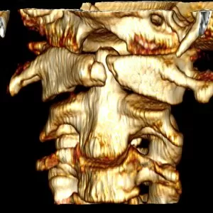
Fractured atlas vertebra, coloured 3D CT (computed tomography) scan. Posterior view of a fractured atlas vertebra (break at upper centre) of a 34 year old man. Part of the skull can be seen at top

Background imageOsteology Collection: Kneecap fracture, X-ray

Kneecap fracture, X-ray
Kneecap fracture. Coloured X-ray of a broken kneecap (patella), as viewed from the side with the knee bent. The kneecap (far left, centre) is a disc of bone that covers the front of the knee joint

Background imageOsteology Collection: Pinned ankle fracture, coloured X-ray

Pinned ankle fracture, coloured X-ray
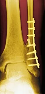
Pinned ankle fracture. Coloured X-ray of the ankle bones of a patient with a pinned ankle fracture. The malleolus (bony projection at the end of a bone) of the fibula (right)

Background imageOsteology Collection: Thalassaemia, X-ray

Thalassaemia, X-ray
Thalassaemia. Coloured hand X-ray of a patient with thalassaemia, also known as Cooleys anaemia. The bones of the hand show demineralization (lighter patches)

Background imageOsteology Collection: Potts disease

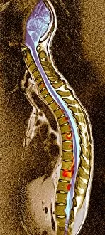
Potts disease. Coloured X-ray of the spine of a patient with Potts disease, or tuberculosis (TB) of the spine. The vertebrae (brown blocks) have become compacted and inflamed (red, yellow)

Background imageOsteology Collection: Healthy skull, coloured X-ray

Healthy skull, coloured X-ray
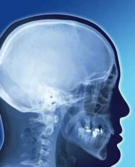
Healthy human skull. Side view X-ray showing the skull and the outline of the head (black). The bones of the cranium are fused together to form a complete structure that protects the brain

Background imageOsteology Collection: Human backbone, artwork

Human backbone, artwork
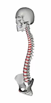
Human backbone, skull and pelvis. Computer artwork of the spine (the backbone or spinal column), seen from an oblique sideways angle, with the front of the spine at left, the skull at top

Background imageOsteology Collection: Bone trabeculae, artwork

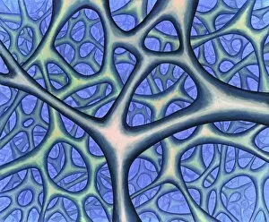
Bone trabeculae, artwork. The trabeculae are the bars of bone forming a honeycombed hollow network. This is the structure of spongy bone, the inner layer of a bone

Background imageOsteology Collection: Bone growth, light micrograph

Bone growth, light micrograph
Bone growth. Light micrograph of actively growing cells in the epiphyseal plate (growth plate) between the diaphysis (shaft) and epiphysis (rounded end) of a long bone

Background imageOsteology Collection: Osteoblasts, TEM

Osteoblasts, TEM
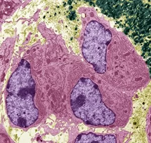
Osteoblasts. Coloured transmission electron micrograph of osteoblasts, bone-producing cells (pink). They contain rough endoplasmic reticulum (RER, dark pink lines), which produces

Background imageOsteology Collection: Bone trabeculae

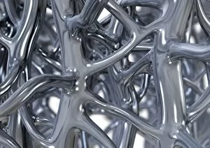
Bone trabeculae. Computer artwork of trabeculae in spongy bone, represented as metal to illustrate its strength. Spongy bone is found inside bones, below a dense shell of hard compact bone

Background imageOsteology Collection: Back deformities

Back deformities, historical anatomical artwork. These patients all have spinal deformities leading to abnormal curvatures of the back

Background imageOsteology Collection: Lower back pain, X-ray artwork

Lower back pain, X-ray artwork
Back pain. Computer X-ray artwork of the rear view of the lower back of a human skeleton. The red area represents lower back pain (lumbago)

Background imageOsteology Collection: Neck pain, X-ray artwork

Neck pain, X-ray artwork
Neck pain. Computer X-ray artwork of a lateral view of a the bones of the neck. The front of the neck is at left. The red area represents neck pain (cervicalgia)

Background imageOsteology Collection: Back pain, X-ray artwork

Back pain, X-ray artwork
Back pain. Computer X-ray artwork of the rear view of a human skeleton. The red area represents back pain (dorsalgia). Back pain usually originates from the muscles, nerves, bones

Background imageOsteology Collection: Arthritic joint

Arthritic joint. Conceptual image of an arthritic joint (red and white) surrounded by healthy joints (white). The healthy joints have smooth, well- defined edges

Background imageOsteology Collection: Osteochondrosis of the neck bones

Osteochondrosis of the neck bones
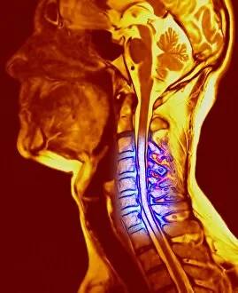
Osteochondrosis of the cervical vertebrae, view from the left side. Computer artwork based on a magnetic resonance imaging (MRI) scan of cervical vertebrae in the neck (blue)

Background imageOsteology Collection: Osteoporotic spongy bone, SEM

Osteoporotic spongy bone, SEM
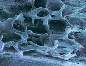
Osteoporotic spongy bone. Coloured scanning electron micrograph (SEM) of the bony network (trabeculae) of spongy (cancellous) bone affected by osteoporosis (brittle bone disease)

Background imageOsteology Collection: Slipped disc, CT scan

Slipped disc, CT scan
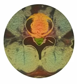
Slipped disc. Coloured computed tomography (CT) scan of an axial section through an inter- vertebral disc (orange) showing herniation

Background imageOsteology Collection: Spinal osteoporosis, X-ray

Spinal osteoporosis, X-ray
Spinal osteoporosis. Coloured X-ray of the lumbar region of the spine of an elderly female patient with osteoporosis. The bones have lost their density and have become porous and fragile

Background imageOsteology Collection: Bone death, X-ray

Bone death, X-ray
Bone death. Coloured X-ray of the shaft of a human long bone showing tissue death (osteonecrosis, brown). This may be caused by a blood clot or embolus that interrupts the blood supply

Background imageOsteology Collection: Arthritic hand

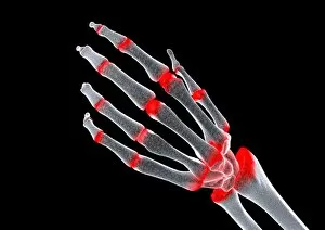
Arthritic hand, computer artwork. The finger and wrist joints are inflamed and swollen. Rheumatoid arthritis is caused by the immune system attacking the bodys own tissues

Background imageOsteology Collection: Arthritic elbow, X-ray

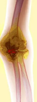
Arthritic elbow, X-ray

Background imageOsteology Collection: Arthritic hand, conceptual artwork

Arthritic hand, conceptual artwork. Arthritis is a degenerative disease that results in the loss of cartilage between joints (red), causing pain and inflammation

Background imageOsteology Collection: Bony spurs in neck, X-ray

Bony spurs in neck, X-ray
Bony spurs in neck, coloured X-ray. Bony spurs (osteophytes, left) grow from the vertebrae as a normal part of the aging process

Background imageOsteology Collection: Normal lower jaw, X-ray

Normal lower jaw, X-ray
Normal lower jaw. Coloured profile X-ray of a human mandible (lower jaw). This is attached to the skull (top) by the temporomandibular joint (TMJ, upper left)
Background imageOsteology Collection: Amputated toe, X-ray

Amputated toe, X-ray
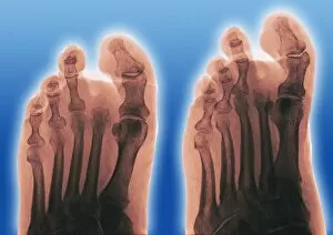
Amputated toe. Coloured frontal (left) and oblique (right) X-rays of a foot of a diabetic, showing an amputated second toe

Background imageOsteology Collection: Vertebra, SEM

Vertebra, SEM
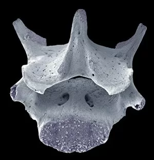
Vertebra. Coloured scanning electron micrograph (SEM) of a single vertebra, part of the vertebral column (spine). The spine is a column of 33 vertebrae (in humans)

Background imageOsteology Collection: Pinned foot bone fracture, X-ray

Pinned foot bone fracture, X-ray
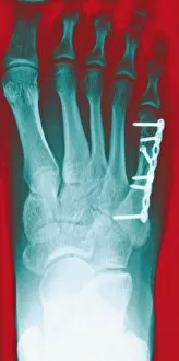
Pinned foot bone fracture. Coloured X-rays of the right foot, showing a metal plate and screws (white) in the foot bone beneath the little toe (upper right)

Background imageOsteology Collection: Temporary ankle immobilisation, X-ray

Temporary ankle immobilisation, X-ray
Temporary ankle immobilisation, coloured profile X-ray. These long pins (white rods) are holding the bone fragments in position until they can be operated on and reset properly

Background imageOsteology Collection: Normal torso, CT scan

Normal torso, CT scan
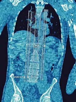
Normal torso, coloured frontal computed tomography (CT) scan. This is a scout image showing the bones of the spine (vertebrae, centre)

Background imageOsteology Collection: Bunion, X-ray

Bunion, X-ray
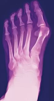
Bunion. Coloured X-ray of a bunion (hallux valgus, upper right), the swelling of the joint between the big toe and the first metatarsal bone in the foot

Background imageOsteology Collection: Pinned wrist fracture, X-ray

Pinned wrist fracture, X-ray
Pinned wrist fracture, coloured frontal X-ray. A pi-plate (white) has been inserted to treat a displaced distal radius (large lower arm bone) fracture

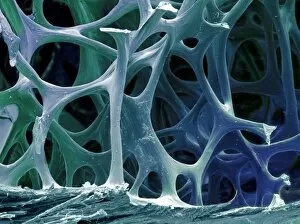
Background imageOsteology Collection: Osteoporotic bone, SEM

Osteoporotic bone, SEM
Osteoporotic bone, coloured scanning electron micrograph (SEM). Osteoporosis (brittle bone disease) causes a reduction in bone density and an increase in porosity

Background imageOsteology Collection: Fractured hip bone, artwork

Fractured hip bone, artwork
Fractured hip bone. Computer artwork of the bones of the hip showing a break (yellow) in the femoral neck

Background imageOsteology Collection: Bird bone tissue, SEM

Bird bone tissue, SEM
Bird bone tissue. Coloured scanning electron micrograph (SEM) of cancellous (spongy) bone from a robin. This tissue, found in the interior of bones

Background imageOsteology Collection: Lumbar spine and sacrum, computer artwork

Lumbar spine and sacrum, computer artwork
Lumbar spine and sacrum, side view, computer artwork. The bones of the spine (vertebrae) are blue and the intervertebral discs are red. The sacrum is at bottom

Background imageOsteology Collection: Lumbar spine, computer artwork

Lumbar spine, computer artwork
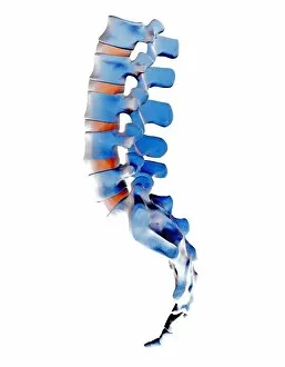
Lumbar spine, side view, computer artwork. The bones of the spine (vertebrae) are blue and the intervertebral discs are red

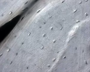
Background imageOsteology Collection: Primate finger bone, SEM

Primate finger bone, SEM

Background imageOsteology Collection: Bone-setting mechanism, 18th century

Bone-setting mechanism, 18th century

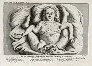
Background imageOsteology Collection: Bone softening, 18th century

Bone softening, 18th century

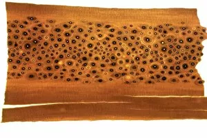
Background imageOsteology Collection: Whale bone sample, light micrograph

Whale bone sample, light micrograph

Background imageOsteology Collection: Bone tissue, light micrograph

Bone tissue, light micrograph


For sale as Licensed Images

Choose your image, Select your licence and Download the media

Osteology, the fascinating study of bones and skeletal structures, unveils a world of intricate details and medical advancements. From total hip replacements to X-ray imaging, this field has revolutionized the way we understand and treat bone-related conditions. In the realm of orthopedics, total hip replacement surgeries have become a game-changer for those suffering from debilitating joint pain. With cutting-edge techniques and prosthetic materials, these procedures restore mobility and improve quality of life. Delving into history, Calots spinal surgery in the 19th century stands as a testament to human resilience. Despite limited resources and knowledge at that time, surgeons courageously ventured into complex spinal operations with remarkable outcomes. Artwork captures both the beauty and vulnerability of our skeletal system. A damaged knee ligament depicted in brushstrokes reminds us of how delicate our bodies can be. Meanwhile, a running skeleton portrayed through art showcases strength and agility even in its bare form. X-ray artwork takes us beneath the surface to reveal hidden stories within our bones. Skeletons captured by this technique offer an ethereal glimpse into their structure while evoking curiosity about their past lives. An X-ray artwork showcasing a skeleton from below adds another dimension to our understanding of anatomy. It invites contemplation on how every angle tells a different story about our body's framework. Sometimes even mundane ailments find artistic expression through X-rays. A headache immortalized as an artwork serves as a reminder that even seemingly minor discomforts can leave traces within us. Fractured ankle X-rays remind us that bones are not invincible; they can break under pressure or unfortunate circumstances. Yet with modern medicine's intervention, healing is possible once again. Microscopic exploration reveals wonders too small for the naked eye to behold. Compact bone viewed under light micrograph exposes its intricate structure like never before – reminding us that there is more than meets the eye when it comes to osteology.