
Research Collection (#96)

"Unveiling the Secrets

Background imageResearch Collection: HGPRTase molecule F006 / 9270

HGPRTase molecule F006 / 9270
HGPRTase. Molecular model of hypoxanthine-guanine phosphoribosyltransferase (HGPRTase) from the malaria parasite Plasmodium falciparum. HGPRTase is involved in the purine salvage pathway

Background imageResearch Collection: Bleomycin hydrolase molecule F006 / 9265

Bleomycin hydrolase molecule F006 / 9265
Bleomycin hydrolase. Molecular model of the cysteine protease bleomycin hydrolase. This enzyme is thought to be a major cause of resistance to the bleomycin chemotherapy drug

Background imageResearch Collection: Gene activator protein F006 / 9269

Gene activator protein F006 / 9269
Gene activator protein. Molecular model of catabolite gene activator protein (CAP, pink and green) bound to a molecule of deoxyribonucleic acid (DNA, across top)

Background imageResearch Collection: TATA box-binding protein and DNA F006 / 9267

TATA box-binding protein and DNA F006 / 9267
TATA box-binding protein and DNA. Molecular model showing a TATA box-binding protein (TBP) complexed with a strand of DNA (deoxyribonucleic acid, red and blue)

Background imageResearch Collection: CD4 protein molecule F006 / 9266

CD4 protein molecule F006 / 9266
CD4 protein. Molecular model of the CD4 T cell (T lymphocyte) receptor protein. CD4 binds to the surface proteins on other cells and pathogenic micro-organisms

Background imageResearch Collection: Calcium-binding protein molecule F006 / 9268

Calcium-binding protein molecule F006 / 9268
Calcium-binding protein. Molecule model of the calcium-binding protein calmodulin (CaM). This protein is found in all eukaryotic cells

Background imageResearch Collection: E coli Holliday junction complex F006 / 9261

E coli Holliday junction complex F006 / 9261
E. coli Holliday junction complex. Molecular model of a RuvA protein (red) in complex with a Holliday junction between homologous strands of DNA (deoxyribonucleic acid, blue) from an E

Background imageResearch Collection: Metabolic enzyme molecule F006 / 9262

Metabolic enzyme molecule F006 / 9262
Metabolic enzyme. Molecular model of the enzyme aconitase, which is involved in the citric acid (or Krebs) cycle. The citric acid cycle is the process by which mitochondria convert glucose to energy

Background imageResearch Collection: Collagen-like molecule F006 / 9264

Collagen-like molecule F006 / 9264
Collagen-like molecule. Molecular model showing the triple helical structure of a collagen-like molecule

Background imageResearch Collection: Carbonic anhydrase II molecule F006 / 9263

Carbonic anhydrase II molecule F006 / 9263
Carbonic anhydrase II, molecular model. This enzyme catalyses the reversible hydration of carbon dioxide

Background imageResearch Collection: Bacteriorhodopsin protein F006 / 9260

Bacteriorhodopsin protein F006 / 9260
Bacteriorhodopsin protein. Molecular model showing the structure of bacteriorhodopsin (bR), a protein found in primitive micro-organisms known as Archaea. This protein acts as a proton pump

Background imageResearch Collection: ATP synthase molecule F006 / 9258

ATP synthase molecule F006 / 9258
ATP synthase molecule. Molecular model showing the structure of ATP synthase (ATPase) subunit A and C. ATPase is an important enzyme that provides energy for cells through the synthesis of adenosine

Background imageResearch Collection: Diels-Alder antibody catalyst molecule F006 / 9259

Diels-Alder antibody catalyst molecule F006 / 9259
Diels-Alder antibody catalyst. Molecular model of an antibody that can be used to catalyse the Diels-Alder reaction

Background imageResearch Collection: Bioluminescent enzyme molecule F006 / 9256

Bioluminescent enzyme molecule F006 / 9256
Bioluminescent enzyme. Molecular model of the bioluminescent enzyme luciferase from the marine bacteria Vibrio harveyi

Background imageResearch Collection: Myosin molecule F006 / 9255

Myosin molecule F006 / 9255
Myosin. Molecular molecule of a smooth muscle myosin. Myosins are a large family of motor proteins that are responsible for muscle contraction in eukaryotic tissues

Background imageResearch Collection: Erythropoietin hormone molecule F006 / 9257

Erythropoietin hormone molecule F006 / 9257
Erythropoietin. Molecular model of the human erythropoietin (EPO) hormone. Erythropoietin regulates blood oxygen levels in the body

Background imageResearch Collection: Eye lens protein molecule F006 / 9253

Eye lens protein molecule F006 / 9253
Eye lens protein. Molecular model of beta-crystallin, a protein found in the lens of the eye. The regular arrangement of the protein in the lens is thought to be responsible for its transparency

Background imageResearch Collection: Beta-galactosidase molecule F006 / 9250

Beta-galactosidase molecule F006 / 9250
Beta-galactosidase, molecular model. This enzyme breaks down sugars containing galactose, such as lactose, into their basic units (monosaccharides)

Background imageResearch Collection: Type II topoisomerase molecule F006 / 9251

Type II topoisomerase molecule F006 / 9251
Type II topoisomerase, molecular model. The topoisomerase enzymes assist in uncoiling DNA (deoxyribonucleic acid). DNA is usually stored in a supercoiled form

Background imageResearch Collection: Human tissue factor molecule F006 / 9254

Human tissue factor molecule F006 / 9254
Human tissue factor. Molecular model of human tissue factor, also known as factor III. Tissue factor is found on the outside of blood vessels and plays a role in the coagulation cascade

Background imageResearch Collection: Nerve growth factor molecule F006 / 9249

Nerve growth factor molecule F006 / 9249
Nerve growth factor (NGF), molecular model. NGF is a small protein, which is involved in the growth, differentiation (specialisation of cells) and survival of particular target neurones (nerve cells)

Background imageResearch Collection: Collagen-like molecule T3-785 F006 / 9252

Collagen-like molecule T3-785 F006 / 9252
Collagen-like molecule. Molecular model showing the triple helical structure of the collagen-like molecule T3-785

Background imageResearch Collection: Hexokinase and glucose complex F006 / 9248

Hexokinase and glucose complex F006 / 9248
Hexokinase and glucose complex. Molecular model of a complex between the enzyme hexokinase and the sugar glucose. Hexokinase promotes the conversion (phosphorylation)

Background imageResearch Collection: T cell receptor B7 molecule F006 / 9247

T cell receptor B7 molecule F006 / 9247
T cell receptor B7. Molecular model of the T cell receptor B7 bound to the viral Tax peptide and MHC class I molecule HLA-A2

Background imageResearch Collection: Myosin fragment molecule F006 / 9245

Myosin fragment molecule F006 / 9245
Myosin fragment. Molecular molecule of a fragment of striated muscle myosin complexed with ADP (adenosine diphosphate). Myosins are a large family of motor proteins that are responsible for muscle

Background imageResearch Collection: Scorpion toxin molecule F006 / 9246

Scorpion toxin molecule F006 / 9246
Scorpion toxin. Molecular model of the neurotoxin BJXTR-IT from the Hottentotta judaicus scorpion. This toxin functions by modulating sodium channels

Background imageResearch Collection: Pit-1 transcription factor bound to DNA F006 / 9242

Pit-1 transcription factor bound to DNA F006 / 9242
Pit-1 transcription factor bound to DNA. Molecular model showing pituitary-specific positive transcription factor 1 (Pit-1) (yellow and pink) bound to a strand of DNA (deoxyribonucleic acid)

Background imageResearch Collection: Leptin molecule F006 / 9243

Leptin molecule F006 / 9243
Leptin molecule. Molecular model of the human hormone leptin. Leptin is a protein produced by adipose (fat) tissue. It interacts with receptors in the brains hypothalamus to signal when a person is

Background imageResearch Collection: Funnel-web spider toxin molecule F006 / 9244

Funnel-web spider toxin molecule F006 / 9244
Funnel-web spider toxin. Molecular model of the atracotxin-HV1 neurotoxin from the Australian funnel-web spider (Hadronyche versuta)

Background imageResearch Collection: Chaperonin protein complex F006 / 9236

Chaperonin protein complex F006 / 9236
Chaperonin protein complex. Molecular model showing the structure of a GroEL GroES (ADP)7 chaperonin complex. Chaperonins are proteins that provide favourable conditions for the correct folding of

Background imageResearch Collection: cAMP-dependent protein kinase molecule F006 / 9240

cAMP-dependent protein kinase molecule F006 / 9240
cAMP-dependent protein kinase. Molecular model of cAMP-dependent protein kinase complexed with a peptide inhibitor and ATP (adenosine triphosphate)

Background imageResearch Collection: Human interferon beta molecule F006 / 9241

Human interferon beta molecule F006 / 9241
Human interferon beta, molecular model. Interferons are proteins produced by white blood cells as part of the immune response to invading pathogens, especially viruses

Background imageResearch Collection: Aspartyl-tRNA synthetase molecule F006 / 9238

Aspartyl-tRNA synthetase molecule F006 / 9238
Aspartyl-tRNA synthetase protein molecule. Molecular model showing bacterial aspartyl-tRNA synthetase complexed with aspartyl tRNA (transfer ribonucleic acid)

Background imageResearch Collection: DNase I complex, molecular model F006 / 9239

DNase I complex, molecular model F006 / 9239
DNase I complex. Molecular model of deoxyribonuclease I complexed with actin

Background imageResearch Collection: Oestrogen sulfotransferase molecule F006 / 9237

Oestrogen sulfotransferase molecule F006 / 9237
Oestrogen sulfotransferase. Molecular model of the enzyme oestrogen sulfotransferase complexed with oestradiol and ADP (adenosine diphosphate)

Background imageResearch Collection: Nucleosome molecule F006 / 9235

Nucleosome molecule F006 / 9235
Nucleosome, molecular model. A nucleosome is a subunit of chromatin, the substance that forms chromosomes. It consists of a short length of DNA (deoxyribonucleic acid)

Background imageResearch Collection: Cellulosome-integrating protein fragment F006 / 9234

Cellulosome-integrating protein fragment F006 / 9234
Cellulosome-integrating protein fragment. Molecular model of the cohesion domain of the cellulosome-integrating protein A (CipA). Cip A acts as a scaffolding protein for the cellulolytic complex

Background imageResearch Collection: Interleukin-6, molecular model F006 / 9232

Interleukin-6, molecular model F006 / 9232
Interleukin-6. Molecular model of the cytokine protein human interleukin-6. This protein is produced in the body and has a wide variety of functions in the immune system

Background imageResearch Collection: DNA gyrase protein segment F006 / 9233

DNA gyrase protein segment F006 / 9233
DNA gyrase protein segment, molecular model. The DNA gyrase enzyme supercoils DNA (deoxyribonucleic acid) for packaging within the cell nucleus

Background imageResearch Collection: Beta glucanase enzyme molecule F006 / 9231

Beta glucanase enzyme molecule F006 / 9231
Beta glucanase enzyme, molecular model. This enzyme hydrolyses the glucose subunits that make up some polysaccharides. This is a circular permeation of the enzyme

Background imageResearch Collection: TATA box-binding protein complex F006 / 9230

TATA box-binding protein complex F006 / 9230
TATA box-binding protein complex. Molecular model showing a yeast TATA box-binding protein (TBP) complexed with a strand of DNA (deoxyribonucleic acid, red and blue) and transcription factor IIB

Background imageResearch Collection: Acetylcholinesterase molecule F006 / 9226

Acetylcholinesterase molecule F006 / 9226
Acetylcholinesterase, molecular model. This enzyme hydrolyses the neurotransmitter acetylcholine, stopping signal transmission

Background imageResearch Collection: Alcohol dehydrogenase molecule F006 / 9228

Alcohol dehydrogenase molecule F006 / 9228
Alcohol dehydrogenase, molecular model. Alcohol dehydrogenase (ADH) is an enzyme that facilitates the break-down of alcohols in the body, which could otherwise be toxic

Background imageResearch Collection: Metabolic enzyme molecule F006 / 9227

Metabolic enzyme molecule F006 / 9227
Metabolic enzyme. Molecular model of the enzyme aconitase, which is involved in the citric acid (or Krebs) cycle. The citric acid cycle is the process by which mitochondria convert glucose to energy

Background imageResearch Collection: Anthrax protective antigen molecule F006 / 9229

Anthrax protective antigen molecule F006 / 9229
Anthrax protective antigen molecule. Computer model showing the structure of a molecule of protective antigen (PA) produced by anthrax (Bacillus anthracis) bacteria

Background imageResearch Collection: Anthrax protective antigen molecule F006 / 9225

Anthrax protective antigen molecule F006 / 9225
Anthrax protective antigen molecule. Computer model showing the structure of a molecule of protective antigen (PA) produced by anthrax (Bacillus anthracis) bacteria

Background imageResearch Collection: Oestrogen receptor and oestradiol F006 / 9222

Oestrogen receptor and oestradiol F006 / 9222
Oestrogen receptor and oestradiol. Molecular model of a n oestrogen receptor bound to the female sex hormone oestradiol. Oestradiol is one of three naturally-occurring oestrogens

Background imageResearch Collection: Tetanus toxin C-fragment molecule F006 / 9223

Tetanus toxin C-fragment molecule F006 / 9223
Tetanus toxin C-fragment. Molecular model of a fragment of the neurotoxin protein produced by the bacterium Clostridium tetani that causes tetanus


For sale as Licensed Images

Choose your image, Select your licence and Download the media

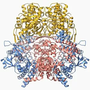
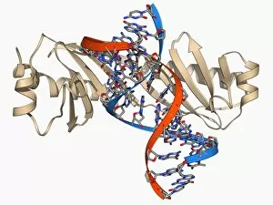
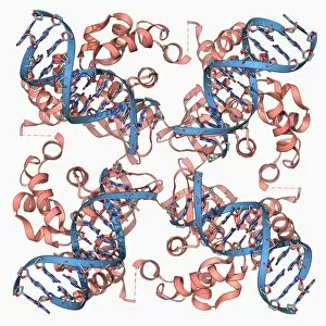
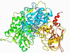
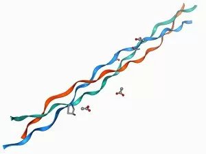
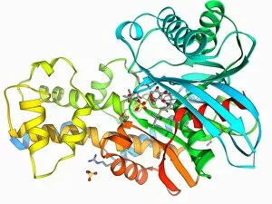
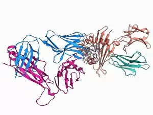
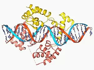
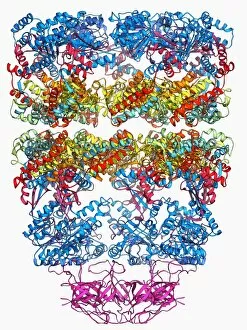
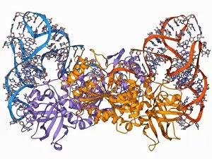
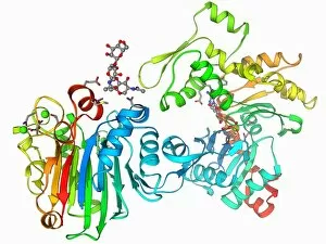
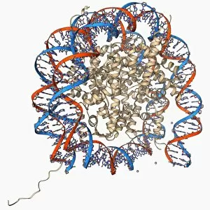
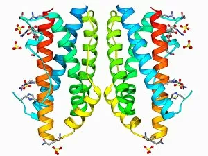
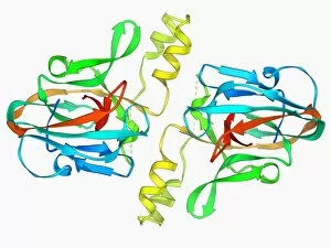
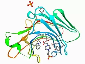
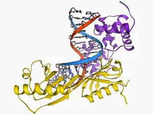
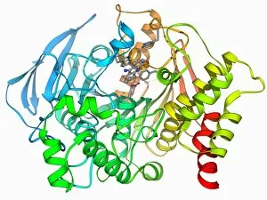
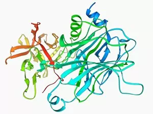
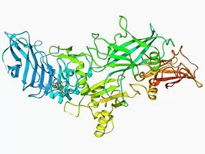
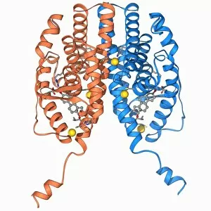
"Unveiling the Secrets: Exploring the Vast Realm of Research" From deciphering the valorous tales behind British Military medals to unraveling the enigmatic patterns in a Rorschach Inkblot Test, research takes us on an intellectual odyssey. It delves into realms beyond our comprehension, like scrutinizing the cosmic microwave background through MAP and decoding intricate histological diagrams of a mammalian retina. Research is not confined to laboratories; it extends to observing nature's wonders firsthand. Jane Goodall, with her unwavering dedication as a British conservationist and zoologist, captured alongside a chimpanzee, epitomizes how research bridges gaps between species. The quest for knowledge transcends boundaries of time and space. Erwin Schrodinger's groundbreaking discoveries in quantum mechanics remind us that research propels humanity forward by challenging conventional wisdom. In particle physics, proton collisions such as C014 / 1797 or witnessing Higgs boson events like C013 / 6892 within ATLAS detectors demonstrate how researchers strive to unlock mysteries at subatomic levels. Pioneers like Marie Curie exemplify relentless pursuit despite adversities faced by women in science. Her revolutionary work with radioactivity continues to inspire generations. It also embarks on daring expeditions akin to HMS Beagle - Darwin's ship that sailed uncharted waters - pushing boundaries of exploration and understanding. Similarly, Apollo 17 astronauts left indelible footprints on the Moon while expanding human horizons beyond Earth's confines. These captivating glimpses into diverse facets highlight its transformative power. It fuels curiosity, challenges assumptions, and empowers us with knowledge that shapes our world. Whether investigating microscopic particles or exploring vast ecosystems – research illuminates paths towards progress and inspires future generations to embark upon their own quests for discovery.