Research Collection (#97)

"Unveiling the Secrets

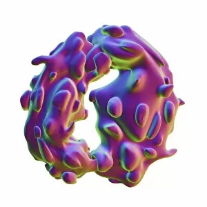
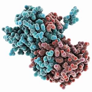
Background imageResearch Collection: Amyloid precursor protein molecule F006 / 9224

Amyloid precursor protein molecule F006 / 9224
Amyloid precursor protein. Molecular model showing the structure of the protease inhibitor domain of an amyloid precursor protein (APP)

Background imageResearch Collection: Type I topoisomerase bound to DNA F006 / 9221

Type I topoisomerase bound to DNA F006 / 9221
Type I topoisomerase bound to DNA. Molecular model showing a type I topoisomerase molecule (khaki) bound to a strand of DNA (deoxyribonucleic acid, red and blue)

Background imageResearch Collection: Type I topoisomerase bound to DNA F006 / 9220

Type I topoisomerase bound to DNA F006 / 9220
Type I topoisomerase bound to DNA. Molecular model showing a type I topoisomerase molecule (khaki) bound to a strand of DNA (deoxyribonucleic acid, red and blue)

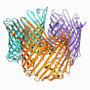
Background imageResearch Collection: Sucrose-specific porin molecule F006 / 9218

Sucrose-specific porin molecule F006 / 9218
Sucrose-specific porin, molecular model. Porins are proteins that span cell membranes and act as a channel through which specific molecules can diffuse

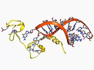
Background imageResearch Collection: HIV nucleocapsid protein molecule F006 / 9219

HIV nucleocapsid protein molecule F006 / 9219
HIV nucleocapsid protein. Molecular model of the nucleocapsid protein (yellow) from HIV-1 (human immunodeficiency virus-type 1) complexed with the Psi RNA (ribonucleic acid) packaging element (orange)

Background imageResearch Collection: ATP-dependent DNA ligase molecule F006 / 9217

ATP-dependent DNA ligase molecule F006 / 9217
ATP dependent DNA ligase. Molecular model of ATP-dependent DNA ligase complexed with ATP (adenosine triphosphate). DNA ligase is an enzyme that binds two strands of DNA (deoxyribonucleic acid)

Background imageResearch Collection: Bacteriophage T4 lysozyme molecule F006 / 9216

Bacteriophage T4 lysozyme molecule F006 / 9216
Bacteriophage T4 lysozyme, molecular model. Lysozymes are enzymes that disrupt the polysaccharide components of bacterial cell walls, leaving them susceptible to destruction

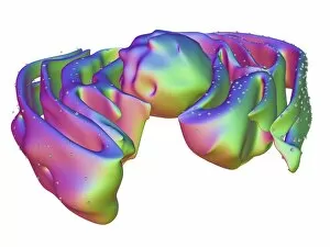
Background imageResearch Collection: Golgi apparatus, artwork F006 / 9211

Golgi apparatus, artwork F006 / 9211
Computer artwork of the Golgi apparatus of the human cell. This organelle functions as a central delivery system for the cell

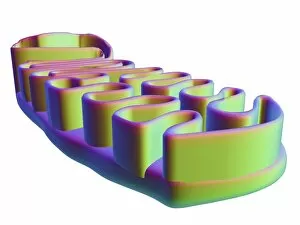
Background imageResearch Collection: Mitochondrial structure, artwork F006 / 9207

Mitochondrial structure, artwork F006 / 9207
Mitochondrial structure. Computer artwork of a mitochondrion, showing a loop of mitochondrial DNA (deoxyribonucleic acid, mtDNA), the organelles genetic material

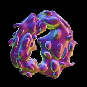
Background imageResearch Collection: Ribosome, artwork F006 / 9206

Ribosome, artwork F006 / 9206
Computer artwork of a ribosome. Ribosomes are protein particles that are found in cell cytoplasm. Each ribosome has a large and a small subunit

Background imageResearch Collection: Neural cells F006 / 9203

Neural cells F006 / 9203
Computer artwork depicting nerve cells and neural communication

Background imageResearch Collection: Nucleus and endoplasmic reticulum F006 / 9201

Nucleus and endoplasmic reticulum F006 / 9201
Computer artwork showing part of a human or eukaryotic cell. In the middle the nucleus which has a membrane with nuclear pores. Inside the nucleus is the DNA

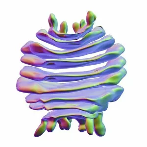
Background imageResearch Collection: Golgi apparatus, artwork F006 / 9199

Golgi apparatus, artwork F006 / 9199
Computer artwork of the Golgi apparatus of the human cell. This organelle functions as a central delivery system for the cell

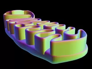
Background imageResearch Collection: Mitochondrial structure, artwork F006 / 9198

Mitochondrial structure, artwork F006 / 9198
Mitochondrial structure. Computer artwork of a mitochondrion, showing a loop of mitochondrial DNA (deoxyribonucleic acid, mtDNA), the organelles genetic material

Background imageResearch Collection: Ribosome, artwork F006 / 9194

Ribosome, artwork F006 / 9194
Computer artwork of a ribosome. Ribosomes are protein particles that are found in cell cytoplasm. Each ribosome has a large and a small subunit

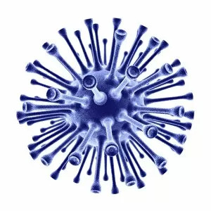
Background imageResearch Collection: H1N1 flu virus particle, artwork F006 / 9186

H1N1 flu virus particle, artwork F006 / 9186
H1N1 flu virus particle. Computer artwork of an H1N1 influenza A (flu) virus particle (virion). In the particles lipid envelope (purple) are two types of protein spike

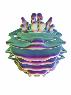
Background imageResearch Collection: Golgi apparatus, artwork F006 / 9189

Golgi apparatus, artwork F006 / 9189
Computer artwork of the Golgi apparatus of the human cell. This organelle functions as a central delivery system for the cell

Background imageResearch Collection: Venus probe, artwork C017 / 0785

Venus probe, artwork C017 / 0785
Venus probe. Computer artwork showing the planet Venus with a space probe (bottom left) in its orbit and a group of landers (lower left) that have been released into the planets atmosphere

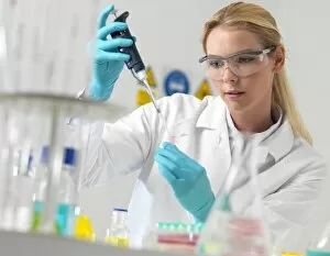
Background imageResearch Collection: Researcher pipetting liquid F006 / 9084

Researcher pipetting liquid F006 / 9084
MODEL RELEASED. Researcher pipetting liquid

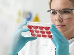
Background imageResearch Collection: Biological research F006 / 9086

Biological research F006 / 9086
MODEL RELEASED. Biological research

Background imageResearch Collection: Researcher pipetting liquid F006 / 9085

Researcher pipetting liquid F006 / 9085
MODEL RELEASED. Researcher pipetting liquid

Background imageResearch Collection: Researcher pipetting liquid F006 / 9083

Researcher pipetting liquid F006 / 9083
MODEL RELEASED. Researcher pipetting liquid

Background imageResearch Collection: Chemistry research F006 / 9082

Chemistry research F006 / 9082
MODEL RELEASED. Chemistry research

Background imageResearch Collection: Chemistry research F006 / 9081

Chemistry research F006 / 9081
MODEL RELEASED. Chemistry research

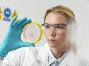
Background imageResearch Collection: Microbiology research F006 / 9080

Microbiology research F006 / 9080
MODEL RELEASED. Microbiology research. Scientist examining cultures in a petri dish

Background imageResearch Collection: Microbiology research F006 / 9079

Microbiology research F006 / 9079
MODEL RELEASED. Microbiology research. Scientist examining cultures in a petri dish

Background imageResearch Collection: Genetic testing of meat, conceptual image F006 / 9070

Genetic testing of meat, conceptual image F006 / 9070
Genetic testing of meat, conceptual image

Background imageResearch Collection: Genetic research, conceptual image F006 / 9068

Genetic research, conceptual image F006 / 9068
Genetic research, conceptual image

Background imageResearch Collection: Genetic research, conceptual image F006 / 9067

Genetic research, conceptual image F006 / 9067
Genetic research, conceptual image

Background imageResearch Collection: Genetic research, conceptual image F006 / 9065

Genetic research, conceptual image F006 / 9065
Genetic research, conceptual image

Background imageResearch Collection: Genetic research, conceptual image F006 / 9066

Genetic research, conceptual image F006 / 9066
Genetic research, conceptual image

Background imageResearch Collection: Light microscope F006 / 9033

Light microscope F006 / 9033
Light microscope. Close up of a slide on a microscope stage

Background imageResearch Collection: Light microscope F006 / 9034

Light microscope F006 / 9034
Light microscope. Close up of a slide on a microscope stage

Background imageResearch Collection: Light microscope F006 / 9032

Light microscope F006 / 9032
Light microscope. Close up of a slide on a microscope stage

Background imageResearch Collection: Haematology lab F006 / 9031

Haematology lab F006 / 9031
MODEL RELEASED. Haematology lab

Background imageResearch Collection: Microbiology research F006 / 9028

Microbiology research F006 / 9028
MODEL RELEASED. Microbiology research

Background imageResearch Collection: Microbiology research F006 / 9027

Microbiology research F006 / 9027
MODEL RELEASED. Microbiology research

Background imageResearch Collection: Scientist using a light microscope F006 / 9029

Scientist using a light microscope F006 / 9029
MODEL RELEASED. Scientist using a light microscope

Background imageResearch Collection: Haematology lab F006 / 9030

Haematology lab F006 / 9030
MODEL RELEASED. Haematology lab

Background imageResearch Collection: Biological research F006 / 9026

Biological research F006 / 9026
MODEL RELEASED. Biological research

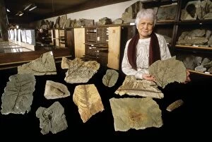
Background imageResearch Collection: Grauvogel-Gall fossil collection, France C018 / 9411

Grauvogel-Gall fossil collection, France C018 / 9411
Grauvogel-Gall fossil collection. French palaeontologist Lea Grauvogel-Stamm (born 1940) with plant, fish and scorpion fossils from the collection she maintains

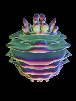
Background imageResearch Collection: SV40 virus capsid, molecular model C018 / 7904

SV40 virus capsid, molecular model C018 / 7904
SV40 virus capsid, molecular model. Simian virus 40 (SV40) is found in monkeys such as Rhesus monkeys and macaques. Potentially tumour-causing, it is used in laboratory research and in vaccines

Background imageResearch Collection: SV40 virus capsid, molecular model C018 / 7903

SV40 virus capsid, molecular model C018 / 7903
SV40 virus capsid, molecular model. Simian virus 40 (SV40) is found in monkeys such as Rhesus monkeys and macaques. Potentially tumour-causing, it is used in laboratory research and in vaccines

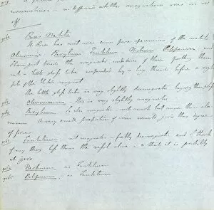
Background imageResearch Collection: Faraday on metals and magnetism, 1847

Faraday on metals and magnetism, 1847
Faraday on metals and magnetism. Page from the notebooks of British physicist Michael Faraday (1791-1867), showing notes and a table of magnetic properties of a range of metals

Background imageResearch Collection: Ebola virus transcription factor fragment

Ebola virus transcription factor fragment. Molecular model of the C-terminal domain (CTD) of Ebola virus transcription factor VP30

Background imageResearch Collection: Faraday on magnetism, 1854

Faraday on magnetism, 1854
Faraday on magnetism. Page from the notebooks of British physicist Michael Faraday (1791-1867), showing notes and a diagram of an experiment with a rotating globe being spun in a magnetic field

Background imageResearch Collection: Nitzan Shipwreck

Nitzan Shipwreck
MV Nitzan was a trawler that was sunk for the purpose of becoming an artificial reef and a diving site. She was sunk on the 8th of April 2012 off the coast of Achziv, Israel

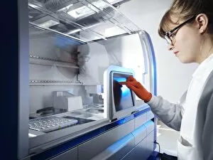
Background imageResearch Collection: Biological sample preparation machine C016 / 9695

Biological sample preparation machine C016 / 9695
MODEL RELEASED. Biological sample preparation machine. This machine is used to purify nucleic acids and proteins from biological samples before further analysis


For sale as Licensed Images

Choose your image, Select your licence and Download the media

"Unveiling the Secrets: Exploring the Vast Realm of Research" From deciphering the valorous tales behind British Military medals to unraveling the enigmatic patterns in a Rorschach Inkblot Test, research takes us on an intellectual odyssey. It delves into realms beyond our comprehension, like scrutinizing the cosmic microwave background through MAP and decoding intricate histological diagrams of a mammalian retina. Research is not confined to laboratories; it extends to observing nature's wonders firsthand. Jane Goodall, with her unwavering dedication as a British conservationist and zoologist, captured alongside a chimpanzee, epitomizes how research bridges gaps between species. The quest for knowledge transcends boundaries of time and space. Erwin Schrodinger's groundbreaking discoveries in quantum mechanics remind us that research propels humanity forward by challenging conventional wisdom. In particle physics, proton collisions such as C014 / 1797 or witnessing Higgs boson events like C013 / 6892 within ATLAS detectors demonstrate how researchers strive to unlock mysteries at subatomic levels. Pioneers like Marie Curie exemplify relentless pursuit despite adversities faced by women in science. Her revolutionary work with radioactivity continues to inspire generations. It also embarks on daring expeditions akin to HMS Beagle - Darwin's ship that sailed uncharted waters - pushing boundaries of exploration and understanding. Similarly, Apollo 17 astronauts left indelible footprints on the Moon while expanding human horizons beyond Earth's confines. These captivating glimpses into diverse facets highlight its transformative power. It fuels curiosity, challenges assumptions, and empowers us with knowledge that shapes our world. Whether investigating microscopic particles or exploring vast ecosystems – research illuminates paths towards progress and inspires future generations to embark upon their own quests for discovery.