Retina Collection (#5)

The retina, a fascinating structure within the human eye, has captivated scientists and artists alike throughout history

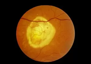
Background imageRetina Collection: Retinal degeneration

Retinal degeneration. Ophthalmoscope view of the retina of a patients eye, showing disciform macular degeneration (yellow patch)

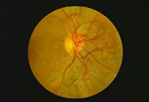
Background imageRetina Collection: Retina damage in diabetes

Retina damage in diabetes
Retinal damage in diabetes. Ophthalmoscope image of diabetic retinopathy, damage to the retina caused by diabetes. Changes to the retinal blood vessels (red) mean that they leak

Background imageRetina Collection: Ophthalmoscopy of disciform macula degeneration

Ophthalmoscopy of disciform macula degeneration
Macula degeneration. Ophthalmoscope view of the retina of a patients eye, showing disciform macula degeneration. At centre right is a large disc-shaped discoloured area covering the macula
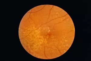
Background imageRetina Collection: Macular degeneration

Macular degeneration, ophthalmoscope image. Numerous yellow drusen spots are seen on the retina, the light-sensitive membrane that lines the back of the eyeball

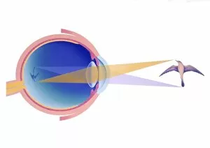
Background imageRetina Collection: Shortsightedness

Shortsightedness. Artwork of section through an eyeball affected by shortsightedness, or myopia. This is a condition in which distant objects appear blurred

Background imageRetina Collection: Light micrograph of monkey retina, high power

Light micrograph of monkey retina, high power
Rods and cones of monkey retina. Light micrograph of a tangential section through the retina of a monkeys eye, showing the rod cells (small) and cone cells (large)

Background imageRetina Collection: Coloured SEM of rod cells in the retina of the eye

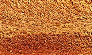
Coloured SEM of rod cells in the retina of the eye
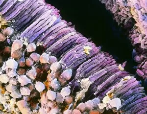
Rod cells. Coloured scanning electron micrograph (SEM) of a group of rod cells in the retina of the eye. Rod cells are light sensitive cells which respond to dim light

Background imageRetina Collection: Optic nerve, light micrograph

Optic nerve, light micrograph
Optic nerve. Coloured light micrograph of a section through the optic nerve. The optic nerve (bright pink) collects impulses from the light sensitive cells of the retina and relays them to the brain

Background imageRetina Collection: Eye structure, light micrograph

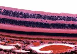
Eye structure, light micrograph
Eye structure. Coloured light micrograph of a section through the inner surface of a human eye. This slice shows the vitreous humour (transparent liquid in the eye, top), the retina (pink)

Background imageRetina Collection: Eye retina

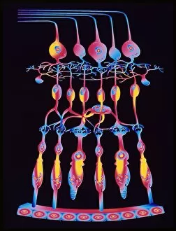
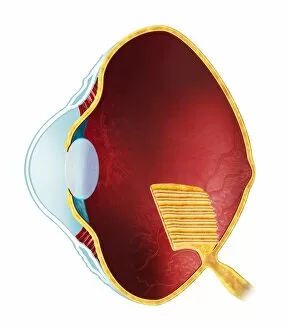
Eye retina
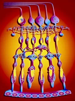
Structure of the retina. Computer artwork showing the structure of the eye retina. The retina is a thin tissue layer on the inner eye responsible for sight. Light strikes from the top

Background imageRetina Collection: Computer artwork of the structure of the retina

Computer artwork of the structure of the retina
Structure of the retina. Computer artwork showing the structure of the eye retina. The retina is a thin tissue layer on the inner eye responsible for sight. Light strikes from the top

Background imageRetina Collection: Blood vessels in eye

Blood vessels in eye
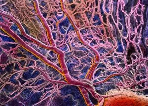
False-colour scanning electron micrograph (SEM) of blood vessels in the choroid of the eye. A branch- ing network of arteries and veins can be seen in this area under the central fovea

Background imageRetina Collection: False-colour SEM of the structure of the retina

False-colour SEM of the structure of the retina
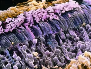
False-colour scanning electron micrograph (SEM) of the eye retina. The retina, which detects visible images, is made up of many layers of cells (seen here)

Background imageRetina Collection: False-colour SEM of rod cells in human retina

False-colour SEM of rod cells in human retina
False-colour scanning electron micrograph (SEM) of rod cells situated in the human retina. Two layers are visible here : one at top (pinkish)

Background imageRetina Collection: Normal retina of eye

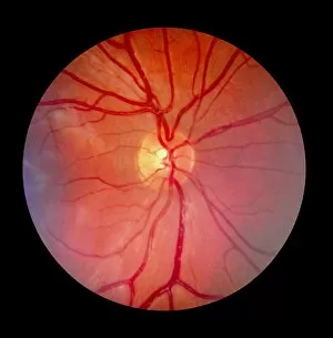
Normal retina of eye
Fundus camera image of the retina of a normal eye, showing the distribution of the retinal veins & arteries: the central retinal artery (a branch of the opthalmic artery)

Background imageRetina Collection: Skate research

Skate research. Skate (Raja erinacea) eye being studied under a microscope at the Woods Hole Marine Biology Laboratory (MBL) in Massachusetts, USA

Background imageRetina Collection: Cell death

Cell death. Computer-enhanced confocal light micrograph of cells in the retina of the eye undergoing programmed cell death (apoptosis)

Background imageRetina Collection: Skates collected for research

Skates collected for research
Skate specimens. Fishing net holding skates (Raja erinacea, with long tails) caught for biological research at the Woods Hole Marine Biology Labora- tory (MBL) in Massachusetts, USA

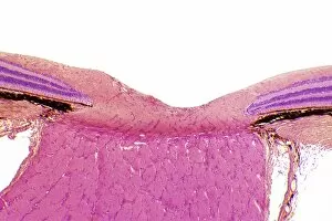
Background imageRetina Collection: Developing pig eye, light micrograph

Developing pig eye, light micrograph. From right the structures seen are: the cornea (brown), the lens (purple), the retina (pink), the choroid (dark brown line) and the sclera (white of the eye)

Background imageRetina Collection: Retina, light micrograph

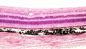
Retina, light micrograph
Retina. Light micrograph of a section through the retina showing its 10 layers. Magnification: x250 when printed at 10 centimetres wide

Background imageRetina Collection: Human eye anatomy, artwork

Human eye anatomy, artwork
Human eye anatomy, computer artwork

Background imageRetina Collection: Bird eye anatomy, artwork

Bird eye anatomy, artwork. The structure of a birds eye is similar to that of humans and other vertebrates, with a clear cornea forming a bulge (left) in front of the lens (oval)

Background imageRetina Collection: Cryptochrome and rhodopsin pigments

Cryptochrome and rhodopsin pigments. Rhodopsin is a pigment found in the rod photoreceptor cells in the retina of the human eye, where it absorbs light falling on the retina

Background imageRetina Collection: Retina blood vessel, SEM

Retina blood vessel, SEM
Retina blood vessel. Coloured scanning electron micrograph (SEM) of a section through a blood vessel in the retina of an eye, showing the red blood cells (red)

Background imageRetina Collection: Retina structure

Retina structure
Retinal structure, artwork. Light falling on the retina passes from top to bottom. At bottom is the choroid layer (red), which lines the inside of the eye

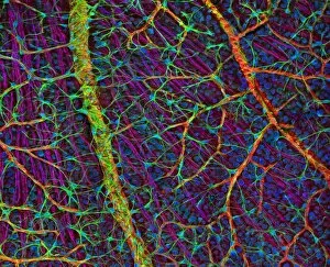
Background imageRetina Collection: Retina blood vessels and nerve cells

Retina blood vessels and nerve cells. Light micrograph showing the blood vessels and nerve cells in the retina of an eye. The retina is the light-sensitive membrane that lines the back of the eyeball

Background imageRetina Collection: Dissected eye, 1844 artwork

Dissected eye, 1844 artwork
Dissected eye. This anatomical artwork is figure 1, plate 78 from volume 3 (1844) of Traite complet de l anatomie de l homme (1831-1854)

Background imageRetina Collection: Retinal rod cell anatomy, diagram

Retinal rod cell anatomy, diagram

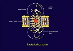
Background imageRetina Collection: Bacteriorhodopsin, diagram

Bacteriorhodopsin, diagram
Bacteriorhodopsin. Diagram of the molecular structure of bacteriorhodopsin, a protein found in primitive micro-organisms known as Archaea

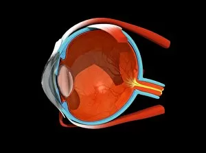
Background imageRetina Collection: Eye anatomy

Eye anatomy. Cutaway artwork passing vertically through a human eye, showing its internal anatomy and structure. The front of the eye is at left, and the structures here include the cornea

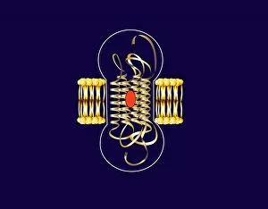
Background imageRetina Collection: Bacteriorhodopsin, artwork

Bacteriorhodopsin, artwork
Bacteriorhodopsin. Artwork of the molecular structure of bacteriorhodopsin, a protein found in primitive micro-organisms known as Archaea

Background imageRetina Collection: Retinal rod cell anatomy, artwork

Retinal rod cell anatomy, artwork
Retinal rod cell anatomy. Artwork of the external (left) and internal (right) anatomical structure of a retinal cell. These cells are found in the retina that lines the back of the eyes

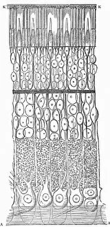
Background imageRetina Collection: Human retina, 19th century artwork

Human retina, 19th century artwork. Artwork from the 1886 ninth edition of Moses and Geology (Samuel Kinns, London). This book was originally published in 1882

Background imageRetina Collection: Retina in glaucoma, artwork

Retina in glaucoma, artwork
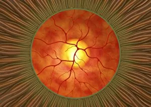
Retina in glaucoma, computer artwork. This view is looking through the pupil (green ring) at the front of the eye, to the retina (orange) at the back of the eye

Background imageRetina Collection: Sensory rhodopsin transducer molecule

Sensory rhodopsin transducer molecule. Computer model showing the molecular structure of sensory rhodopsin transducer (SRT)

Background imageRetina Collection: Gecko retina, SEM

Gecko retina, SEM
Gecko retina. Coloured scanning electron micrograph (SEM) of an oblique freeze-fracture through the retina of a gecko (family Gekkonidae)

Background imageRetina Collection: Vitamin A (retinal) molecule

Vitamin A (retinal) molecule
Vitamin A. Molecular model of the retinal form of vitamin A. Retinal is one of three forms of vitamin A; the other two are retinol and retinoic acid

Background imageRetina Collection: Rhodopsin protein molecule

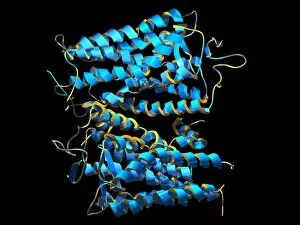
Rhodopsin protein molecule. Computer model of a molecule of the protein opsin that forms part of the rhodopsin complex. Rhodopsin is found in the rod photoreceptor cells in the retina of the human

Background imageRetina Collection: Dogfish head, transverse section

Dogfish head, transverse section
Dogfish head. Light micrograph of a transverse section through the head of a lesser spotted dogfish (Scyliorhinus canicula), in the region of the eyes (upper left and upper right)

Background imageRetina Collection: Rabbit eye, longitudinal section

Rabbit eye, longitudinal section
Rabbit eye. Light micrograph of a longitudinal section through the eye of a rabbit (Oryctolagus cuniculus). The rabbit has a typical mammalian eye structure

Background imageRetina Collection: Recording Eye Prints

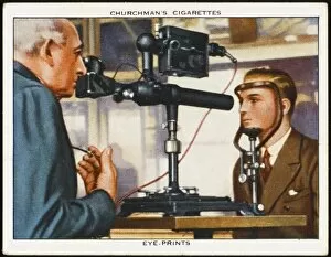
Recording Eye Prints
An invention by two doctors: the eye print; recording the pattern made by veins on the retina, which differs for every individual. Science aiding the criminologist


For sale as Licensed Images

Choose your image, Select your licence and Download the media

The retina, a fascinating structure within the human eye, has captivated scientists and artists alike throughout history. From histological diagrams to intricate artwork, the intricacies of its anatomy have been meticulously studied and depicted. In the 17th century, Descartes' optics theory shed light on how light interacts with the retina. This groundbreaking understanding paved the way for further exploration into this complex organ. An engraving from 1899 beautifully illustrates the external anatomy of the human eye, showcasing how it works in harmony with the retina. The rod and cone cells of the eye are highlighted in another image taken through a scanning electron microscope (SEM), revealing their delicate structures responsible for vision. Advancements in technology have allowed us to delve deeper into studying retinas through biometric scans. These scans provide valuable insights into individual characteristics and contribute to fields like artificial intelligence. A captivating false-color SEM image showcases a central fovea within a retina—an area crucial for sharp vision—highlighting its intricate details that make it an essential part of our visual system. At a molecular level, rhodopsin protein molecules play a vital role in converting light signals received by retinal cells into electrical impulses sent to our brain—a process critical for sight perception. As we explore further, we discover not only its anatomical features but also its network of blood vessels that supply oxygen and nutrients to sustain this remarkable organ's functionality. From ancient theories to modern scientific discoveries, artful depictions or microscopic examinations—the study of retinas continues to intrigue us as we unravel more about one of nature's most incredible creations: our eyes.