Retina Collection (#2)

The retina, a fascinating structure within the human eye, has captivated scientists and artists alike throughout history

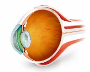
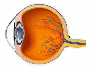
Background imageRetina Collection: Anatomy of human eye, perspective

Anatomy of human eye, perspective

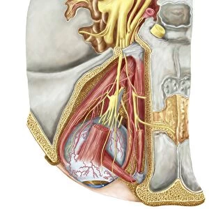
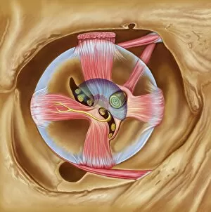
Background imageRetina Collection: Orbital cut showing abducent nerve with ciliary ganglion and oculomotor nerve

Orbital cut showing abducent nerve with ciliary ganglion and oculomotor nerve

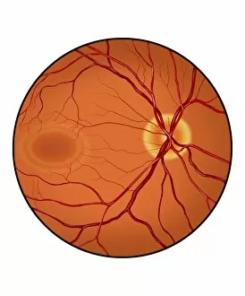
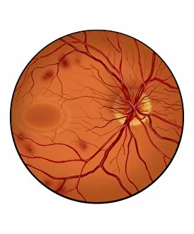
Background imageRetina Collection: Retina of a normal eye

Retina of a normal eye

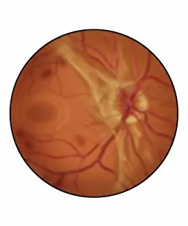
Background imageRetina Collection: Retina with advanced diabetic retinopathy

Retina with advanced diabetic retinopathy, showing increased scar tissue, hemorrhaging and diffuse vitreous blood (blurred appearance)

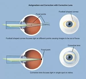
Background imageRetina Collection: Astigmatism and correction with corrective lens

Astigmatism and correction with corrective lens

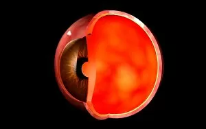
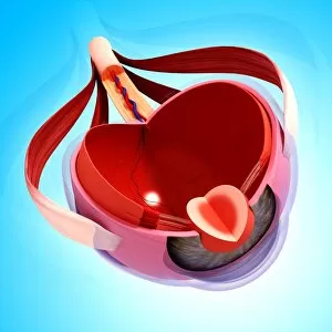
Background imageRetina Collection: Conceptual image of human eye cross section

Conceptual image of human eye cross section

Background imageRetina Collection: Anatomy of human eye

Anatomy of human eye

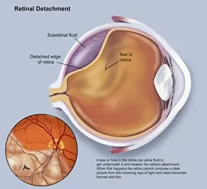
Background imageRetina Collection: Diagram of a retinal detachment

Diagram of a retinal detachment

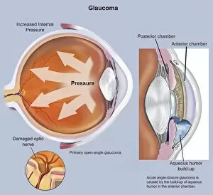
Background imageRetina Collection: Retina of eye with glaucoma

Retina of eye with glaucoma

Background imageRetina Collection: Retina with neovascularization

Retina with neovascularization showing retinal hemorrhaging

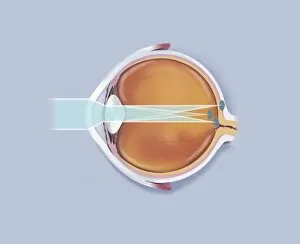
Background imageRetina Collection: Anatomy of human eye showing focal points

Anatomy of human eye showing focal points

Background imageRetina Collection: Abducent nerve in the orbit with lateral rectus muscle

Abducent nerve in the orbit with lateral rectus muscle

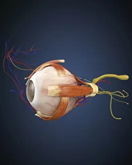
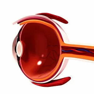
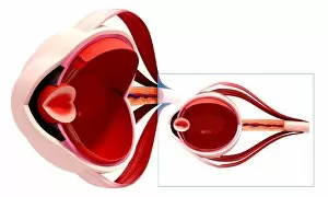
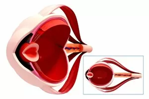
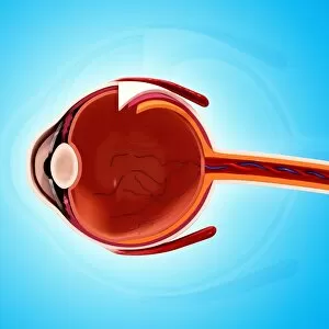
Background imageRetina Collection: Human eye with muscles and circulatory system

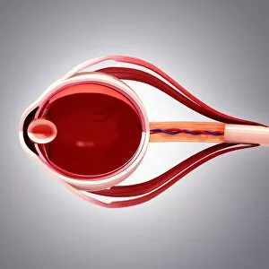
Human eye with muscles and circulatory system

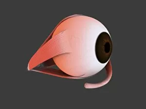
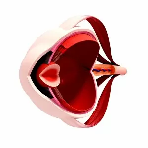
Background imageRetina Collection: Conceptual image of human eye anatomy

Conceptual image of human eye anatomy

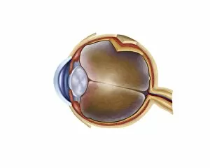
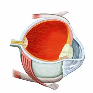
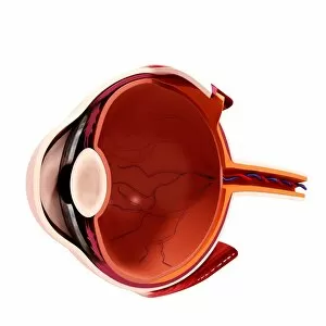
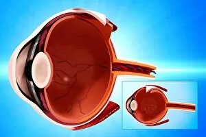
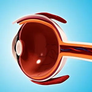
Background imageRetina Collection: Cross section of human eye

Cross section of human eye

Background imageRetina Collection: Picture No. 10876996

Picture No. 10876996
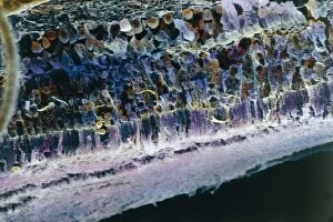
Scanning Electron Micrograph (SEM): Human Retina showing photoreceptors rod and cone cells Date:

Background imageRetina Collection: Picture No. 10876993

Picture No. 10876993
Scanning Electron Micrograph (SEM): Human Retina showing photoreceptors rod and cone cells Date:

Background imageRetina Collection: Picture No. 10876995

Picture No. 10876995
Scanning Electron Micrograph (SEM): Human Retina showing photoreceptors rod and cone cells Date:

Background imageRetina Collection: Picture No. 10876994

Picture No. 10876994
Scanning Electron Micrograph (SEM): Human Retina showing photoreceptors rod and cone cells Date:

Background imageRetina Collection: Descartes explanation of vision, showing light rays being passed through the eye

Descartes explanation of vision, showing light rays being passed through the eye, being focused by lens (I) and forming images T, S, R on retina

Background imageRetina Collection: Visual system, Eye, Retina, Sensory cells under microscope

Visual system, Eye, Retina, Sensory cells under microscope
Medicine - Human Histology - Visual system - Eye - Retina - Sensory cells under microscope

Background imageRetina Collection: Sharks eye with a slit pupil, close up

Sharks eye with a slit pupil, close up

Background imageRetina Collection: Tablet computer with biometric eye scan F006 / 6308

Tablet computer with biometric eye scan F006 / 6308
Tablet computer showing conceptual artwork of a biometric eye scan. Biometrics is the identification of individuals by their body parts

Background imageRetina Collection: Eye anatomy, artwork F007 / 6126

Eye anatomy, artwork F007 / 6126
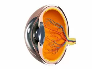
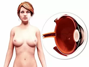
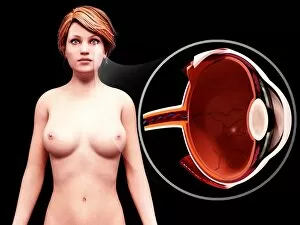
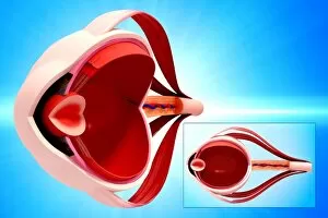
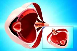
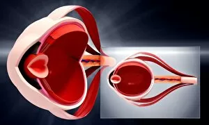
Eye anatomy, computer artwork

Background imageRetina Collection: Eye anatomy, artwork F007 / 6127

Eye anatomy, artwork F007 / 6127
Eye anatomy, computer artwork

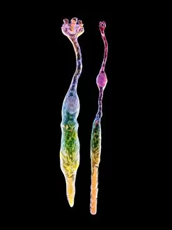
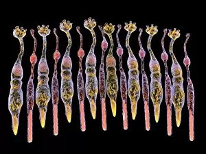
Background imageRetina Collection: Eye retina F008 / 0713

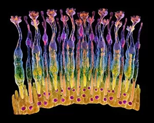
Eye retina F008 / 0713
The retina, inside the eye, contains a light-sensitive membranous layer of cells. These are specialized nerve cells: elongated rods (flower like shapes) and cone-tipped cells, that produce vision
Background imageRetina Collection: Human eye anatomy, artwork F008 / 0234

Human eye anatomy, artwork F008 / 0234
Human eye anatomy, computer artwork

Background imageRetina Collection: Human eye anatomy, artwork F008 / 1075

Human eye anatomy, artwork F008 / 1075
Human eye anatomy, computer artwork

Background imageRetina Collection: Human eye anatomy, artwork F008 / 0233

Human eye anatomy, artwork F008 / 0233
Human eye anatomy, computer artwork

Background imageRetina Collection: Human eye anatomy, artwork F008 / 0240

Human eye anatomy, artwork F008 / 0240
Human eye anatomy, computer artwork

Background imageRetina Collection: Human eye anatomy, artwork F008 / 0231

Human eye anatomy, artwork F008 / 0231
Human eye anatomy, computer artwork

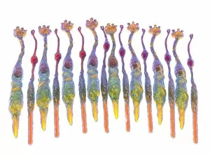
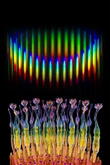
Background imageRetina Collection: Eye retina, rod and cone F008 / 0719

Eye retina, rod and cone F008 / 0719
The retina, inside the eye, contains a light-sensitive membranous layer of cells. These are specialized nerve cells: elongated rods (flower like shape) and cone-tipped cells, that produce vision
Background imageRetina Collection: Human eye anatomy, artwork F008 / 1072

Human eye anatomy, artwork F008 / 1072
Human eye anatomy, computer artwork

Background imageRetina Collection: Human eye anatomy, artwork F008 / 1073

Human eye anatomy, artwork F008 / 1073
Human eye anatomy, computer artwork

Background imageRetina Collection: Human eye anatomy, artwork F008 / 0232

Human eye anatomy, artwork F008 / 0232
Human eye anatomy, computer artwork

Background imageRetina Collection: Eye retina F008 / 0714

Eye retina F008 / 0714
The retina, inside the eye, contains a light-sensitive membranous layer of cells. These are specialized nerve cells: elongated rods (flower like shapes) and cone-tipped cells, that produce vision

Background imageRetina Collection: Eye retina F008 / 0712

Eye retina F008 / 0712
The retina, inside the eye, contains a light-sensitive membranous layer of cells. These are specialized nerve cells: elongated rods (flower like shapes) and cone-tipped cells, that produce vision

Background imageRetina Collection: Eye retina F008 / 0715

Eye retina F008 / 0715
The retina, inside the eye, contains a light-sensitive membranous layer of cells. These are specialized nerve cells: elongated rods (flower like shapes) and cone-tipped cells, that produce vision

Background imageRetina Collection: Human eye anatomy, artwork F008 / 1196

Human eye anatomy, artwork F008 / 1196
Human eye anatomy, computer artwork

Background imageRetina Collection: Human eye anatomy, artwork F008 / 1076

Human eye anatomy, artwork F008 / 1076
Human eye anatomy, computer artwork

Background imageRetina Collection: Human eye anatomy, artwork F008 / 0239

Human eye anatomy, artwork F008 / 0239
Human eye anatomy, computer artwork

Background imageRetina Collection: Human eye anatomy, artwork F008 / 0244

Human eye anatomy, artwork F008 / 0244
Human eye anatomy, computer artwork

Background imageRetina Collection: Human eye anatomy, artwork F008 / 1194

Human eye anatomy, artwork F008 / 1194
Human eye anatomy, computer artwork

Background imageRetina Collection: Human eye anatomy, artwork F008 / 0246

Human eye anatomy, artwork F008 / 0246
Human eye anatomy, computer artwork

Background imageRetina Collection: Human eye anatomy, artwork F008 / 1071

Human eye anatomy, artwork F008 / 1071
Human eye anatomy, computer artwork

Background imageRetina Collection: Human eye anatomy, artwork F008 / 0243

Human eye anatomy, artwork F008 / 0243
Human eye anatomy, computer artwork

Background imageRetina Collection: Human eye anatomy, artwork F008 / 1068

Human eye anatomy, artwork F008 / 1068
Human eye anatomy, computer artwork

Background imageRetina Collection: Human eye anatomy, artwork F008 / 1074

Human eye anatomy, artwork F008 / 1074
Human eye anatomy, computer artwork


For sale as Licensed Images

Choose your image, Select your licence and Download the media

The retina, a fascinating structure within the human eye, has captivated scientists and artists alike throughout history. From histological diagrams to intricate artwork, the intricacies of its anatomy have been meticulously studied and depicted. In the 17th century, Descartes' optics theory shed light on how light interacts with the retina. This groundbreaking understanding paved the way for further exploration into this complex organ. An engraving from 1899 beautifully illustrates the external anatomy of the human eye, showcasing how it works in harmony with the retina. The rod and cone cells of the eye are highlighted in another image taken through a scanning electron microscope (SEM), revealing their delicate structures responsible for vision. Advancements in technology have allowed us to delve deeper into studying retinas through biometric scans. These scans provide valuable insights into individual characteristics and contribute to fields like artificial intelligence. A captivating false-color SEM image showcases a central fovea within a retina—an area crucial for sharp vision—highlighting its intricate details that make it an essential part of our visual system. At a molecular level, rhodopsin protein molecules play a vital role in converting light signals received by retinal cells into electrical impulses sent to our brain—a process critical for sight perception. As we explore further, we discover not only its anatomical features but also its network of blood vessels that supply oxygen and nutrients to sustain this remarkable organ's functionality. From ancient theories to modern scientific discoveries, artful depictions or microscopic examinations—the study of retinas continues to intrigue us as we unravel more about one of nature's most incredible creations: our eyes.