Virions Collection (#5)

Virions, the tiny particles that cause infections, come in various forms and can be seen under a transmission electron microscope (TEM

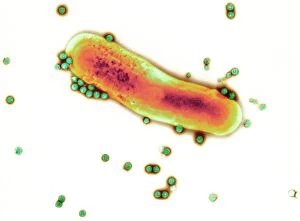
Background imageVirions Collection: Adenovirus particles and bacterium, TEM

Adenovirus particles and bacterium, TEM
Adenovirus particles and bacterium. Coloured transmission electron micrograph (TEM) of adenovirus particles (green) with a gut bacterium (orange)

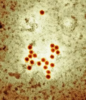
Background imageVirions Collection: Avian flu virus, TEM

Avian flu virus, TEM
Avian flu. Coloured transmission electron micrograph (TEM) of influenza A virus particles (red). This is strain H5N1, which usually infects poultry, but began infecting humans in Hong Kong in 1997

Background imageVirions Collection: Coxsackie virus particles, TEM

Coxsackie virus particles, TEM
Coxsackie virus particles, coloured transmission electron micrograph (TEM). Coxsackie viruses were named after the town in the USA where they were isolated

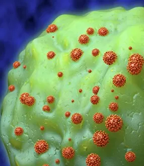
Background imageVirions Collection: Norwalk virus infection

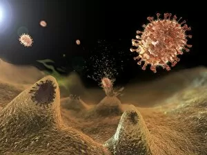
Norwalk virus infection. Computer artwork of a norwalk virus and a human gut. The stomach is at top, the large intestine goes around the outside of the virus

Background imageVirions Collection: Ducks and bird flu virus particles

Ducks and bird flu virus particles
Ducks and avian influenza virus particles, composite image. The virus particles (brown) have been imaged using a transmission electron microscope (TEM)

Background imageVirions Collection: Respiratory syncytial virus, TEM

Respiratory syncytial virus, TEM
Respiratory syncytial virus (RSV) particles, coloured transmission electron micrograph (TEM). This pneumovirus, a type of paramyxovirus

Background imageVirions Collection: Hepatitis A viruses, TEM

Hepatitis A viruses, TEM
Hepatitis A viruses. Coloured transmission electron micrograph (TEM) of hepatitis A viruses, a cause of liver inflammation. Hepatitis A is transmitted through infected food or drink

Background imageVirions Collection: HIV particles exiting a cell

HIV particles exiting a cell. Computer artwork of HIV (human immunodeficiency virus) particles exiting a T-lymphocyte (white blood cell). HIV causes AIDS (acquired immune deficiency syndrome)

Background imageVirions Collection: Astrovirus particles, TEM

Astrovirus particles, TEM
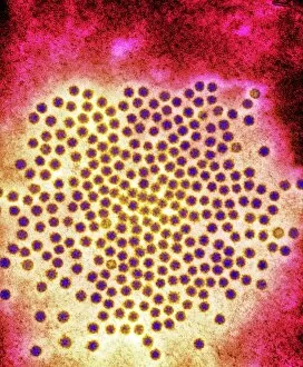
Astrovirus particles. Coloured transmission electron micrograph (TEM) of a cluster of astrovirus particles (purple). These are small viruses, measuring approximately 28 nanometres in diameter

Background imageVirions Collection: Viral throat infection, artwork

Viral throat infection, artwork
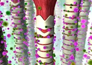
Viral throat infection. Artwork of several tracheae (windpipes) being infected by viruses (spiky balls). The common cold is a viral infection that attacks the throat

Background imageVirions Collection: SARS virus particles, TEM

SARS virus particles, TEM
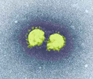
SARS virus particles. Coloured transmission electron micrograph (TEM) of two SARS virus particles (green). Severe acute respiratory syndrome (SARS)

Background imageVirions Collection: AIDS virus particles, computer artwork

AIDS virus particles, computer artwork
HIV particles, computer artwork. HIV (human immunodeficiency virus) causes AIDS (acquired immune deficiency syndrome). The outer layer of the virus particle is a membrane envelope (black)

Background imageVirions Collection: Influenzavirus A, TEM

Influenzavirus A, TEM
Influenzavirus A. Coloured transmission electron micrograph (TEM) of the influenza A virus, which can infect multiple species and is the most virulent human pathogen of the three influenza types

Background imageVirions Collection: Norwalk virus particles

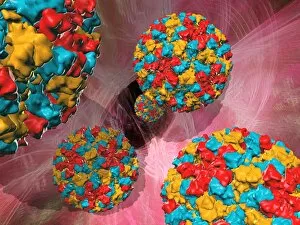
Norwalk virus particles, computer artwork. This small virus consists of an RNA (ribonucleic acid) core surrounded by a protein capsid (multicoloured)

Background imageVirions Collection: Influenzavirus C, TEM

Influenzavirus C, TEM
Influenzavirus C. Coloured transmission electron micrograph (TEM) of the influenza C virus, which is the cause of influenza (flu) in humans and pigs

Background imageVirions Collection: Virus research, artwork

Virus research, artwork
Virus research. Conceptual computer artwork representing research into viruses, showing a virus particle (lower left) and a computer screen displaying data on the virus

Background imageVirions Collection: Shingles

Shingles. Artwork of varicella-zoster virus particles (virions, blue) during shingles. Each viron comprises an icosahedral (20-sided) capsid containing viral DNA

Background imageVirions Collection: Vaccinia virus particles, SEM

Vaccinia virus particles, SEM
Vaccinia virus particles. Coloured scanning electron micrograph (SEM) of vaccinia virus particles (red) budding from a cell (blue and green) after replicating

Background imageVirions Collection: Insect viruses, TEM

Insect viruses, TEM
Insect viruses. Coloured transmission electron micrograph (TEM) of unidentified isometric insect viruses. These viruses most commonly infect their insect hosts when they feed. Magnification unknown

Background imageVirions Collection: HIV particles, computer artwork

HIV particles, computer artwork. HIV (human immunodeficiency virus) causes AIDS (acquired immune deficiency syndrome). The virus consists of an RNA genome

Background imageVirions Collection: Moth virus

Moth virus

Background imageVirions Collection: Rotavirus particle, artwork and TEM

Rotavirus particle, artwork and TEM
Rotavirus particle. Computer artwork (left) and coloured transmission electron micrograph (TEM) of the geometric structure of a rotavirus particles capsid shell

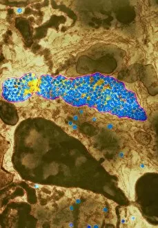
Background imageVirions Collection: Porcine endogenous retrovirus, TEM

Porcine endogenous retrovirus, TEM

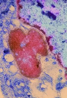
Background imageVirions Collection: Ebola virus replication, TEM

Ebola virus replication, TEM

Background imageVirions Collection: Dengue fever virus particles, TEM

Dengue fever virus particles, TEM

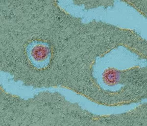
Background imageVirions Collection: Dengue fever virus replication, TEM

Dengue fever virus replication, TEM

Background imageVirions Collection: Mosaic plant viruses, TEM

Mosaic plant viruses, TEM
Turnip yellow mosaic viruses. Coloured transmission electron micrograph (TEM) of turnip yellow mosaic viruses (TYMV). This virus infects crucifer plants such as turnip, swede and cauliflower

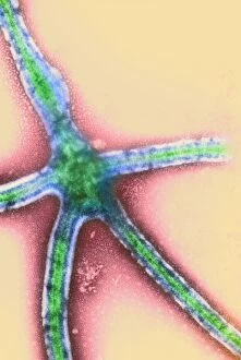
Background imageVirions Collection: Potato X virus, TEM

Potato X virus, TEM
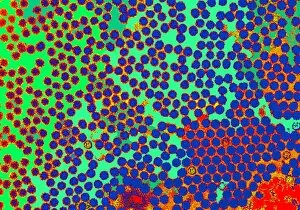
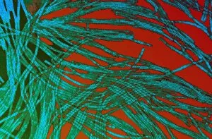
Potato X virus. Coloured transmission electron micrograph (TEM) of particles (green/blue) of the potato X virus. Potato X is a member of the potexvirus group that infects potato plants

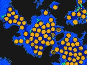
Background imageVirions Collection: Hepatitis A virus particles, TEM

Hepatitis A virus particles, TEM
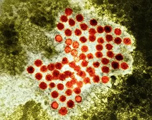
Hepatitis A virus. Coloured transmission electron micrograph (TEM) of a cluster of Hepatitis A virus particles, the cause of infectious hepatitis

Background imageVirions Collection: Influenzavirus B, TEM

Influenzavirus B, TEM
Influenzavirus B. Coloured transmission electron micrograph (TEM) of particles of the influenza B virus, which is the cause of influenza (flu) in humans

Background imageVirions Collection: Flu viruses

Flu viruses. Influenza viruses (brown) budding from a host cell. Each virus consists of a core of RNA (ribonucleic acid) genetic material surrounded by a protein coat (brown)


For sale as Licensed Images

Choose your image, Select your licence and Download the media

Virions, the tiny particles that cause infections, come in various forms and can be seen under a transmission electron microscope (TEM). Norovirus particles, responsible for stomach bugs often spread through sneezing or contaminated surfaces, are captured in artwork C013 / 5949. Similarly, coronavirus particles can also be observed using TEM and have recently gained attention due to their role in the ongoing pandemic. Influenza virus particles, another common cause of respiratory illnesses, appear distinct under TEM as well. Hepatitis C viruses and Rift Valley fever virus are other examples that have been visualized using this powerful microscopy technique. Herpes virus particles are beautifully depicted through computer artwork, showcasing their intricate structure. Paramyxovirus particles and herpes simplex viruses are two more types that have been studied extensively with TEM. The images reveal their unique shapes and features. One striking image shows AIDS viruses budding from a cell under TEM observation. This highlights the devastating impact of these particular virions on human health. Lastly, dengue fever virus particles can also be observed using TEM technology. These tiny entities play a significant role in spreading dengue fever through mosquito bites.